 Movie
Movie Controller

[English] 日本語
 Yorodumi
- EMDB-2930: Cryo-electron microscopy map of La Crosse orthobunyavirus polymer... -
+ Open data

- Basic information
| Entry | Database: EMDB / ID: EMD-2930 | |||||||||
|---|---|---|---|---|---|---|---|---|---|---|
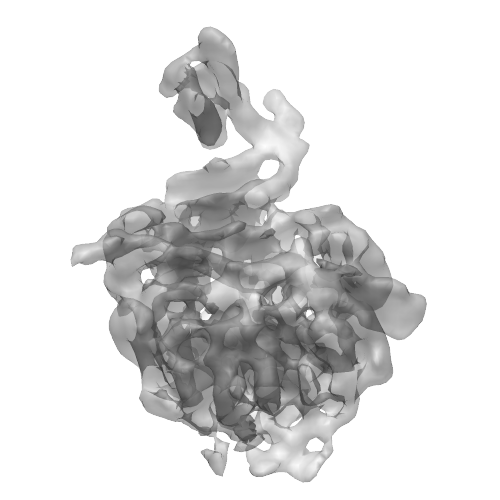
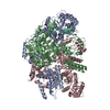
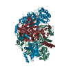
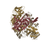
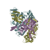
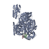
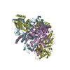
| Title | Cryo-electron microscopy map of La Crosse orthobunyavirus polymerase (apo form, residues 1-1750) | |||||||||
 Map data Map data | Cryo-electron microscopy map of La Crosse orthobunyavirus polymerase (apo form, residues 1-1750) | |||||||||
 Sample Sample |
| |||||||||
 Keywords Keywords | RNA dependent RNA polymerase / segmented negative stranded RNA virus / replication / transcription | |||||||||
| Function / homology |  Function and homology information Function and homology informationhost cell endoplasmic reticulum / virion component / host cell endoplasmic reticulum-Golgi intermediate compartment / Hydrolases; Acting on ester bonds / host cell Golgi apparatus / RNA-directed RNA polymerase / viral RNA genome replication / nucleotide binding / hydrolase activity / RNA-directed RNA polymerase activity ...host cell endoplasmic reticulum / virion component / host cell endoplasmic reticulum-Golgi intermediate compartment / Hydrolases; Acting on ester bonds / host cell Golgi apparatus / RNA-directed RNA polymerase / viral RNA genome replication / nucleotide binding / hydrolase activity / RNA-directed RNA polymerase activity / DNA-templated transcription / RNA binding / metal ion binding Similarity search - Function | |||||||||
| Biological species |  La Crosse virus La Crosse virus | |||||||||
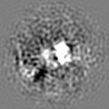
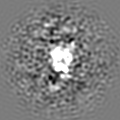
| Method | single particle reconstruction / cryo EM / Resolution: 8.3 Å | |||||||||
 Authors Authors | Gerlach P / Malet H / Cusack S / Reguera J | |||||||||
 Citation Citation | Journal: Cell / Year: 2015 Title: Structural Insights into Bunyavirus Replication and Its Regulation by the vRNA Promoter. Authors: Piotr Gerlach / Hélène Malet / Stephen Cusack / Juan Reguera /  Abstract: Segmented negative-strand RNA virus (sNSV) polymerases transcribe and replicate the viral RNA (vRNA) within a ribonucleoprotein particle (RNP). We present cryo-EM and X-ray structures of, ...Segmented negative-strand RNA virus (sNSV) polymerases transcribe and replicate the viral RNA (vRNA) within a ribonucleoprotein particle (RNP). We present cryo-EM and X-ray structures of, respectively, apo- and vRNA bound La Crosse orthobunyavirus (LACV) polymerase that give atomic-resolution insight into how such RNPs perform RNA synthesis. The complementary 3' and 5' vRNA extremities are sequence specifically bound in separate sites on the polymerase. The 5' end binds as a stem-loop, allosterically structuring functionally important polymerase active site loops. Identification of distinct template and product exit tunnels allows proposal of a detailed model for template-directed replication with minimal disruption to the circularised RNP. The similar overall architecture and vRNA binding of monomeric LACV to heterotrimeric influenza polymerase, despite high sequence divergence, suggests that all sNSV polymerases have a common evolutionary origin and mechanism of RNA synthesis. These results will aid development of replication inhibitors of diverse, serious human pathogenic viruses. | |||||||||
| History |
|
- Structure visualization
| Movie |
Movie viewer |
|---|---|
| Structure viewer | EM map: SurfViewMolmilJmol/JSmol |
| Supplemental images |
- Downloads & links
-EMDB archive
| Map data | emd_2930.map.gz | 6.2 MB | EMDB map data format | |
|---|---|---|---|---|
| Header (meta data) | emd-2930-v30.xmlemd-2930.xml | 10.1 KB 10.1 KB | Display Display | EMDB header |
| Images |  emd_2930.png emd_2930.png | 115.2 KB | ||
| Archive directory |  http://ftp.pdbj.org/pub/emdb/structures/EMD-2930ftp://ftp.pdbj.org/pub/emdb/structures/EMD-2930 http://ftp.pdbj.org/pub/emdb/structures/EMD-2930ftp://ftp.pdbj.org/pub/emdb/structures/EMD-2930 | HTTPS FTP |
-Related structure data

-Links
| EMDB pages | EMDB (EBI/PDBe) / EMDataResource |
|---|
-Map
| File | Download / File: emd_2930.map.gz / Format: CCP4 / Size: 6.4 MB / Type: IMAGE STORED AS FLOATING POINT NUMBER (4 BYTES) | ||||||||||||||||||||||||||||||||||||||||||||||||||||||||||||||||||||
|---|---|---|---|---|---|---|---|---|---|---|---|---|---|---|---|---|---|---|---|---|---|---|---|---|---|---|---|---|---|---|---|---|---|---|---|---|---|---|---|---|---|---|---|---|---|---|---|---|---|---|---|---|---|---|---|---|---|---|---|---|---|---|---|---|---|---|---|---|---|
| Annotation | Cryo-electron microscopy map of La Crosse orthobunyavirus polymerase (apo form, residues 1-1750) | ||||||||||||||||||||||||||||||||||||||||||||||||||||||||||||||||||||
| Projections & slices | Image control
Images are generated by Spider. | ||||||||||||||||||||||||||||||||||||||||||||||||||||||||||||||||||||
| Voxel size | X=Y=Z: 2.02 Å | ||||||||||||||||||||||||||||||||||||||||||||||||||||||||||||||||||||
| Density |
| ||||||||||||||||||||||||||||||||||||||||||||||||||||||||||||||||||||
| Symmetry | Space group: 1 | ||||||||||||||||||||||||||||||||||||||||||||||||||||||||||||||||||||
| Details | EMDB XML:
CCP4 map header:
| ||||||||||||||||||||||||||||||||||||||||||||||||||||||||||||||||||||
 Z (Sec.)
 Y (Row.)
 X (Col.)


-Supplemental data
- Sample components
-Entire : La Crosse orthobunyavirus polymerase (residues 1-1750, apo form)
| Entire | Name: La Crosse orthobunyavirus polymerase (residues 1-1750, apo form) |
|---|---|
| Components |
|
-Supramolecule #1000: La Crosse orthobunyavirus polymerase (residues 1-1750, apo form)
| Supramolecule | Name: La Crosse orthobunyavirus polymerase (residues 1-1750, apo form) type: sample / ID: 1000 / Number unique components: 1 |
|---|---|
| Molecular weight | Experimental: 203.5 KDa / Theoretical: 203.5 KDa |
-Macromolecule #1: La Crosse RNA-dependent RNA polymerase
| Macromolecule | Name: La Crosse RNA-dependent RNA polymerase / type: protein_or_peptide / ID: 1 / Number of copies: 1 / Oligomeric state: monomer / Recombinant expression: Yes |
|---|---|
| Source (natural) | Organism: La Crosse virus / Strain: LACV/mosquito/1978 / Location in cell: cytoplasm |
| Molecular weight | Experimental: 20.35 KDa / Theoretical: 20.35 KDa |
| Recombinant expression | Organism:  Trichoplusia ni (cabbage looper) / Recombinant cell: Hi5 / Recombinant plasmid: pFastBac Trichoplusia ni (cabbage looper) / Recombinant cell: Hi5 / Recombinant plasmid: pFastBac |
| Sequence | UniProtKB: RNA-directed RNA polymerase L / GO: RNA-directed RNA polymerase activity / InterPro: RNA-dependent RNA polymerase, bunyaviral |
-Experimental details
-Structure determination
| Method | cryo EM |
|---|---|
 Processing Processing | single particle reconstruction |
| Aggregation state | particle |
-Sample preparation
| Concentration | 0.2 mg/mL |
|---|---|
| Buffer | pH: 8 / Details: 20 mM Tris-HCl, 150 mM NaCl, 2 mM TCEP, pH 8 |
| Grid | Details: Quantifoil grid 400 mesh 2/1 |
| Vitrification | Cryogen name: ETHANE / Chamber humidity: 100 % / Chamber temperature: 120 K / Instrument: FEI VITROBOT MARK III / Method: Blot for 2s before plunging |
- Electron microscopy
| Microscope | FEI TITAN KRIOS |
|---|---|
| Temperature | Average: 100 K |
| Details | Binning factor 2 used for data collection |
| Date | Mar 11, 2013 |
| Image recording | Category: CCD / Film or detector model: FEI FALCON II (4k x 4k) / Digitization - Sampling interval: 28 µm / Number real images: 6129 / Average electron dose: 14 e/Å2 |
| Tilt angle min | 0 |
| Tilt angle max | 0 |
| Electron beam | Acceleration voltage: 80 kV / Electron source:  FIELD EMISSION GUN FIELD EMISSION GUN |
| Electron optics | Calibrated magnification: 138129 / Illumination mode: FLOOD BEAM / Imaging mode: BRIGHT FIELD / Cs: 2.7 mm / Nominal defocus max: 2.0 µm / Nominal defocus min: 0.5 µm / Nominal magnification: 138129 |
| Sample stage | Specimen holder model: FEI TITAN KRIOS AUTOGRID HOLDER |
| Experimental equipment |  Model: Titan Krios / Image courtesy: FEI Company |
-Image processing
| Details | CTF correction using CTFFIND3 Manual picking in EMAN boxer 2D classification in IMAGIC5 and RELION1.3 Automated picking using the fast projection matching algorithm Refinement and 3D classification in RELION1.3 |
|---|---|
| Final reconstruction | Applied symmetry - Point group: C1 (asymmetric) / Algorithm: OTHER / Resolution.type: BY AUTHOR / Resolution: 8.3 Å / Resolution method: OTHER / Software - Name: RELION1.3 / Number images used: 178983 |
-Atomic model buiding 1
| Initial model | PDB ID: Chain - Chain ID: A |
|---|---|
| Software | Name: Chimera |
| Details | Two rigid bodies were used: 1-184 185-1745 |
| Refinement | Space: REAL / Protocol: RIGID BODY FIT |
