 Movie
Movie Controller

[English] 日本語
 Yorodumi
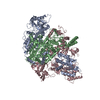
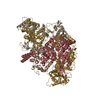
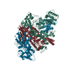
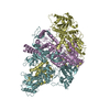
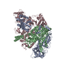
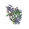
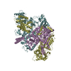
- PDB-5amr: Structure of the La Crosse Bunyavirus polymerase in complex with ... -
+ Open data

- Basic information
| Entry | Database: PDB / ID: 5amr | ||||||
|---|---|---|---|---|---|---|---|
| Title | Structure of the La Crosse Bunyavirus polymerase in complex with the 3' viral RNA | ||||||
 Components Components |
| ||||||
 Keywords Keywords | HYDROLASE / POLYMERASE / RNADRNAPOL / BUNYAVIRUS / RNA | ||||||
| Function / homology |  Function and homology information Function and homology informationhost cell endoplasmic reticulum / virion component / host cell endoplasmic reticulum-Golgi intermediate compartment / Hydrolases; Acting on ester bonds / host cell Golgi apparatus / RNA-directed RNA polymerase / viral RNA genome replication / nucleotide binding / hydrolase activity / RNA-directed RNA polymerase activity ...host cell endoplasmic reticulum / virion component / host cell endoplasmic reticulum-Golgi intermediate compartment / Hydrolases; Acting on ester bonds / host cell Golgi apparatus / RNA-directed RNA polymerase / viral RNA genome replication / nucleotide binding / hydrolase activity / RNA-directed RNA polymerase activity / DNA-templated transcription / RNA binding / metal ion binding Similarity search - Function | ||||||
| Biological species |  BUNYAVIRUS LA CROSSE BUNYAVIRUS LA CROSSE | ||||||
| Method |  X-RAY DIFFRACTION / SYNCHROTRON / MIRAS / Resolution: 2.57 Å X-RAY DIFFRACTION / SYNCHROTRON / MIRAS / Resolution: 2.57 Å | ||||||
 Authors Authors | Reguera, J. / Gerlach, P. / Cusack, S. | ||||||
 Citation Citation | Journal: Cell / Year: 2015 Title: Structural Insights into Bunyavirus Replication and Its Regulation by the vRNA Promoter. Authors: Piotr Gerlach / Hélène Malet / Stephen Cusack / Juan Reguera /  Abstract: Segmented negative-strand RNA virus (sNSV) polymerases transcribe and replicate the viral RNA (vRNA) within a ribonucleoprotein particle (RNP). We present cryo-EM and X-ray structures of, ...Segmented negative-strand RNA virus (sNSV) polymerases transcribe and replicate the viral RNA (vRNA) within a ribonucleoprotein particle (RNP). We present cryo-EM and X-ray structures of, respectively, apo- and vRNA bound La Crosse orthobunyavirus (LACV) polymerase that give atomic-resolution insight into how such RNPs perform RNA synthesis. The complementary 3' and 5' vRNA extremities are sequence specifically bound in separate sites on the polymerase. The 5' end binds as a stem-loop, allosterically structuring functionally important polymerase active site loops. Identification of distinct template and product exit tunnels allows proposal of a detailed model for template-directed replication with minimal disruption to the circularised RNP. The similar overall architecture and vRNA binding of monomeric LACV to heterotrimeric influenza polymerase, despite high sequence divergence, suggests that all sNSV polymerases have a common evolutionary origin and mechanism of RNA synthesis. These results will aid development of replication inhibitors of diverse, serious human pathogenic viruses. | ||||||
| History |
|
- Structure visualization
| Structure viewer | Molecule: MolmilJmol/JSmol |
|---|
- Downloads & links
-Download
| PDBx/mmCIF format | 5amr.cif.gz | 377.3 KB | Display | PDBx/mmCIF format |
|---|---|---|---|---|
| PDB format | pdb5amr.ent.gz | 291.9 KB | Display | PDB format |
| PDBx/mmJSON format | 5amr.json.gz | Tree view | PDBx/mmJSON format | |
| Others |  Other downloads Other downloads |
-Validation report
| Arichive directory | https://data.pdbj.org/pub/pdb/validation_reports/am/5amrftp://data.pdbj.org/pub/pdb/validation_reports/am/5amr | HTTPS FTP |
|---|
-Related structure data


-Links
 PDBj
PDBj


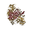
- Assembly
| Deposited unit | 
| ||||||||
|---|---|---|---|---|---|---|---|---|---|
| 1 |
| ||||||||
| Unit cell |
|
-Components
| #1: Protein | Mass: 263429.594 Da / Num. of mol.: 1 Source method: isolated from a genetically manipulated source Source: (gene. exp.) BUNYAVIRUS LA CROSSE / Plasmid: PFASTBAC / Cell line (production host): High Five / Production host:  TRICHOPLUSIA NI (cabbage looper) / References: UniProt: A5HC98, RNA-directed RNA polymerase TRICHOPLUSIA NI (cabbage looper) / References: UniProt: A5HC98, RNA-directed RNA polymerase |
|---|---|
| #2: RNA chain | Mass: 5060.023 Da / Num. of mol.: 1 Source method: isolated from a genetically manipulated source Source: (gene. exp.) BUNYAVIRUS LA CROSSE / Plasmid: PFASTBAC / Cell line (production host): High Five / Production host: TRICHOPLUSIA NI (cabbage looper) |
| #3: RNA chain | Mass: 2509.577 Da / Num. of mol.: 1 Source method: isolated from a genetically manipulated source Source: (gene. exp.) BUNYAVIRUS LA CROSSE / Plasmid: PFASTBAC / Cell line (production host): High Five / Production host: TRICHOPLUSIA NI (cabbage looper) |
| #4: Water | ChemComp-HOH /  Mass: 18.015 Da / Num. of mol.: 92 / Source method: isolated from a natural source / Formula: H2O Mass: 18.015 Da / Num. of mol.: 92 / Source method: isolated from a natural source / Formula: H2O |
| Sequence details | MDYQEYQQFL |
-Experimental details
-Experiment
| Experiment | Method: X-RAY DIFFRACTION / Number of used crystals: 1 |
|---|
- Sample preparation
| Crystal | Density Matthews: 2.9 Å3/Da / Density % sol: 62 % / Description: NONE |
|---|---|
| Crystal grow | pH: 7 Details: 0.1M HEPES PH 7, 0.2M SODIUM CITRATE DIHYDRATE, 0.3M AMMONIUM SULPHATE, 15% PEG 3350. |
-Data collection
| Diffraction | Mean temperature: 77 K |
|---|---|
| Diffraction source | Source: SYNCHROTRON / Site: ESRF / Beamline: ID23-1 / Wavelength: 0.99187 |
| Detector | Type: DECTRIS PILATUS 6M / Detector: PIXEL / Date: Jul 27, 2014 |
| Radiation | Protocol: SINGLE WAVELENGTH / Monochromatic (M) / Laue (L): M / Scattering type: x-ray |
| Radiation wavelength | Wavelength: 0.99187 Å / Relative weight: 1 |
| Reflection | Resolution: 2.57→50 Å / Num. obs: 71095 / % possible obs: 94.7 % / Observed criterion σ(I): 3 / Redundancy: 3.55 % / Rmerge(I) obs: 0.01 / Net I/σ(I): 11.2 |
| Reflection shell | Resolution: 2.57→2.6 Å / Redundancy: 3.56 % / Rmerge(I) obs: 0.09 / Mean I/σ(I) obs: 1.28 / % possible all: 97.4 |
- Processing
| Software |
| |||||||||||||||||||||||||||||||||||||||||||||||||||||||||||||||||||||||||||||||||||||||||||||||||||||||||||||||||||||||||||||||||||||||||||||||||||||||||||||||||||||||||||||||||||||||||||||||||||||||||||||||||||||||||
|---|---|---|---|---|---|---|---|---|---|---|---|---|---|---|---|---|---|---|---|---|---|---|---|---|---|---|---|---|---|---|---|---|---|---|---|---|---|---|---|---|---|---|---|---|---|---|---|---|---|---|---|---|---|---|---|---|---|---|---|---|---|---|---|---|---|---|---|---|---|---|---|---|---|---|---|---|---|---|---|---|---|---|---|---|---|---|---|---|---|---|---|---|---|---|---|---|---|---|---|---|---|---|---|---|---|---|---|---|---|---|---|---|---|---|---|---|---|---|---|---|---|---|---|---|---|---|---|---|---|---|---|---|---|---|---|---|---|---|---|---|---|---|---|---|---|---|---|---|---|---|---|---|---|---|---|---|---|---|---|---|---|---|---|---|---|---|---|---|---|---|---|---|---|---|---|---|---|---|---|---|---|---|---|---|---|---|---|---|---|---|---|---|---|---|---|---|---|---|---|---|---|---|---|---|---|---|---|---|---|---|---|---|---|---|---|---|---|---|
| Refinement | Method to determine structure: MIRAS Starting model: NONE Resolution: 2.57→48.657 Å / SU ML: 0.43 / σ(F): 1.35 / Phase error: 28.76 / Stereochemistry target values: ML
| |||||||||||||||||||||||||||||||||||||||||||||||||||||||||||||||||||||||||||||||||||||||||||||||||||||||||||||||||||||||||||||||||||||||||||||||||||||||||||||||||||||||||||||||||||||||||||||||||||||||||||||||||||||||||
| Solvent computation | Shrinkage radii: 0.9 Å / VDW probe radii: 1.11 Å / Solvent model: FLAT BULK SOLVENT MODEL | |||||||||||||||||||||||||||||||||||||||||||||||||||||||||||||||||||||||||||||||||||||||||||||||||||||||||||||||||||||||||||||||||||||||||||||||||||||||||||||||||||||||||||||||||||||||||||||||||||||||||||||||||||||||||
| Refinement step | Cycle: LAST / Resolution: 2.57→48.657 Å
| |||||||||||||||||||||||||||||||||||||||||||||||||||||||||||||||||||||||||||||||||||||||||||||||||||||||||||||||||||||||||||||||||||||||||||||||||||||||||||||||||||||||||||||||||||||||||||||||||||||||||||||||||||||||||
| Refine LS restraints |
| |||||||||||||||||||||||||||||||||||||||||||||||||||||||||||||||||||||||||||||||||||||||||||||||||||||||||||||||||||||||||||||||||||||||||||||||||||||||||||||||||||||||||||||||||||||||||||||||||||||||||||||||||||||||||
| LS refinement shell |
|
