National Institutes of Health/National Heart, Lung, and Blood Institute (NIH/NHLBI)
HL049413
United States
National Institutes of Health/National Heart, Lung, and Blood Institute (NIH/NHLBI)
HL139554
United States
National Institutes of Health/National Heart, Lung, and Blood Institute (NIH/NHLBI)
HL147821
United States
Citation
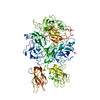
Journal: Blood / Year: 2023 Title: Cryo-EM structure of coagulation factor V short. Authors: Bassem M Mohammed / Leslie A Pelc / Michael J Rau / Enrico Di Cera / Abstract: Coagulation factor V (fV) is the precursor of activated fV (fVa), an essential component of the prothrombinase complex required for the rapid activation of prothrombin in the penultimate step of the ...Coagulation factor V (fV) is the precursor of activated fV (fVa), an essential component of the prothrombinase complex required for the rapid activation of prothrombin in the penultimate step of the coagulation cascade. In addition, fV regulates the tissue factor pathway inhibitor α (TFPIα) and protein C pathways that inhibit the coagulation response. A recent cryogenic electron microscopy (cryo-EM) structure of fV has revealed the architecture of its A1-A2-B-A3-C1-C2 assembly but left the mechanism that keeps fV in its inactive state unresolved because of an intrinsic disorder in the B domain. A splice variant of fV, fV short, carries a large deletion of the B domain that produces constitutive fVa-like activity and unmasks epitopes for the binding of TFPIα. The cryo-EM structure of fV short was solved at 3.2 Å resolution and revealed the arrangement of the entire A1-A2-B-A3-C1-C2 assembly. The shorter B domain stretches across the entire width of the protein, making contacts with the A1, A2, and A3 domains but suspended over the C1 and C2 domains. In the portion distal to the splice site, several hydrophobic clusters and acidic residues provide a potential binding site for the basic C-terminal end of TFPIα. In fV, these epitopes may bind intramolecularly to the basic region of the B domain. The cryo-EM structure reported in this study advances our understanding of the mechanism that keeps fV in its inactive state, provides new targets for mutagenesis and facilitates future structural analysis of fV short in complex with TFPIα, protein S, and fXa.
Entire : Recombinantly expressed coagulation factor V short full length wi...
Entire
Name: Recombinantly expressed coagulation factor V short full length with a c-terminus HPC4 tag
Components
Tissue: Recombinantly expressed coagulation factor V short full length with a c-terminus HPC4 tag
Protein or peptide: Coagulation factor V
-
Supramolecule #1: Recombinantly expressed coagulation factor V short full length wi...
Supramolecule
Name: Recombinantly expressed coagulation factor V short full length with a c-terminus HPC4 tag type: tissue / ID: 1 / Parent: 0 / Macromolecule list: all
Source (natural)
Organism: Homo sapiens (human) / Organ: Liver / Tissue: Blood
-
Macromolecule #1: Coagulation factor V
Macromolecule
Name: Coagulation factor V / type: protein_or_peptide / ID: 1 / Number of copies: 1 / Enantiomer: LEVO
Source (natural)
Organism: Homo sapiens (human) / Organ: Liver / Tissue: Blood
In the structure databanks used in Yorodumi, some data are registered as the other names, "COVID-19 virus" and "2019-nCoV". Here are the details of the virus and the list of structure data.
Jan 31, 2019. EMDB accession codes are about to change! (news from PDBe EMDB page)
EMDB accession codes are about to change! (news from PDBe EMDB page)
The allocation of 4 digits for EMDB accession codes will soon come to an end. Whilst these codes will remain in use, new EMDB accession codes will include an additional digit and will expand incrementally as the available range of codes is exhausted. The current 4-digit format prefixed with “EMD-” (i.e. EMD-XXXX) will advance to a 5-digit format (i.e. EMD-XXXXX), and so on. It is currently estimated that the 4-digit codes will be depleted around Spring 2019, at which point the 5-digit format will come into force.
The EM Navigator/Yorodumi systems omit the EMD- prefix.
Related info.:Q: What is EMD? / ID/Accession-code notation in Yorodumi/EM Navigator
Yorodumi is a browser for structure data from EMDB, PDB, SASBDB, etc.
This page is also the successor to EM Navigator detail page, and also detail information page/front-end page for Omokage search.
The word "yorodu" (or yorozu) is an old Japanese word meaning "ten thousand". "mi" (miru) is to see.
Related info.:EMDB / PDB / SASBDB / Comparison of 3 databanks / Yorodumi Search / Aug 31, 2016. New EM Navigator & Yorodumi / Yorodumi Papers / Jmol/JSmol / Function and homology information / Changes in new EM Navigator and Yorodumi
 Movie
Movie Controller


 Open data
 Basic information
 Map data
 Sample
 Keywords
 Function and homology information
 Homo sapiens (human)
 Authors
 United States, 3 items
  Citation
 Structure visualization
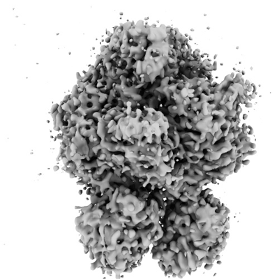
 Downloads & links
 emd_29011.png
 http://ftp.pdbj.org/pub/emdb/structures/EMD-29011
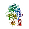


 X (Sec.)
 Y (Row.)
 Z (Col.)


 Sample components
 Processing
 Electron microscopy
 FIELD EMISSION GUN