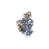
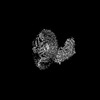
- EMDB-28376: Structure of a human EMC:human Cav1.2 channel complex in GDN dete... -
+
データを開く
IDまたはキーワード:
読み込み中...
-
基本情報
登録情報
データベース: EMDB / ID: EMD-28376
タイトル
Structure of a human EMC:human Cav1.2 channel complex in GDN detergent (ECAB Map 3)
マップデータ
Structure of a human EMC:human Cav1.2 channel complex in GDN detergent (ECAB Map 3)
試料
複合体: Ternary complex of the human ER membrane protein complex (EMC) with human CaV alpha1C and rabbit CaV beta3
複合体: Human CaV alpha1C
タンパク質・ペプチド: x 1種
複合体: ER membrane protein complex subunit 1
タンパク質・ペプチド: x 1種
複合体: Rabbit CaV beta3
タンパク質・ペプチド: x 1種
タンパク質・ペプチド: x 8種
リガンド: x 2種
キーワード
endoplasmic reticulum membrane protein complex / voltage-gated calcium channel / holdase / biogenesis / MEMBRANE PROTEIN
機能・相同性
機能・相同性情報
extrinsic component of endoplasmic reticulum membrane / : / EMC complex / positive regulation of high voltage-gated calcium channel activity / omegasome membrane / protein insertion into ER membrane by stop-transfer membrane-anchor sequence / voltage-gated calcium channel activity involved in AV node cell action potential / voltage-gated calcium channel activity involved in cardiac muscle cell action potential / immune system development / magnesium ion transport ...extrinsic component of endoplasmic reticulum membrane / : / EMC complex / positive regulation of high voltage-gated calcium channel activity / omegasome membrane / protein insertion into ER membrane by stop-transfer membrane-anchor sequence / voltage-gated calcium channel activity involved in AV node cell action potential / voltage-gated calcium channel activity involved in cardiac muscle cell action potential / immune system development / magnesium ion transport / positive regulation of adenylate cyclase activity / membrane depolarization during atrial cardiac muscle cell action potential / calcium ion transmembrane transport via high voltage-gated calcium channel / tail-anchored membrane protein insertion into ER membrane / Miscellaneous transport and binding events / Phase 2 - plateau phase / cobalt ion transmembrane transporter activity / ferrous iron transmembrane transporter activity / membrane depolarization during AV node cell action potential / high voltage-gated calcium channel activity / copper ion transport / cardiac conduction / L-type voltage-gated calcium channel complex / magnesium ion transmembrane transporter activity / membrane depolarization during cardiac muscle cell action potential / positive regulation of muscle contraction / cell communication by electrical coupling involved in cardiac conduction / regulation of ventricular cardiac muscle cell action potential / NCAM1 interactions / camera-type eye development / cardiac muscle cell action potential involved in contraction / embryonic forelimb morphogenesis / calcium ion transport into cytosol / voltage-gated calcium channel complex / Phase 0 - rapid depolarisation / alpha-actinin binding / regulation of heart rate by cardiac conduction / calcium ion import across plasma membrane / RHOA GTPase cycle / autophagosome assembly / regulation of cardiac muscle contraction by regulation of the release of sequestered calcium ion / voltage-gated calcium channel activity / positive regulation of endothelial cell proliferation / positive regulation of endothelial cell migration / calcium channel regulator activity / Regulation of insulin secretion / postsynaptic density membrane / calcium ion transmembrane transport / Z disc / positive regulation of angiogenesis / Adrenaline,noradrenaline inhibits insulin secretion / heart development / positive regulation of cytosolic calcium ion concentration / carbohydrate binding / early endosome membrane / angiogenesis / perikaryon / calmodulin binding / early endosome / postsynaptic density / cilium / Golgi membrane / apoptotic process / dendrite / endoplasmic reticulum membrane / endoplasmic reticulum / Golgi apparatus / protein-containing complex / extracellular region / nucleoplasm / metal ion binding / membrane / plasma membrane / cytoplasm 類似検索 - 分子機能
EMC1 N-terminal beta-propeller domain / ER membrane protein complex subunit 8/9 / : / Uncharacterised protein family (UPF0172) / EMC2 TPR-like repeat domain / TMEM85/ER membrane protein complex subunit 4 / ER membrane protein complex subunit 4 / ER membrane protein complex subunit 7, beta-sandwich domain / ER membrane protein complex subunit 7 / ER membrane protein complex subunit 7, beta-sandwich domain ...EMC1 N-terminal beta-propeller domain / ER membrane protein complex subunit 8/9 / : / Uncharacterised protein family (UPF0172) / EMC2 TPR-like repeat domain / TMEM85/ER membrane protein complex subunit 4 / ER membrane protein complex subunit 4 / ER membrane protein complex subunit 7, beta-sandwich domain / ER membrane protein complex subunit 7 / ER membrane protein complex subunit 7, beta-sandwich domain / ER membrane protein complex subunit 6 / ER membrane protein complex subunit 3 / ER membrane protein complex subunit 1, C-terminal / Membrane magnesium transporter / ER membrane protein complex subunit 1 / ER membrane protein complex subunit 6-like / EMC6 / ER membrane protein complex subunit 1, C-terminal / Membrane magnesium transporter / ER membrane protein complex subunit 10 / ER membrane protein complex subunit 2-like / Integral membrane protein EMC3/TMCO1-like / Integral membrane protein EMC3/TMCO1-like / Integral membrane protein DUF106 / Voltage-dependent calcium channel, L-type, beta-3 subunit / Voltage-dependent L-type calcium channel subunit beta-3, SH3 domain / Voltage-dependent calcium channel, L-type, alpha-1C subunit / Voltage-dependent calcium channel, L-type, beta subunit / Voltage-dependent L-type calcium channel subunit beta-1-4, N-terminal A domain / Voltage gated calcium channel subunit beta domain 4Aa N terminal / Voltage-gated calcium channel subunit alpha, C-terminal / Voltage-gated calcium channel subunit alpha, C-term / Voltage-dependent calcium channel, L-type, alpha-1 subunit / Voltage-dependent calcium channel, alpha-1 subunit, IQ domain / Voltage gated calcium channel IQ domain / Voltage gated calcium channel IQ domain / Voltage-dependent calcium channel, alpha-1 subunit / Carbohydrate-binding-like fold / Voltage-dependent L-type calcium channel, IQ-associated domain / Voltage-dependent L-type calcium channel, IQ-associated / : / Guanylate kinase/L-type calcium channel beta subunit / Guanylate kinase / Guanylate kinase homologues. / Quinoprotein alcohol dehydrogenase-like superfamily / Voltage-dependent channel domain superfamily / TPR repeat region circular profile. / MPN domain / MPN domain profile. / TPR repeat profile. / Tetratricopeptide repeats / Tetratricopeptide repeat / SH3-like domain superfamily / Src homology 3 (SH3) domain profile. / SH3 domain / Ion transport domain / Ion transport protein / Tetratricopeptide-like helical domain superfamily / WD40/YVTN repeat-like-containing domain superfamily / P-loop containing nucleoside triphosphate hydrolase 類似検索 - ドメイン・相同性
ER membrane protein complex subunit 8 / Voltage-dependent L-type calcium channel subunit beta-3 / Voltage-dependent L-type calcium channel subunit alpha-1C / ER membrane protein complex subunit 2 / ER membrane protein complex subunit 4 / ER membrane protein complex subunit 10 / ER membrane protein complex subunit 5 / ER membrane protein complex subunit 1 / ER membrane protein complex subunit 6 / Endoplasmic reticulum membrane protein complex subunit 7 / ER membrane protein complex subunit 3 類似検索 - 構成要素
National Institutes of Health/National Heart, Lung, and Blood Institute (NIH/NHLBI)
HL080050
米国
National Institutes of Health/National Institute on Deafness and Other Communication Disorders (NIH/NIDCD)
DC007664
米国
引用
ジャーナル: Nature / 年: 2023 タイトル: EMC chaperone-Ca structure reveals an ion channel assembly intermediate. 著者: Zhou Chen / Abhisek Mondal / Fayal Abderemane-Ali / Seil Jang / Sangeeta Niranjan / José L Montaño / Balyn W Zaro / Daniel L Minor / 要旨: Voltage-gated ion channels (VGICs) comprise multiple structural units, the assembly of which is required for function. Structural understanding of how VGIC subunits assemble and whether chaperone ...Voltage-gated ion channels (VGICs) comprise multiple structural units, the assembly of which is required for function. Structural understanding of how VGIC subunits assemble and whether chaperone proteins are required is lacking. High-voltage-activated calcium channels (Cas) are paradigmatic multisubunit VGICs whose function and trafficking are powerfully shaped by interactions between pore-forming Ca1 or Ca2 Caα (ref. ), and the auxiliary Caβ and Caαδ subunits. Here we present cryo-electron microscopy structures of human brain and cardiac Ca1.2 bound with Caβ to a chaperone-the endoplasmic reticulum membrane protein complex (EMC)-and of the assembled Ca1.2-Caβ-Caαδ-1 channel. These structures provide a view of an EMC-client complex and define EMC sites-the transmembrane (TM) and cytoplasmic (Cyto) docks; interaction between these sites and the client channel causes partial extraction of a pore subunit and splays open the Caαδ-interaction site. The structures identify the Caαδ-binding site for gabapentinoid anti-pain and anti-anxiety drugs, show that EMC and Caαδ interactions with the channel are mutually exclusive, and indicate that EMC-to-Caαδ hand-off involves a divalent ion-dependent step and Ca1.2 element ordering. Disruption of the EMC-Ca complex compromises Ca function, suggesting that the EMC functions as a channel holdase that facilitates channel assembly. Together, the structures reveal a Ca assembly intermediate and EMC client-binding sites that could have wide-ranging implications for the biogenesis of VGICs and other membrane proteins.
超分子 #1: Ternary complex of the human ER membrane protein complex (EMC) wi...
超分子
名称: Ternary complex of the human ER membrane protein complex (EMC) with human CaV alpha1C and rabbit CaV beta3 タイプ: complex / ID: 1 / 親要素: 0 / 含まれる分子: #1-#11
 ムービー
ムービー コントローラー


 データを開く
 基本情報
基本情報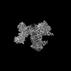
 マップデータ
 試料
試料 キーワード
キーワード 機能・相同性情報
 Homo sapiens (ヒト) /
 データ登録者
 米国, 2件
  引用
引用 構造の表示
構造の表示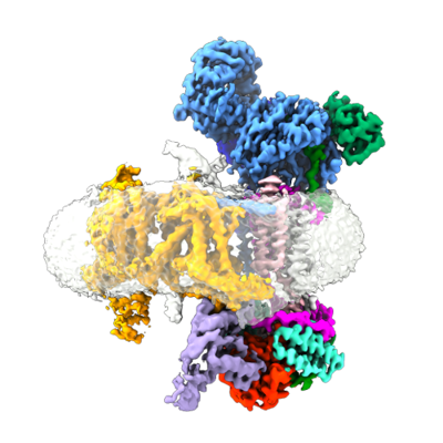
 ダウンロードとリンク
 emd_28376.png
 http://ftp.pdbj.org/pub/emdb/structures/EMD-28376
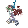


 X (Sec.)
 Y (Row.)
 Z (Col.)


 試料の構成要素
 解析
解析 電子顕微鏡法
 FIELD EMISSION GUN