 Movie
Movie Controller


+ Open data

- Basic information
| Entry | 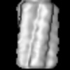 | ||||||||||||
|---|---|---|---|---|---|---|---|---|---|---|---|---|---|
| Title | Erwinia phage vB_EamM_RAY (RAY) Putative PhuZ Filament | ||||||||||||
 Map data Map data | |||||||||||||
 Sample Sample |
| ||||||||||||
 Keywords Keywords | Bacteriophage / structural protein / viral tubulin / VIRAL PROTEIN | ||||||||||||
| Biological species |  Erwinia phage vB_EamM_RAY (virus) Erwinia phage vB_EamM_RAY (virus) | ||||||||||||
| Method | subtomogram averaging / cryo EM / Resolution: 25.0 Å | ||||||||||||
 Authors Authors | Laughlin TG / Villa E | ||||||||||||
| Funding support |  United States, 3 items United States, 3 items
| ||||||||||||
 Citation Citation | Journal: bioRxiv / Year: 2023 Title: Identifying the core genome of the nucleus-forming bacteriophage family and characterization of phage RAY. Abstract: We recently discovered that some bacteriophages establish a nucleus-like replication compartment (phage nucleus), but the core genes that define nucleus-based phage replication and their phylogenetic ...We recently discovered that some bacteriophages establish a nucleus-like replication compartment (phage nucleus), but the core genes that define nucleus-based phage replication and their phylogenetic distribution were unknown. By studying phages that encode the major phage nucleus protein chimallin, including previously sequenced yet uncharacterized phages, we discovered that chimallin-encoding phages share a set of 72 highly conserved genes encoded within seven distinct gene blocks. Of these, 21 core genes are unique to this group, and all but one of these unique genes encode proteins of unknown function. We propose that phages with this core genome comprise a novel viral family we term Chimalliviridae. Fluorescence microscopy and cryo-electron tomography studies of phage vB_EamM_RAY confirm that many of the key steps of nucleus-based replication encoded in the core genome are conserved among diverse chimalliviruses, and reveal that non-core components can confer intriguing variations on this replication mechanism. For instance, unlike previously studied nucleus-forming phages, RAY doesn't degrade the host genome, and its PhuZ homolog appears to form a five-stranded filament with a lumen. This work expands our understanding of phage nucleus and PhuZ spindle diversity and function, providing a roadmap for identifying key mechanisms underlying nucleus-based phage replication. | ||||||||||||
| History |
|
- Structure visualization
| Supplemental images |
|---|

- Downloads & links
-EMDB archive
| Map data | emd_28008.map.gz | 503.1 KB |  EMDB map data format EMDB map data format | |
|---|---|---|---|---|
| Header (meta data) | emd-28008-v30.xmlemd-28008.xml | 18.5 KB 18.5 KB | Display Display | EMDB header |
| FSC (resolution estimation) | emd_28008_fsc.xml | 2 KB | Display | FSC data file |
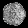
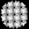
| Images | 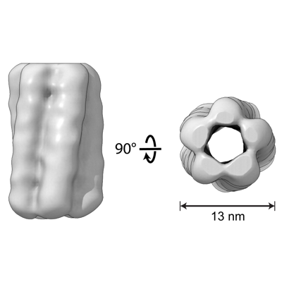 emd_28008.png emd_28008.png | 41.7 KB | ||
| Masks | emd_28008_msk_1.map | 550.3 KB | Mask map | |
| Filedesc metadata | emd-28008.cif.gz | 4.8 KB | ||
| Others | emd_28008_half_map_1.map.gzemd_28008_half_map_2.map.gz | 404 KB 405.1 KB | ||
| Archive directory |  http://ftp.pdbj.org/pub/emdb/structures/EMD-28008ftp://ftp.pdbj.org/pub/emdb/structures/EMD-28008 http://ftp.pdbj.org/pub/emdb/structures/EMD-28008ftp://ftp.pdbj.org/pub/emdb/structures/EMD-28008 | HTTPS FTP |
-Related structure data
| Related structure data | C: citing same article ( |
|---|
-Links
| EMDB pages | EMDB (EBI/PDBe) / EMDataResource |
|---|
-Map
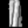
| File | Download / File: emd_28008.map.gz / Format: CCP4 / Size: 549.8 KB / Type: IMAGE STORED AS FLOATING POINT NUMBER (4 BYTES) | ||||||||||||||||||||||||||||||||||||
|---|---|---|---|---|---|---|---|---|---|---|---|---|---|---|---|---|---|---|---|---|---|---|---|---|---|---|---|---|---|---|---|---|---|---|---|---|---|
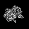
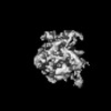
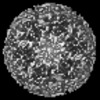
| Projections & slices | Image control
Images are generated by Spider. | ||||||||||||||||||||||||||||||||||||
| Voxel size | X=Y=Z: 4.265 Å | ||||||||||||||||||||||||||||||||||||
| Density |
| ||||||||||||||||||||||||||||||||||||
| Symmetry | Space group: 1 | ||||||||||||||||||||||||||||||||||||
| Details | EMDB XML:
|
 Z (Sec.)
 Y (Row.)
 X (Col.)


-Supplemental data
-Mask #1
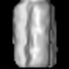
| File | emd_28008_msk_1.map | ||||||||||||
|---|---|---|---|---|---|---|---|---|---|---|---|---|---|
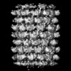
| Projections & Slices |
| ||||||||||||
| Density Histograms |


-Half map: #1
| File | emd_28008_half_map_1.map | ||||||||||||
|---|---|---|---|---|---|---|---|---|---|---|---|---|---|
| Projections & Slices |
| ||||||||||||
| Density Histograms |


-Half map: #2
| File | emd_28008_half_map_2.map | ||||||||||||
|---|---|---|---|---|---|---|---|---|---|---|---|---|---|
| Projections & Slices |
| ||||||||||||
| Density Histograms |


- Sample components
-Entire : Erwinia phage vB_EamM_RAY (RAY) Putative PhuZ Filament
| Entire | Name: Erwinia phage vB_EamM_RAY (RAY) Putative PhuZ Filament |
|---|---|
| Components |
|
-Supramolecule #1: Erwinia phage vB_EamM_RAY (RAY) Putative PhuZ Filament
| Supramolecule | Name: Erwinia phage vB_EamM_RAY (RAY) Putative PhuZ Filament type: complex / ID: 1 / Parent: 0 |
|---|---|
| Source (natural) | Organism: Erwinia phage vB_EamM_RAY (virus) |
-Experimental details
-Structure determination
| Method | cryo EM |
|---|---|
 Processing Processing | subtomogram averaging |
| Aggregation state | cell |
-Sample preparation
| Buffer | pH: 7.5 / Details: Lysogeny Broth containing 5% trehalose |
|---|---|
| Grid | Model: Quantifoil R2/1 / Material: COPPER / Mesh: 200 / Support film - Material: CARBON / Support film - topology: HOLEY / Pretreatment - Type: GLOW DISCHARGE / Pretreatment - Time: 60 sec. / Pretreatment - Atmosphere: OTHER / Pretreatment - Pressure: 0.019 kPa / Details: 20 mA in PELCO EasiGLO |
| Vitrification | Cryogen name: ETHANE-PROPANE / Instrument: HOMEMADE PLUNGER |
| Details | cell suspension |
- Electron microscopy
| Microscope | TFS KRIOS |
|---|---|
| Specialist optics | Energy filter - Name: GIF Quantum LS / Energy filter - Slit width: 20 eV |
| Image recording | Film or detector model: GATAN K2 SUMMIT (4k x 4k) / Detector mode: COUNTING / Number grids imaged: 2 / Average electron dose: 2.4 e/Å2 |
| Electron beam | Acceleration voltage: 300 kV / Electron source:  FIELD EMISSION GUN FIELD EMISSION GUN |
| Electron optics | C2 aperture diameter: 70.0 µm / Illumination mode: FLOOD BEAM / Imaging mode: BRIGHT FIELD / Cs: 2.7 mm / Nominal defocus max: 6.0 µm / Nominal defocus min: 4.0 µm / Nominal magnification: 33000 |
| Sample stage | Specimen holder model: FEI TITAN KRIOS AUTOGRID HOLDER / Cooling holder cryogen: NITROGEN |
| Experimental equipment |  Model: Titan Krios / Image courtesy: FEI Company |
-Image processing
| Final reconstruction | Number classes used: 1 / Applied symmetry - Point group: C1 (asymmetric) / Resolution.type: BY AUTHOR / Resolution: 25.0 Å / Resolution method: FSC 0.143 CUT-OFF / Software - Name: RELION (ver. 3.1.3) / Number subtomograms used: 3080 |
|---|---|
| Extraction | Number tomograms: 26 / Number images used: 5000 / Method: manual tracing and over-sampling Software: (Name: IMOD, Dynamo (ver. 1.1514), MATLAB (ver. 2019b))Details: Manually picked the start and end points of filaments using IMOD and generated over-sampled cropping models using Dynamo. |
| Final angle assignment | Type: MAXIMUM LIKELIHOOD / Software - Name: RELION (ver. 3.1.3) |
| FSC plot (resolution estimation) |  |
