 ムービー
ムービー コントローラー


+ データを開く

- 基本情報
基本情報
| 登録情報 | データベース: EMDB / ID: EMD-2790 | |||||||||
|---|---|---|---|---|---|---|---|---|---|---|
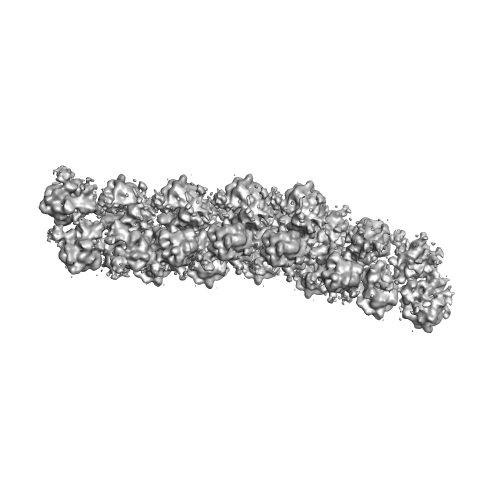
| タイトル | The molecular structure of the left-handed supra- molecular helix of eukaryotic polyribosomes | |||||||||
 マップデータ マップデータ | 3D poly-ribosome structure from Wheat Germ in vitro cell free system | |||||||||
 試料 試料 |
| |||||||||
 キーワード キーワード | cryo-ET / polyribosome / sub-tomogram averaging | |||||||||
| 機能・相同性 |  機能・相同性情報 機能・相同性情報cytoplasmic translational elongation / translational elongation / protein kinase activator activity / ribonucleoprotein complex binding / cytosolic ribosome / maturation of LSU-rRNA from tricistronic rRNA transcript (SSU-rRNA, 5.8S rRNA, LSU-rRNA) / large ribosomal subunit / small ribosomal subunit / cytosolic small ribosomal subunit / large ribosomal subunit rRNA binding ...cytoplasmic translational elongation / translational elongation / protein kinase activator activity / ribonucleoprotein complex binding / cytosolic ribosome / maturation of LSU-rRNA from tricistronic rRNA transcript (SSU-rRNA, 5.8S rRNA, LSU-rRNA) / large ribosomal subunit / small ribosomal subunit / cytosolic small ribosomal subunit / large ribosomal subunit rRNA binding / cytosolic large ribosomal subunit / cytoplasmic translation / negative regulation of translation / structural constituent of ribosome / ribosome / translation / ribonucleoprotein complex / mRNA binding / signal transduction / RNA binding / zinc ion binding 類似検索 - 分子機能 | |||||||||
| 生物種 |  | |||||||||
| 手法 | サブトモグラム平均法 / クライオ電子顕微鏡法 / 解像度: 34.0 Å | |||||||||
 データ登録者 データ登録者 | Myasnikov AG / Afonina ZHA / Menetret J-F / Shirokov VA / Spirin AS / Klaholz BP | |||||||||
 引用 引用 | ジャーナル: Nat Commun / 年: 2014 タイトル: The molecular structure of the left-handed supra-molecular helix of eukaryotic polyribosomes. 著者: Alexander G Myasnikov / Zhanna A Afonina / Jean-François Ménétret / Vladimir A Shirokov / Alexander S Spirin / Bruno P Klaholz /   要旨: During protein synthesis, several ribosomes bind to a single messenger RNA (mRNA) forming large macromolecular assemblies called polyribosomes. Here we report the detailed molecular structure of a ...During protein synthesis, several ribosomes bind to a single messenger RNA (mRNA) forming large macromolecular assemblies called polyribosomes. Here we report the detailed molecular structure of a 100 MDa eukaryotic poly-ribosome complex derived from cryo electron tomography, sub-tomogram averaging and pseudo-atomic modelling by crystal structure fitting. The structure allowed the visualization of the three functional parts of the polysome assembly, the central core region that forms a rather compact left-handed supra-molecular helix, and the more open regions that harbour the initiation and termination sites at either ends. The helical region forms a continuous mRNA channel where the mRNA strand bridges neighbouring exit and entry sites of the ribosomes and prevents mRNA looping between ribosomes. This structure provides unprecedented insights into protein- and RNA-mediated inter-ribosome contacts that involve conserved sites through 40S subunits and long protruding RNA expansion segments, suggesting a role in stabilizing the overall polyribosomal assembly. | |||||||||
| 履歴 |
|
- 構造の表示
構造の表示
| ムービー |
ムービービューア |
|---|---|
| 構造ビューア | EMマップ: SurfViewMolmilJmol/JSmol |
| 添付画像 |
- ダウンロードとリンク
-EMDBアーカイブ
| マップデータ | emd_2790.map.gz | 32.5 MB | EMDBマップデータ形式 | |
|---|---|---|---|---|
| ヘッダ (付随情報) | emd-2790-v30.xmlemd-2790.xml | 12.9 KB 12.9 KB | 表示 表示 | EMDBヘッダ |
| 画像 |  EMD-2790.png EMD-2790.png | 70.4 KB | ||
| アーカイブディレクトリ |  http://ftp.pdbj.org/pub/emdb/structures/EMD-2790ftp://ftp.pdbj.org/pub/emdb/structures/EMD-2790 http://ftp.pdbj.org/pub/emdb/structures/EMD-2790ftp://ftp.pdbj.org/pub/emdb/structures/EMD-2790 | HTTPS FTP |
-検証レポート
| 文書・要旨 | emd_2790_validation.pdf.gz | 191.6 KB | 表示 | EMDB検証レポート |
|---|---|---|---|---|
| 文書・詳細版 | emd_2790_full_validation.pdf.gz | 190.7 KB | 表示 | |
| XML形式データ | emd_2790_validation.xml.gz | 4.6 KB | 表示 | |
| アーカイブディレクトリ | https://ftp.pdbj.org/pub/emdb/validation_reports/EMD-2790ftp://ftp.pdbj.org/pub/emdb/validation_reports/EMD-2790 | HTTPS FTP |
-関連構造データ

-リンク
| EMDBのページ | EMDB (EBI/PDBe) / EMDataResource |
|---|---|
| 「今月の分子」の関連する項目 |


-マップ
| ファイル | ダウンロード / ファイル: emd_2790.map.gz / 形式: CCP4 / 大きさ: 48.8 MB / タイプ: IMAGE STORED AS FLOATING POINT NUMBER (4 BYTES) | ||||||||||||||||||||||||||||||||||||||||||||||||||||||||||||||||||||
|---|---|---|---|---|---|---|---|---|---|---|---|---|---|---|---|---|---|---|---|---|---|---|---|---|---|---|---|---|---|---|---|---|---|---|---|---|---|---|---|---|---|---|---|---|---|---|---|---|---|---|---|---|---|---|---|---|---|---|---|---|---|---|---|---|---|---|---|---|---|
| 注釈 | 3D poly-ribosome structure from Wheat Germ in vitro cell free system | ||||||||||||||||||||||||||||||||||||||||||||||||||||||||||||||||||||
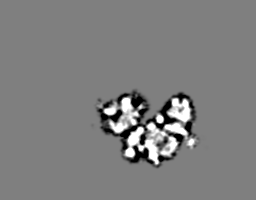
| 投影像・断面図 | 画像のコントロール
画像は Spider により作成 これらの図は立方格子座標系で作成されたものです | ||||||||||||||||||||||||||||||||||||||||||||||||||||||||||||||||||||
| ボクセルのサイズ | X=Y=Z: 6.8 Å | ||||||||||||||||||||||||||||||||||||||||||||||||||||||||||||||||||||
| 密度 |
| ||||||||||||||||||||||||||||||||||||||||||||||||||||||||||||||||||||
| 対称性 | 空間群: 1 | ||||||||||||||||||||||||||||||||||||||||||||||||||||||||||||||||||||
| 詳細 | EMDB XML:
CCP4マップ ヘッダ情報:
| ||||||||||||||||||||||||||||||||||||||||||||||||||||||||||||||||||||
 Z (Sec.)
 Y (Row.)
 X (Col.)


-添付データ
- 試料の構成要素
-全体 : poly-ribosome from in vitro wheat germ system
| 全体 | 名称: poly-ribosome from in vitro wheat germ system |
|---|---|
| 要素 |
|
-超分子 #1000: poly-ribosome from in vitro wheat germ system
| 超分子 | 名称: poly-ribosome from in vitro wheat germ system / タイプ: sample / ID: 1000 / 集合状態: 23-meric / Number unique components: 1 |
|---|---|
| 分子量 | 理論値: 100 MDa |
-超分子 #1: Wheat germ poly-ribosome
| 超分子 | 名称: Wheat germ poly-ribosome / タイプ: complex / ID: 1 / 組換発現: No / データベース: NCBI / Ribosome-details: ribosome-eukaryote: ALL |
|---|---|
| 由来(天然) | 生物種: |
| 分子量 | 理論値: 100 MDa |
-実験情報
-構造解析
| 手法 | クライオ電子顕微鏡法 |
|---|---|
 解析 解析 | サブトモグラム平均法 |
| 試料の集合状態 | helical array |
-試料調製
| 濃度 | 0.5 mg/mL |
|---|---|
| 緩衝液 | pH: 7.6 詳細: 25mM HEPES-KOH, 3mM Mg(OAc)2, 85mM KOAc, 1.6mM DTT, 0.25mM spermidine |
| グリッド | 詳細: 3ul of sample applied on 300 mesh holy carbon Quantifoil 2/2 grid. Blotting was done in Vitrobot Mark IV |
| 凍結 | 凍結剤: ETHANE / チャンバー内湿度: 95 % / チャンバー内温度: 120 K / 装置: FEI VITROBOT MARK IV 手法: 3ul of sample applied on 300 mesh holy carbon Quantifoil 2/2 grid. Blotting was done in Vitrobot Mark IV, blot time 0.5 sec, blot force 5 |
- 電子顕微鏡法
| 顕微鏡 | FEI TECNAI F30 |
|---|---|
| 温度 | 最低: 80 K / 最高: 100 K / 平均: 90 K |
| アライメント法 | Legacy - 非点収差: Objective lens astigmatism was corrected at 120,000 times magnification Legacy - Electron beam tilt params: 0 |
| 日付 | 2013年1月1日 |
| 撮影 | カテゴリ: CCD フィルム・検出器のモデル: FEI FALCON I (4k x 4k) 平均電子線量: 30 e/Å2 / ビット/ピクセル: 16 |
| 電子線 | 加速電圧: 150 kV / 電子線源:  FIELD EMISSION GUN FIELD EMISSION GUN |
| 電子光学系 | 倍率(補正後): 41176 / 照射モード: FLOOD BEAM / 撮影モード: BRIGHT FIELD / Cs: 2 mm / 最大 デフォーカス(公称値): 4.0 µm / 最小 デフォーカス(公称値): 2.0 µm / 倍率(公称値): 39000 |
| 試料ステージ | 試料ホルダーモデル: GATAN HELIUM / Tilt series - Axis1 - Min angle: -70 ° / Tilt series - Axis1 - Max angle: 70 ° |
| 実験機器 |  モデル: Tecnai F30 / 画像提供: FEI Company |
-画像解析
| 詳細 | The subtomograms were selected in imod manually. The averaging was done in xmipp program by using ml_tomo subroutine. |
|---|---|
| 最終 再構成 | 想定した対称性 - らせんパラメータ - Δz: 83 Å 想定した対称性 - らせんパラメータ - ΔΦ: 90 ° 想定した対称性 - らせんパラメータ - 軸対称性: C4 (4回回転対称) アルゴリズム: OTHER / 解像度のタイプ: BY AUTHOR / 解像度: 34.0 Å / 解像度の算出法: OTHER / ソフトウェア - 名称: Imod, Xmipp, Imagic / 使用したサブトモグラム数: 106 |
| 最終 3次元分類 | クラス数: 1 |
-原子モデル構築 1
| 初期モデル | PDB ID:  3iz6 |
|---|---|
| ソフトウェア | 名称: Chimera |
| 詳細 | 40S and 60S was fitted separately |
| 精密化 | 空間: REAL / プロトコル: RIGID BODY FIT |
| 得られたモデル | 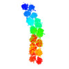 PDB-4v3p: |
-原子モデル構築 2
| 初期モデル | PDB ID: 3iz7 |
|---|---|
| ソフトウェア | 名称: Chimera |
| 詳細 | 40S and 60S was fitted separately |
| 精密化 | 空間: REAL / プロトコル: RIGID BODY FIT |
| 得られたモデル | PDB-4v3p: |
-原子モデル構築 3
| 初期モデル | PDB ID: 3izr |
|---|---|
| ソフトウェア | 名称: Chimera |
| 詳細 | 40S and 60S was fitted separately |
| 精密化 | 空間: REAL / プロトコル: RIGID BODY FIT |
| 得られたモデル | PDB-4v3p: |
