Movie
Movie Controller


+ Open data

- Basic information
| Entry |  | |||||||||
|---|---|---|---|---|---|---|---|---|---|---|
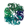
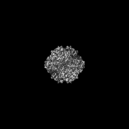
| Title | Human Brain Glyceraldehyde 3-phosphate dehydrogenase | |||||||||
 Map data Map data | ||||||||||
 Sample Sample |
| |||||||||
 Keywords Keywords | human brain / GAPDH / OXIDOREDUCTASE | |||||||||
| Function / homology |  Function and homology information Function and homology informationpeptidyl-cysteine S-trans-nitrosylation / Transferases; Transferring nitrogenous groups; Transferring other nitrogenous groups / negative regulation of endopeptidase activity / glyceraldehyde-3-phosphate dehydrogenase (phosphorylating) / : / aspartic-type endopeptidase inhibitor activity / glyceraldehyde-3-phosphate dehydrogenase (NAD+) (phosphorylating) activity / Gluconeogenesis / Glycolysis / GAIT complex ...peptidyl-cysteine S-trans-nitrosylation / Transferases; Transferring nitrogenous groups; Transferring other nitrogenous groups / negative regulation of endopeptidase activity / glyceraldehyde-3-phosphate dehydrogenase (phosphorylating) / : / aspartic-type endopeptidase inhibitor activity / glyceraldehyde-3-phosphate dehydrogenase (NAD+) (phosphorylating) activity / Gluconeogenesis / Glycolysis / GAIT complex / canonical glycolysis / peptidyl-cysteine S-nitrosylase activity / regulation of macroautophagy / defense response to fungus / positive regulation of type I interferon production / lipid droplet / positive regulation of cytokine production / glycolytic process / cellular response to type II interferon / microtubule cytoskeleton organization / NAD binding / disordered domain specific binding / NADP binding / antimicrobial humoral immune response mediated by antimicrobial peptide / microtubule cytoskeleton / neuron apoptotic process / nuclear membrane / microtubule binding / killing of cells of another organism / vesicle / positive regulation of canonical NF-kappaB signal transduction / negative regulation of translation / protein stabilization / ribonucleoprotein complex / perinuclear region of cytoplasm / extracellular exosome / membrane / identical protein binding / nucleus / plasma membrane / cytoplasm / cytosol Similarity search - Function | |||||||||
| Biological species |  Homo sapiens (human) Homo sapiens (human) | |||||||||
| Method | single particle reconstruction / cryo EM / Resolution: 3.22 Å | |||||||||
 Authors Authors | Tringides ML | |||||||||
| Funding support |  United States, 1 items United States, 1 items
| |||||||||
 Citation Citation | Journal: Life Sci Alliance / Year: 2023 Title: A cryo-electron microscopic approach to elucidate protein structures from human brain microsomes. Authors: Marios L Tringides / Zhemin Zhang / Christopher E Morgan / Chih-Chia Su / Edward W Yu / Abstract: We recently developed a "Build and Retrieve" cryo-electron microscopy (cryo-EM) methodology, which is capable of simultaneously producing near-atomic resolution cryo-EM maps for several individual ...We recently developed a "Build and Retrieve" cryo-electron microscopy (cryo-EM) methodology, which is capable of simultaneously producing near-atomic resolution cryo-EM maps for several individual proteins from a heterogeneous, multiprotein sample. Here we report the use of "Build and Retrieve" to define the composition of a raw human brain microsomal lysate. From this sample, we simultaneously identify and solve cryo-EM structures of five different brain enzymes whose functions affect neurotransmitter recycling, iron metabolism, glycolysis, axonal development, energy homeostasis, and retinoic acid biosynthesis. Interestingly, malfunction of these important proteins has been directly linked to several neurodegenerative disorders, such as Alzheimer's, Huntington's, and Parkinson's diseases. Our work underscores the importance of cryo-EM in facilitating tissue and organ proteomics at the atomic level. | |||||||||
| History |
|
- Structure visualization
| Supplemental images |
|---|

- Downloads & links
-EMDB archive
| Map data | emd_27579.map.gz | 4.1 MB | EMDB map data format | |
|---|---|---|---|---|
| Header (meta data) | emd-27579-v30.xmlemd-27579.xml | 15.7 KB 15.7 KB | Display Display | EMDB header |
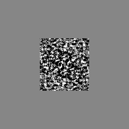
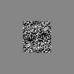
| Images |  emd_27579.png emd_27579.png | 460.4 KB | ||
| Filedesc metadata | emd-27579.cif.gz | 5.5 KB | ||
| Others | emd_27579_half_map_1.map.gzemd_27579_half_map_2.map.gz | 59.1 MB 59.1 MB | ||
| Archive directory |  http://ftp.pdbj.org/pub/emdb/structures/EMD-27579ftp://ftp.pdbj.org/pub/emdb/structures/EMD-27579 http://ftp.pdbj.org/pub/emdb/structures/EMD-27579ftp://ftp.pdbj.org/pub/emdb/structures/EMD-27579 | HTTPS FTP |
-Related structure data
| Related structure data |  8dnsMC  8dnmC 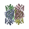 8dnoC  8dnpC 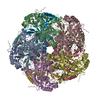 8dnuC M: atomic model generated by this map C: citing same article ( |
|---|---|
| Similar structure data |


-Links
| EMDB pages | EMDB (EBI/PDBe) / EMDataResource |
|---|---|
| Related items in Molecule of the Month |


-Map
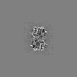
| File | Download / File: emd_27579.map.gz / Format: CCP4 / Size: 64 MB / Type: IMAGE STORED AS FLOATING POINT NUMBER (4 BYTES) | ||||||||||||||||||||||||||||||||||||
|---|---|---|---|---|---|---|---|---|---|---|---|---|---|---|---|---|---|---|---|---|---|---|---|---|---|---|---|---|---|---|---|---|---|---|---|---|---|
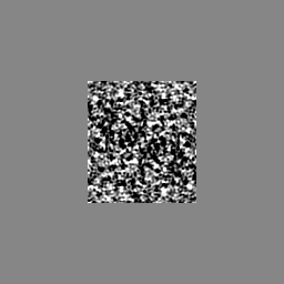
| Projections & slices | Image control
Images are generated by Spider. | ||||||||||||||||||||||||||||||||||||
| Voxel size | X=Y=Z: 1.07 Å | ||||||||||||||||||||||||||||||||||||
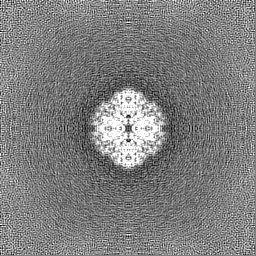
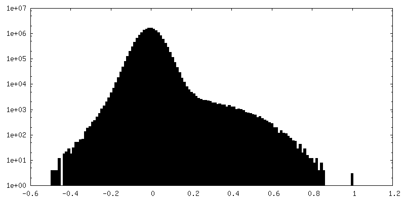
| Density |
| ||||||||||||||||||||||||||||||||||||
| Symmetry | Space group: 1 | ||||||||||||||||||||||||||||||||||||
| Details | EMDB XML:
|
 X (Sec.)
 Y (Row.)
 Z (Col.)


-Supplemental data
-Half map: #1
| File | emd_27579_half_map_1.map | ||||||||||||
|---|---|---|---|---|---|---|---|---|---|---|---|---|---|
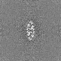
| Projections & Slices |
| ||||||||||||
| Density Histograms |


-Half map: #2
| File | emd_27579_half_map_2.map | ||||||||||||
|---|---|---|---|---|---|---|---|---|---|---|---|---|---|
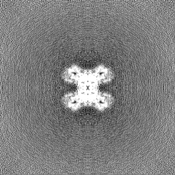
| Projections & Slices |
| ||||||||||||
| Density Histograms |


- Sample components
-Entire : Glyceraldehyde 3-phosphate dehydrogenase
| Entire | Name: Glyceraldehyde 3-phosphate dehydrogenase |
|---|---|
| Components |
|
-Supramolecule #1: Glyceraldehyde 3-phosphate dehydrogenase
| Supramolecule | Name: Glyceraldehyde 3-phosphate dehydrogenase / type: complex / ID: 1 / Parent: 0 / Macromolecule list: all |
|---|---|
| Source (natural) | Organism: Homo sapiens (human) |
-Macromolecule #1: Glyceraldehyde-3-phosphate dehydrogenase
| Macromolecule | Name: Glyceraldehyde-3-phosphate dehydrogenase / type: protein_or_peptide / ID: 1 / Number of copies: 4 / Enantiomer: LEVO EC number: glyceraldehyde-3-phosphate dehydrogenase (phosphorylating) |
|---|---|
| Source (natural) | Organism: Homo sapiens (human) |
| Molecular weight | Theoretical: 36.099168 KDa |
| Sequence | String: MGKVKVGVNG FGRIGRLVTR AAFNSGKVDI VAINDPFIDL NYMVYMFQYD STHGKFHGTV KAENGKLVIN GNPITIFQER DPSKIKWGD AGAEYVVEST GVFTTMEKAG AHLQGGAKRV IISAPSADAP MFVMGVNHEK YDNSLKIISN ASCTTNCLAP L AKVIHDNF ...String: MGKVKVGVNG FGRIGRLVTR AAFNSGKVDI VAINDPFIDL NYMVYMFQYD STHGKFHGTV KAENGKLVIN GNPITIFQER DPSKIKWGD AGAEYVVEST GVFTTMEKAG AHLQGGAKRV IISAPSADAP MFVMGVNHEK YDNSLKIISN ASCTTNCLAP L AKVIHDNF GIVEGLMTTV HAITATQKTV DGPSGKLWRD GRGALQNIIP ASTGAAKAVG KVIPELNGKL TGMAFRVPTA NV SVVDLTC RLEKPAKYDD IKKVVKQASE GPLKGILGYT EHQVVSSDFN SDTHSSTFDA GAGIALNDHF VKLISWYDNE FGY SNRVVD LMAHMASKE UniProtKB: Glyceraldehyde-3-phosphate dehydrogenase |
-Experimental details
-Structure determination
| Method | cryo EM |
|---|---|
 Processing Processing | single particle reconstruction |
| Aggregation state | tissue |
-Sample preparation
| Buffer | pH: 7.5 |
|---|---|
| Vitrification | Cryogen name: ETHANE |
- Electron microscopy
| Microscope | TFS KRIOS |
|---|---|
| Image recording | Film or detector model: GATAN K3 BIOQUANTUM (6k x 4k) / Average electron dose: 36.5 e/Å2 |
| Electron beam | Acceleration voltage: 300 kV / Electron source:  FIELD EMISSION GUN FIELD EMISSION GUN |
| Electron optics | Illumination mode: FLOOD BEAM / Imaging mode: BRIGHT FIELD / Nominal defocus max: 3.706 µm / Nominal defocus min: 1.212 µm |
| Experimental equipment |  Model: Titan Krios / Image courtesy: FEI Company |