+ データを開く
- 基本情報
基本情報
| 登録情報 |  | ||||||||||||
|---|---|---|---|---|---|---|---|---|---|---|---|---|---|
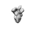
| タイトル | nsEM map of beta coronavirus HKU1 spike complexed with polyclonal Fab samples from individual 182322 at day 0 using Sulfo-SDAD crosslinker | ||||||||||||
 マップデータ マップデータ | nsEM map of beta coronavirus HKU1 spike complexed with polyclonal Fab samples from individual 182322 at day 0 using Sulfo-SDAD crosslinker | ||||||||||||
 試料 試料 |
| ||||||||||||
 キーワード キーワード | beta coronaviruses / HKU1 / nsEM / EMPEM / VIRAL PROTEIN | ||||||||||||
| 生物種 |  Homo sapiens (ヒト) Homo sapiens (ヒト) | ||||||||||||
| 手法 | 単粒子再構成法 / ネガティブ染色法 / 解像度: 25.0 Å | ||||||||||||
 データ登録者 データ登録者 | Torrents de la Pena A / Sewall LM / Ward AB | ||||||||||||
| 資金援助 |  米国, 3件 米国, 3件
| ||||||||||||
 引用 引用 |  ジャーナル: Cell Rep Methods / 年: 2023 ジャーナル: Cell Rep Methods / 年: 2023タイトル: Increasing sensitivity of antibody-antigen interactions using photo-cross-linking. 著者: Alba Torrents de la Peña / Leigh M Sewall / Rebeca de Paiva Froes Rocha / Abigail M Jackson / Payal P Pratap / Sandhya Bangaru / Christopher A Cottrell / Subhasis Mohanty / Albert C Shaw / Andrew B Ward /  要旨: Understanding antibody-antigen interactions in a polyclonal immune response in humans and animal models is critical for rational vaccine design. Current approaches typically characterize antibodies ...Understanding antibody-antigen interactions in a polyclonal immune response in humans and animal models is critical for rational vaccine design. Current approaches typically characterize antibodies that are functionally relevant or highly abundant. Here, we use photo-cross-linking and single-particle electron microscopy to increase antibody detection and unveil epitopes of low-affinity and low-abundance antibodies, leading to a broader structural characterization of polyclonal immune responses. We employed this approach across three different viral glycoproteins and showed increased sensitivity of detection relative to currently used methods. Results were most noticeable in early and late time points of a polyclonal immune response. Additionally, the use of photo-cross-linking revealed intermediate antibody binding states and demonstrated a distinctive way to study antibody binding mechanisms. This technique can be used to structurally characterize the landscape of a polyclonal immune response of patients in vaccination or post-infection studies at early time points, allowing for rapid iterative design of vaccine immunogens. | ||||||||||||
| 履歴 |
|
- 構造の表示
構造の表示
| 添付画像 |
|---|
- ダウンロードとリンク
-EMDBアーカイブ
| マップデータ |  emd_27377.map.gz emd_27377.map.gz | 32.7 MB |  EMDBマップデータ形式 EMDBマップデータ形式 | |
|---|---|---|---|---|
| ヘッダ (付随情報) |  emd-27377-v30.xml emd-27377-v30.xml emd-27377.xml emd-27377.xml | 15.1 KB 15.1 KB | 表示 表示 |  EMDBヘッダ EMDBヘッダ |
| 画像 |  emd_27377.png emd_27377.png | 19.7 KB | ||
| その他 |  emd_27377_half_map_1.map.gz emd_27377_half_map_1.map.gz emd_27377_half_map_2.map.gz emd_27377_half_map_2.map.gz | 33 MB 33 MB | ||
| アーカイブディレクトリ |  http://ftp.pdbj.org/pub/emdb/structures/EMD-27377 http://ftp.pdbj.org/pub/emdb/structures/EMD-27377 ftp://ftp.pdbj.org/pub/emdb/structures/EMD-27377 ftp://ftp.pdbj.org/pub/emdb/structures/EMD-27377 | HTTPS FTP |
-検証レポート
| 文書・要旨 |  emd_27377_validation.pdf.gz emd_27377_validation.pdf.gz | 566.3 KB | 表示 |  EMDB検証レポート EMDB検証レポート |
|---|---|---|---|---|
| 文書・詳細版 |  emd_27377_full_validation.pdf.gz emd_27377_full_validation.pdf.gz | 565.9 KB | 表示 | |
| XML形式データ |  emd_27377_validation.xml.gz emd_27377_validation.xml.gz | 11.5 KB | 表示 | |
| CIF形式データ |  emd_27377_validation.cif.gz emd_27377_validation.cif.gz | 13.5 KB | 表示 | |
| アーカイブディレクトリ |  https://ftp.pdbj.org/pub/emdb/validation_reports/EMD-27377 https://ftp.pdbj.org/pub/emdb/validation_reports/EMD-27377 ftp://ftp.pdbj.org/pub/emdb/validation_reports/EMD-27377 ftp://ftp.pdbj.org/pub/emdb/validation_reports/EMD-27377 | HTTPS FTP |
-関連構造データ
| 関連構造データ | C: 同じ文献を引用 ( |
|---|
- リンク
リンク
| EMDBのページ |  EMDB (EBI/PDBe) / EMDB (EBI/PDBe) /  EMDataResource EMDataResource |
|---|
- マップ
マップ
| ファイル |  ダウンロード / ファイル: emd_27377.map.gz / 形式: CCP4 / 大きさ: 42.9 MB / タイプ: IMAGE STORED AS FLOATING POINT NUMBER (4 BYTES) ダウンロード / ファイル: emd_27377.map.gz / 形式: CCP4 / 大きさ: 42.9 MB / タイプ: IMAGE STORED AS FLOATING POINT NUMBER (4 BYTES) | ||||||||||||||||||||||||||||||||||||
|---|---|---|---|---|---|---|---|---|---|---|---|---|---|---|---|---|---|---|---|---|---|---|---|---|---|---|---|---|---|---|---|---|---|---|---|---|---|
| 注釈 | nsEM map of beta coronavirus HKU1 spike complexed with polyclonal Fab samples from individual 182322 at day 0 using Sulfo-SDAD crosslinker | ||||||||||||||||||||||||||||||||||||
| 投影像・断面図 | 画像のコントロール
画像は Spider により作成 | ||||||||||||||||||||||||||||||||||||
| ボクセルのサイズ | X=Y=Z: 2.06 Å | ||||||||||||||||||||||||||||||||||||
| 密度 |
| ||||||||||||||||||||||||||||||||||||
| 対称性 | 空間群: 1 | ||||||||||||||||||||||||||||||||||||
| 詳細 | EMDB XML:
|
-添付データ
-ハーフマップ: nsEM map of beta coronavirus HKU1 spike complexed...
| ファイル | emd_27377_half_map_1.map | ||||||||||||
|---|---|---|---|---|---|---|---|---|---|---|---|---|---|
| 注釈 | nsEM map of beta coronavirus HKU1 spike complexed with polyclonal Fab samples from individual 182322 at day 0 using Sulfo-SDAD crosslinker | ||||||||||||
| 投影像・断面図 |
| ||||||||||||
| 密度ヒストグラム |
-ハーフマップ: nsEM map of beta coronavirus HKU1 spike complexed...
| ファイル | emd_27377_half_map_2.map | ||||||||||||
|---|---|---|---|---|---|---|---|---|---|---|---|---|---|
| 注釈 | nsEM map of beta coronavirus HKU1 spike complexed with polyclonal Fab samples from individual 182322 at day 0 using Sulfo-SDAD crosslinker | ||||||||||||
| 投影像・断面図 |
| ||||||||||||
| 密度ヒストグラム |
- 試料の構成要素
-全体 : nsEM map of hemagglutinin H1 A/Mich/045/15 complexed with polyclo...
| 全体 | 名称: nsEM map of hemagglutinin H1 A/Mich/045/15 complexed with polyclonal Fab samples from individual 182419 at day 0 |
|---|---|
| 要素 |
|
-超分子 #1: nsEM map of hemagglutinin H1 A/Mich/045/15 complexed with polyclo...
| 超分子 | 名称: nsEM map of hemagglutinin H1 A/Mich/045/15 complexed with polyclonal Fab samples from individual 182419 at day 0 タイプ: complex / ID: 1 / 親要素: 0 詳細: HA recombinantly expressed in HEK 293F cells Fab samples isolated from several individuals at different timepoints |
|---|---|
| 由来(天然) | 生物種:  Homo sapiens (ヒト) Homo sapiens (ヒト) |
-実験情報
-構造解析
| 手法 | ネガティブ染色法 |
|---|---|
 解析 解析 | 単粒子再構成法 |
| 試料の集合状態 | particle |
- 試料調製
試料調製
| 濃度 | 0.02 mg/mL |
|---|---|
| 緩衝液 | pH: 7.4 / 構成要素 - 濃度: 0.02 0.020 / 構成要素 - 式: TBS / 構成要素 - 名称: Tris-buffered saline |
| 染色 | タイプ: NEGATIVE / 材質: Uranyl Formate |
| グリッド | モデル: EMS Lacey Carbon / 材質: COPPER / メッシュ: 400 / 支持フィルム - 材質: CARBON / 支持フィルム - トポロジー: CONTINUOUS |
| 詳細 | 15 ug of H1 A/Mich15/045/15 was complexed overnight with 500 ug of polyclonal Fab samples and SEC purified. The complex peak was concentrated to ~0.02 mg/ml and imaged |
- 電子顕微鏡法
| 顕微鏡 | FEI TECNAI SPIRIT |
|---|---|
| 撮影 | フィルム・検出器のモデル: FEI EAGLE (4k x 4k) / 撮影したグリッド数: 1 / 平均電子線量: 50.0 e/Å2 |
| 電子線 | 加速電圧: 120 kV / 電子線源: LAB6 |
| 電子光学系 | C2レンズ絞り径: 70.0 µm / 照射モード: FLOOD BEAM / 撮影モード: BRIGHT FIELD / 最大 デフォーカス(公称値): 2.0 µm / 最小 デフォーカス(公称値): 1.5 µm / 倍率(公称値): 52000 |
| 試料ステージ | ホルダー冷却材: NITROGEN |
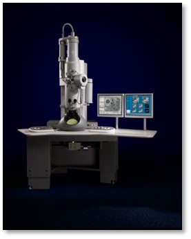
| 実験機器 |  モデル: Tecnai Spirit / 画像提供: FEI Company |
 ムービー
ムービー コントローラー


 Z (Sec.)
 Y (Row.)
 X (Col.)


