Movie
Movie Controller

[English] 日本語
 Yorodumi
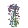
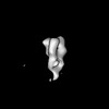
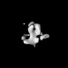
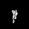
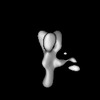
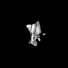
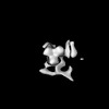
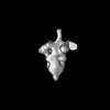
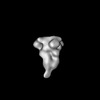
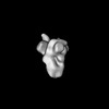
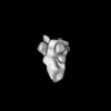
- EMDB-27358: nsEM map of hemagglutinin H3 A/Sing/INFIMH/16 complexed with poly... -
+ Open data

- Basic information
| Entry | 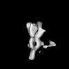 | ||||||||||||
|---|---|---|---|---|---|---|---|---|---|---|---|---|---|
| Title | nsEM map of hemagglutinin H3 A/Sing/INFIMH/16 complexed with polyclonal Fab samples from individual 182419 at day 2 using Sulfo-SDAD photo-cross-linker | ||||||||||||
 Map data Map data | nsEM map of hemagglutinin H3 A/Sing/INFIMH/16 complexed with polyclonal Fab samples from individual 182419 at day 2 using Sulfo-SDAD photo-cross-linker | ||||||||||||
 Sample Sample |
| ||||||||||||
 Keywords Keywords | influenza / H3 / nsEM / EMPEM / VIRAL PROTEIN | ||||||||||||
| Biological species |  Homo sapiens (human) Homo sapiens (human) | ||||||||||||
| Method | single particle reconstruction / negative staining / Resolution: 25.0 Å | ||||||||||||
 Authors Authors | Torrents de la Pena A / Sewall LM / Ward AB | ||||||||||||
| Funding support |  United States, 3 items United States, 3 items
| ||||||||||||
 Citation Citation | Journal: Cell Rep Methods / Year: 2023 Title: Increasing sensitivity of antibody-antigen interactions using photo-cross-linking. Authors: Alba Torrents de la Peña / Leigh M Sewall / Rebeca de Paiva Froes Rocha / Abigail M Jackson / Payal P Pratap / Sandhya Bangaru / Christopher A Cottrell / Subhasis Mohanty / Albert C Shaw / Andrew B Ward / Abstract: Understanding antibody-antigen interactions in a polyclonal immune response in humans and animal models is critical for rational vaccine design. Current approaches typically characterize antibodies ...Understanding antibody-antigen interactions in a polyclonal immune response in humans and animal models is critical for rational vaccine design. Current approaches typically characterize antibodies that are functionally relevant or highly abundant. Here, we use photo-cross-linking and single-particle electron microscopy to increase antibody detection and unveil epitopes of low-affinity and low-abundance antibodies, leading to a broader structural characterization of polyclonal immune responses. We employed this approach across three different viral glycoproteins and showed increased sensitivity of detection relative to currently used methods. Results were most noticeable in early and late time points of a polyclonal immune response. Additionally, the use of photo-cross-linking revealed intermediate antibody binding states and demonstrated a distinctive way to study antibody binding mechanisms. This technique can be used to structurally characterize the landscape of a polyclonal immune response of patients in vaccination or post-infection studies at early time points, allowing for rapid iterative design of vaccine immunogens. | ||||||||||||
| History |
|
- Structure visualization
| Supplemental images |
|---|
- Downloads & links
-EMDB archive
| Map data | emd_27358.map.gz | 23.5 MB |  EMDB map data format EMDB map data format | |
|---|---|---|---|---|
| Header (meta data) | emd-27358-v30.xmlemd-27358.xml | 15.1 KB 15.1 KB | Display Display | EMDB header |
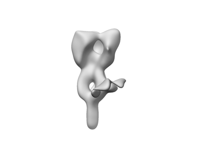
| Images |  emd_27358.png emd_27358.png | 17.2 KB | ||
| Others | emd_27358_half_map_1.map.gzemd_27358_half_map_2.map.gz | 23.5 MB 23.5 MB | ||
| Archive directory |  http://ftp.pdbj.org/pub/emdb/structures/EMD-27358ftp://ftp.pdbj.org/pub/emdb/structures/EMD-27358 http://ftp.pdbj.org/pub/emdb/structures/EMD-27358ftp://ftp.pdbj.org/pub/emdb/structures/EMD-27358 | HTTPS FTP |
-Related structure data
| Related structure data | C: citing same article ( |
|---|


-Links
| EMDB pages | EMDB (EBI/PDBe) / EMDataResource |
|---|
-Map
| File | Download / File: emd_27358.map.gz / Format: CCP4 / Size: 30.5 MB / Type: IMAGE STORED AS FLOATING POINT NUMBER (4 BYTES) | ||||||||||||||||||||||||||||||||||||
|---|---|---|---|---|---|---|---|---|---|---|---|---|---|---|---|---|---|---|---|---|---|---|---|---|---|---|---|---|---|---|---|---|---|---|---|---|---|
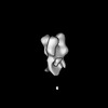
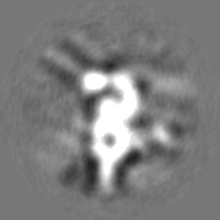
| Annotation | nsEM map of hemagglutinin H3 A/Sing/INFIMH/16 complexed with polyclonal Fab samples from individual 182419 at day 2 using Sulfo-SDAD photo-cross-linker | ||||||||||||||||||||||||||||||||||||
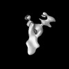
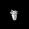
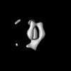
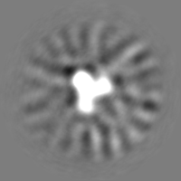
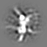
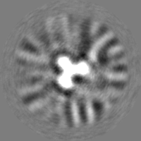
| Projections & slices | Image control
Images are generated by Spider. | ||||||||||||||||||||||||||||||||||||
| Voxel size | X=Y=Z: 1.77 Å | ||||||||||||||||||||||||||||||||||||
| Density |
| ||||||||||||||||||||||||||||||||||||
| Symmetry | Space group: 1 | ||||||||||||||||||||||||||||||||||||
| Details | EMDB XML:
|
 Z (Sec.)
 Y (Row.)
 X (Col.)


-Supplemental data
-Half map: nsEM map of hemagglutinin H3 A/Sing/INFIMH/16 complexed with...
| File | emd_27358_half_map_1.map | ||||||||||||
|---|---|---|---|---|---|---|---|---|---|---|---|---|---|
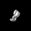
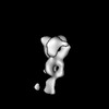
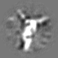
| Annotation | nsEM map of hemagglutinin H3 A/Sing/INFIMH/16 complexed with polyclonal Fab samples from individual 182419 at day 2 using Sulfo-SDAD photo-cross-linker | ||||||||||||
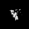
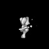
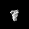
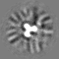
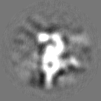
| Projections & Slices |
| ||||||||||||
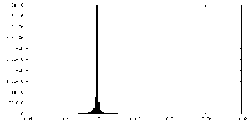
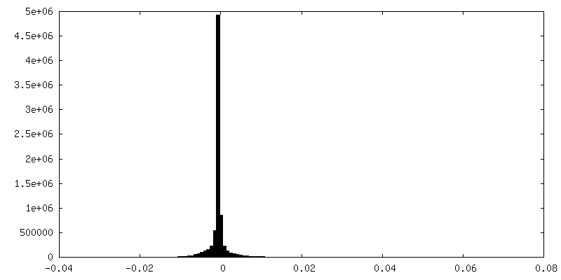
| Density Histograms |

-Half map: nsEM map of hemagglutinin H3 A/Sing/INFIMH/16 complexed with...
| File | emd_27358_half_map_2.map | ||||||||||||
|---|---|---|---|---|---|---|---|---|---|---|---|---|---|
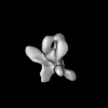
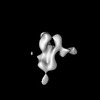
| Annotation | nsEM map of hemagglutinin H3 A/Sing/INFIMH/16 complexed with polyclonal Fab samples from individual 182419 at day 2 using Sulfo-SDAD photo-cross-linker | ||||||||||||
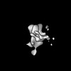
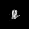
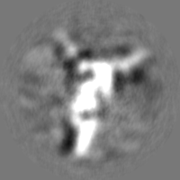
| Projections & Slices |
| ||||||||||||
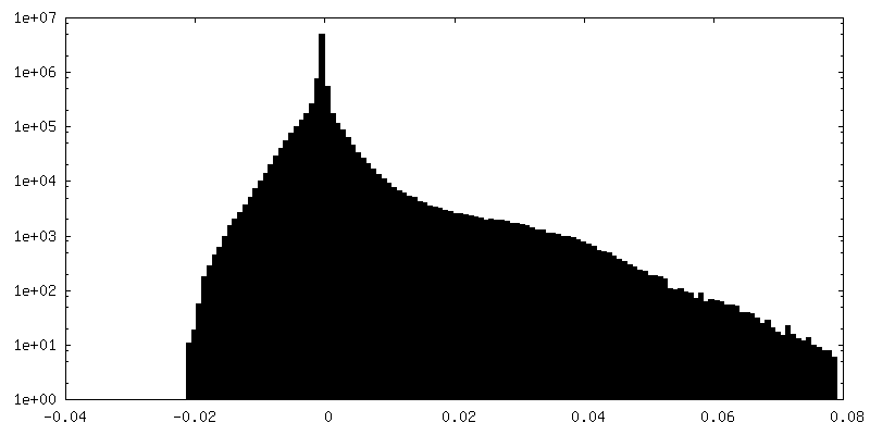
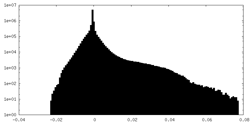
| Density Histograms |


- Sample components
-Entire : nsEM map of hemagglutinin H1 A/Mich/045/15 complexed with polyclo...
| Entire | Name: nsEM map of hemagglutinin H1 A/Mich/045/15 complexed with polyclonal Fab samples from individual 182419 at day 0 |
|---|---|
| Components |
|
-Supramolecule #1: nsEM map of hemagglutinin H1 A/Mich/045/15 complexed with polyclo...
| Supramolecule | Name: nsEM map of hemagglutinin H1 A/Mich/045/15 complexed with polyclonal Fab samples from individual 182419 at day 0 type: complex / ID: 1 / Parent: 0 Details: HA recombinantly expressed in HEK 293F cells Fab samples isolated from several individuals at different timepoints |
|---|---|
| Source (natural) | Organism: Homo sapiens (human) |
-Experimental details
-Structure determination
| Method | negative staining |
|---|---|
 Processing Processing | single particle reconstruction |
| Aggregation state | particle |
-Sample preparation
| Concentration | 0.02 mg/mL |
|---|---|
| Buffer | pH: 7.4 / Component - Concentration: 0.02 0.020 / Component - Formula: TBS / Component - Name: Tris-buffered saline |
| Staining | Type: NEGATIVE / Material: Uranyl Formate |
| Grid | Model: EMS Lacey Carbon / Support film - Material: CARBON / Support film - topology: CONTINUOUS |
| Details | 15 ug of H1 A/Mich15/045/15 was complexed overnight with 500 ug of polyclonal Fab samples and SEC purified. The complex peak was concentrated to ~0.02 mg/ml and imaged |
- Electron microscopy
| Microscope | FEI TECNAI SPIRIT |
|---|---|
| Image recording | Film or detector model: FEI EAGLE (4k x 4k) / Number grids imaged: 1 / Average electron dose: 50.0 e/Å2 |
| Electron beam | Acceleration voltage: 120 kV / Electron source: LAB6 |
| Electron optics | C2 aperture diameter: 70.0 µm / Illumination mode: FLOOD BEAM / Imaging mode: BRIGHT FIELD / Nominal defocus max: 2.0 µm / Nominal defocus min: 1.5 µm / Nominal magnification: 52000 |
| Sample stage | Cooling holder cryogen: NITROGEN |
| Experimental equipment |  Model: Tecnai Spirit / Image courtesy: FEI Company |