 Movie
Movie Controller


+ Open data

- Basic information
| Entry |  | |||||||||
|---|---|---|---|---|---|---|---|---|---|---|
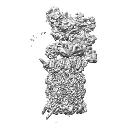
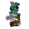
| Title | Human 19S-20S proteasome, state SD3/EC2 | |||||||||
 Map data Map data | ||||||||||
 Sample Sample |
| |||||||||
| Biological species |  Homo sapiens (human) Homo sapiens (human) | |||||||||
| Method | single particle reconstruction / cryo EM / Resolution: 3.3 Å | |||||||||
 Authors Authors | Zhao J | |||||||||
| Funding support | 1 items
| |||||||||
 Citation Citation | Journal: Proc Natl Acad Sci U S A / Year: 2022 Title: Structural insights into the human PA28-20S proteasome enabled by efficient tagging and purification of endogenous proteins. Authors: Jianhua Zhao / Suraj Makhija / Chenyu Zhou / Hanxiao Zhang / YongQiang Wang / Monita Muralidharan / Bo Huang / Yifan Cheng /  Abstract: The ability to produce folded and functional proteins is a necessity for structural biology and many other biological sciences. This task is particularly challenging for numerous biomedically ...The ability to produce folded and functional proteins is a necessity for structural biology and many other biological sciences. This task is particularly challenging for numerous biomedically important targets in human cells, including membrane proteins and large macromolecular assemblies, hampering mechanistic studies and drug development efforts. Here we describe a method combining CRISPR-Cas gene editing and fluorescence-activated cell sorting to rapidly tag and purify endogenous proteins in HEK cells for structural characterization. We applied this approach to study the human proteasome from HEK cells and rapidly determined cryogenic electron microscopy structures of major proteasomal complexes, including a high-resolution structure of intact human PA28αβ-20S. Our structures reveal that PA28 with a subunit stoichiometry of 3α/4β engages tightly with the 20S proteasome. Addition of a hydrophilic peptide shows that polypeptides entering through PA28 are held in the antechamber of 20S prior to degradation in the proteolytic chamber. This study provides critical insights into an important proteasome complex and demonstrates key methodologies for the tagging of proteins from endogenous sources. | |||||||||
| History |
|
- Structure visualization
| Supplemental images |
|---|
- Downloads & links
-EMDB archive
| Map data | emd_27019.map.gz | 127.3 MB |  EMDB map data format EMDB map data format | |
|---|---|---|---|---|
| Header (meta data) | emd-27019-v30.xmlemd-27019.xml | 12.9 KB 12.9 KB | Display Display | EMDB header |
| FSC (resolution estimation) | emd_27019_fsc.xml | 20.8 KB | Display | FSC data file |
| Images |  emd_27019.png emd_27019.png | 90.7 KB | ||
| Others | emd_27019_half_map_1.map.gzemd_27019_half_map_2.map.gz | 474.4 MB 474.4 MB | ||
| Archive directory |  http://ftp.pdbj.org/pub/emdb/structures/EMD-27019ftp://ftp.pdbj.org/pub/emdb/structures/EMD-27019 http://ftp.pdbj.org/pub/emdb/structures/EMD-27019ftp://ftp.pdbj.org/pub/emdb/structures/EMD-27019 | HTTPS FTP |
-Related structure data


-Links
| EMDB pages | EMDB (EBI/PDBe) / EMDataResource |
|---|
-Map
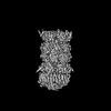
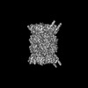
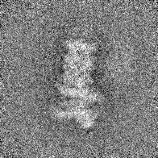
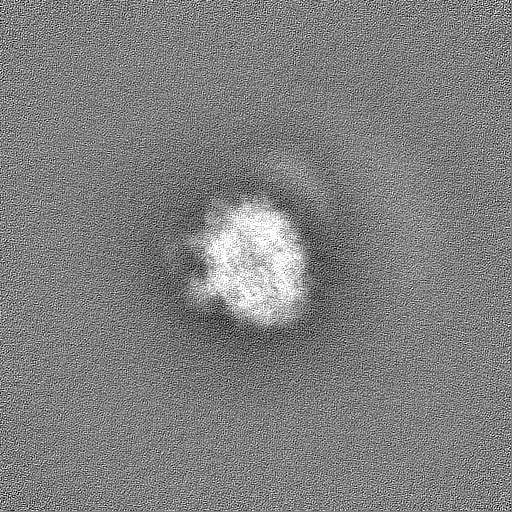
| File | Download / File: emd_27019.map.gz / Format: CCP4 / Size: 149.9 MB / Type: IMAGE STORED AS FLOATING POINT NUMBER (4 BYTES) | ||||||||||||||||||||||||||||||||||||
|---|---|---|---|---|---|---|---|---|---|---|---|---|---|---|---|---|---|---|---|---|---|---|---|---|---|---|---|---|---|---|---|---|---|---|---|---|---|
| Projections & slices | Image control
Images are generated by Spider. | ||||||||||||||||||||||||||||||||||||
| Voxel size | X=Y=Z: 1.06 Å | ||||||||||||||||||||||||||||||||||||
| Density |
| ||||||||||||||||||||||||||||||||||||
| Symmetry | Space group: 1 | ||||||||||||||||||||||||||||||||||||
| Details | EMDB XML:
|
 Z (Sec.)
 Y (Row.)
 X (Col.)


-Supplemental data
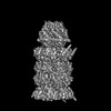
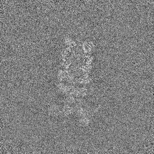
-Half map: #1
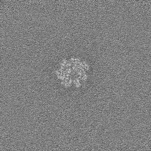
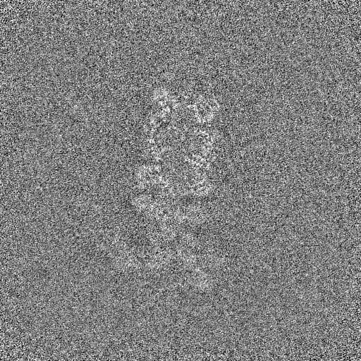
| File | emd_27019_half_map_1.map | ||||||||||||
|---|---|---|---|---|---|---|---|---|---|---|---|---|---|
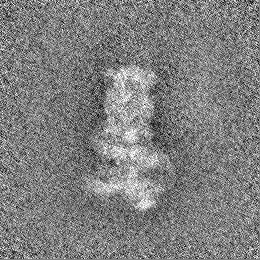
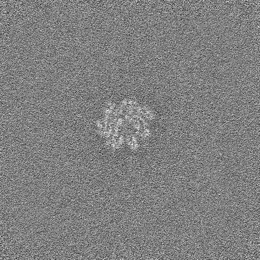
| Projections & Slices |
| ||||||||||||
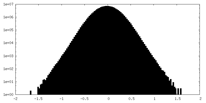
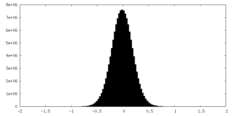
| Density Histograms |


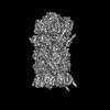
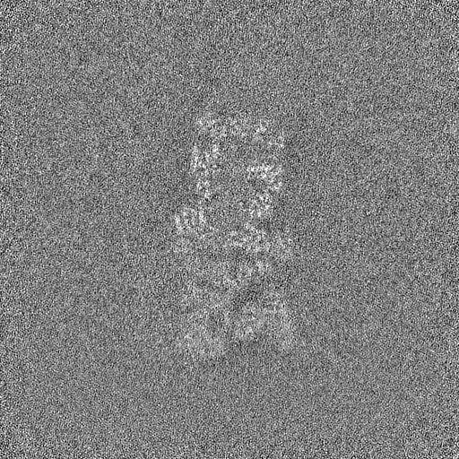
-Half map: #2
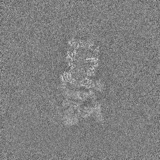
| File | emd_27019_half_map_2.map | ||||||||||||
|---|---|---|---|---|---|---|---|---|---|---|---|---|---|
| Projections & Slices |
| ||||||||||||
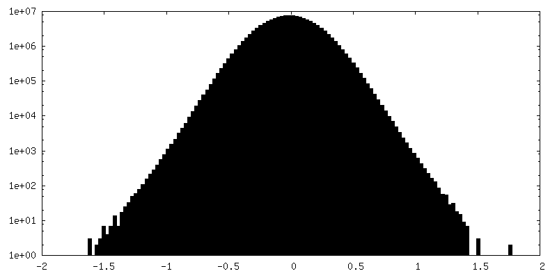
| Density Histograms |

- Sample components
-Entire : human 19S-20S proteasome complex
| Entire | Name: human 19S-20S proteasome complex |
|---|---|
| Components |
|
-Supramolecule #1: human 19S-20S proteasome complex
| Supramolecule | Name: human 19S-20S proteasome complex / type: complex / Chimera: Yes / ID: 1 / Parent: 0 |
|---|---|
| Source (natural) | Organism: Homo sapiens (human) |
-Experimental details
-Structure determination
| Method | cryo EM |
|---|---|
 Processing Processing | single particle reconstruction |
| Aggregation state | particle |
-Sample preparation
| Buffer | pH: 7.5 |
|---|---|
| Vitrification | Cryogen name: ETHANE |
- Electron microscopy
| Microscope | FEI TITAN KRIOS |
|---|---|
| Image recording | Film or detector model: GATAN K3 (6k x 4k) / Average electron dose: 30.0 e/Å2 |
| Electron beam | Acceleration voltage: 300 kV / Electron source:  FIELD EMISSION GUN FIELD EMISSION GUN |
| Electron optics | Illumination mode: FLOOD BEAM / Imaging mode: BRIGHT FIELD / Nominal defocus max: 2.0 µm / Nominal defocus min: 1.0 µm |
| Experimental equipment |  Model: Titan Krios / Image courtesy: FEI Company |
-Image processing
| Final reconstruction | Resolution.type: BY AUTHOR / Resolution: 3.3 Å / Resolution method: FSC 0.143 CUT-OFF / Number images used: 48612 |
|---|---|
| Initial angle assignment | Type: PROJECTION MATCHING |
| Final angle assignment | Type: PROJECTION MATCHING |
| FSC plot (resolution estimation) | 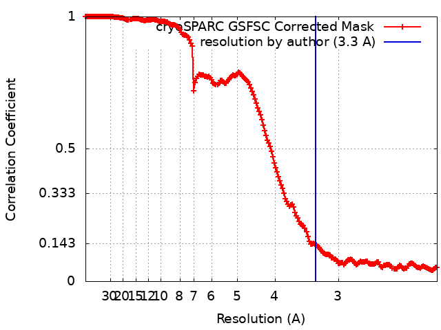 |
