 ムービー
ムービー コントローラー


+ データを開く

- 基本情報
基本情報
| 登録情報 |  | |||||||||
|---|---|---|---|---|---|---|---|---|---|---|
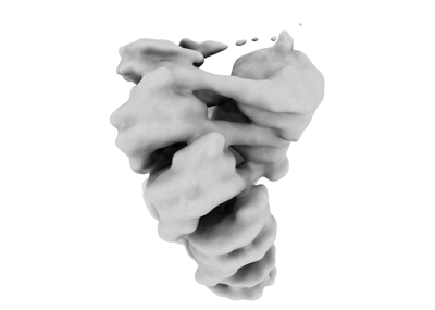
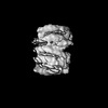
| タイトル | Cryo-EM structure of centromeric chromatin array shaped by the kinetochore protein CENP-N | |||||||||
 マップデータ マップデータ | ||||||||||
 試料 試料 |
| |||||||||
| 生物種 |  Homo sapiens (ヒト) Homo sapiens (ヒト) | |||||||||
| 手法 | 単粒子再構成法 / クライオ電子顕微鏡法 / 解像度: 12.5 Å | |||||||||
 データ登録者 データ登録者 | Zhou K / Luger K | |||||||||
| 資金援助 |  米国, 1件 米国, 1件
| |||||||||
 引用 引用 | ジャーナル: Nat Struct Mol Biol / 年: 2022 タイトル: CENP-N promotes the compaction of centromeric chromatin. 著者: Keda Zhou / Magdalena Gebala / Dustin Woods / Kousik Sundararajan / Garrett Edwards / Dan Krzizike / Jeff Wereszczynski / Aaron F Straight / Karolin Luger / 要旨: The histone variant CENP-A is the epigenetic determinant for the centromere, where it is interspersed with canonical H3 to form a specialized chromatin structure that nucleates the kinetochore. How ...The histone variant CENP-A is the epigenetic determinant for the centromere, where it is interspersed with canonical H3 to form a specialized chromatin structure that nucleates the kinetochore. How nucleosomes at the centromere arrange into higher order structures is unknown. Here we demonstrate that the human CENP-A-interacting protein CENP-N promotes the stacking of CENP-A-containing mononucleosomes and nucleosomal arrays through a previously undefined interaction between the α6 helix of CENP-N with the DNA of a neighboring nucleosome. We describe the cryo-EM structures and biophysical characterization of such CENP-N-mediated nucleosome stacks and nucleosomal arrays and demonstrate that this interaction is responsible for the formation of densely packed chromatin at the centromere in the cell. Our results provide first evidence that CENP-A, together with CENP-N, promotes specific chromatin higher order structure at the centromere. | |||||||||
| 履歴 |
|
- 構造の表示
構造の表示
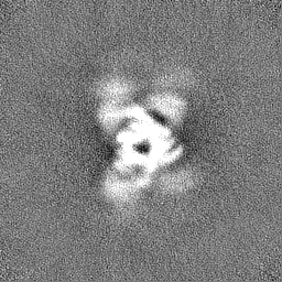
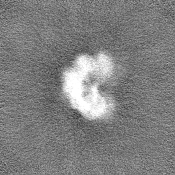
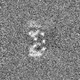
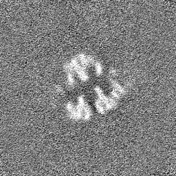
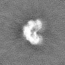
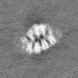
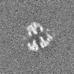
| 添付画像 |
|---|
- ダウンロードとリンク
-EMDBアーカイブ
| マップデータ | emd_26333.map.gz | 59.1 MB |  EMDBマップデータ形式 EMDBマップデータ形式 | |
|---|---|---|---|---|
| ヘッダ (付随情報) | emd-26333-v30.xmlemd-26333.xml | 11.9 KB 11.9 KB | 表示 表示 | EMDBヘッダ |
| FSC (解像度算出) | emd_26333_fsc.xml | 9.8 KB | 表示 | FSCデータファイル |
| 画像 |  emd_26333.png emd_26333.png | 30.2 KB | ||
| マスクデータ | emd_26333_msk_1.map | 64 MB | マスクマップ | |
| その他 | emd_26333_half_map_1.map.gzemd_26333_half_map_2.map.gz | 59.4 MB 59.4 MB | ||
| アーカイブディレクトリ |  http://ftp.pdbj.org/pub/emdb/structures/EMD-26333ftp://ftp.pdbj.org/pub/emdb/structures/EMD-26333 http://ftp.pdbj.org/pub/emdb/structures/EMD-26333ftp://ftp.pdbj.org/pub/emdb/structures/EMD-26333 | HTTPS FTP |
-検証レポート
| 文書・要旨 | emd_26333_validation.pdf.gz | 919.1 KB | 表示 | EMDB検証レポート |
|---|---|---|---|---|
| 文書・詳細版 | emd_26333_full_validation.pdf.gz | 918.6 KB | 表示 | |
| XML形式データ | emd_26333_validation.xml.gz | 16.3 KB | 表示 | |
| CIF形式データ | emd_26333_validation.cif.gz | 21.6 KB | 表示 | |
| アーカイブディレクトリ | https://ftp.pdbj.org/pub/emdb/validation_reports/EMD-26333ftp://ftp.pdbj.org/pub/emdb/validation_reports/EMD-26333 | HTTPS FTP |
-関連構造データ
-リンク
| EMDBのページ | EMDB (EBI/PDBe) / EMDataResource |
|---|
-マップ
| ファイル | ダウンロード / ファイル: emd_26333.map.gz / 形式: CCP4 / 大きさ: 64 MB / タイプ: IMAGE STORED AS FLOATING POINT NUMBER (4 BYTES) | ||||||||||||||||||||||||||||||||||||
|---|---|---|---|---|---|---|---|---|---|---|---|---|---|---|---|---|---|---|---|---|---|---|---|---|---|---|---|---|---|---|---|---|---|---|---|---|---|
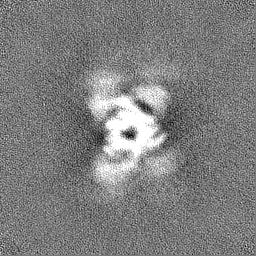
| 投影像・断面図 | 画像のコントロール
画像は Spider により作成 | ||||||||||||||||||||||||||||||||||||
| ボクセルのサイズ | X=Y=Z: 2.48 Å | ||||||||||||||||||||||||||||||||||||
| 密度 |
| ||||||||||||||||||||||||||||||||||||
| 対称性 | 空間群: 1 | ||||||||||||||||||||||||||||||||||||
| 詳細 | EMDB XML:
|
 Z (Sec.)
 Y (Row.)
 X (Col.)


-添付データ
-マスク #1
| ファイル | emd_26333_msk_1.map | ||||||||||||
|---|---|---|---|---|---|---|---|---|---|---|---|---|---|
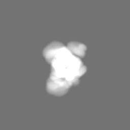
| 投影像・断面図 |
| ||||||||||||
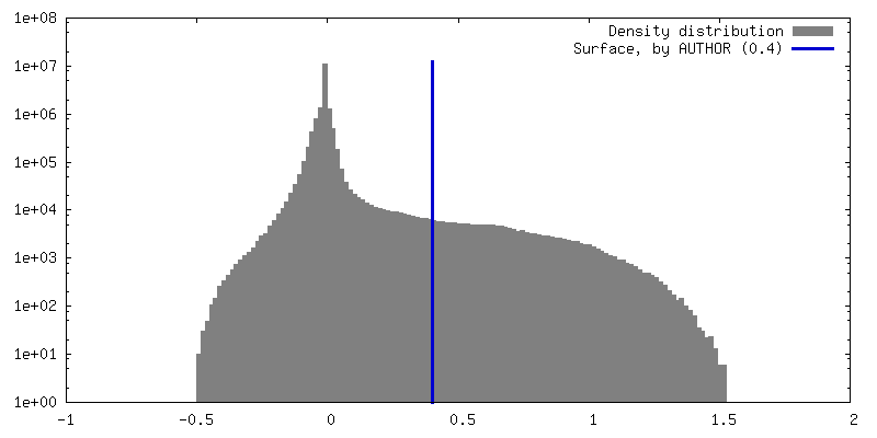
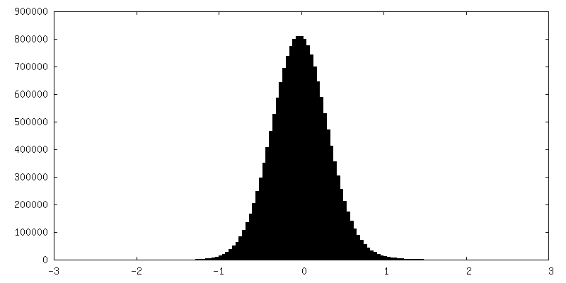
| 密度ヒストグラム |


-ハーフマップ: #1
| ファイル | emd_26333_half_map_1.map | ||||||||||||
|---|---|---|---|---|---|---|---|---|---|---|---|---|---|
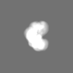
| 投影像・断面図 |
| ||||||||||||
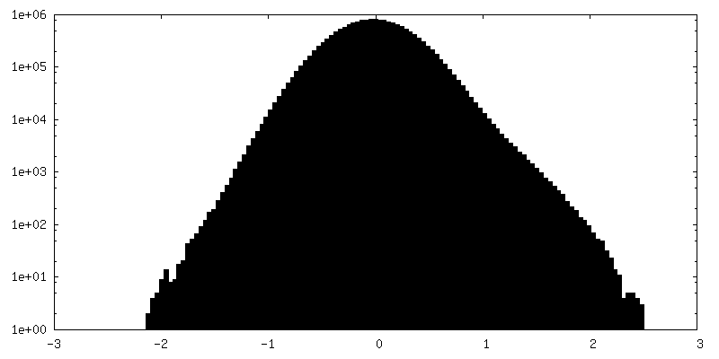
| 密度ヒストグラム |


-ハーフマップ: #2
| ファイル | emd_26333_half_map_2.map | ||||||||||||
|---|---|---|---|---|---|---|---|---|---|---|---|---|---|
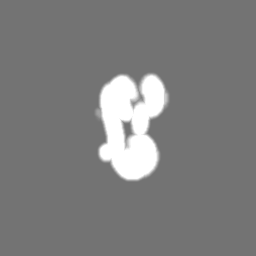
| 投影像・断面図 |
| ||||||||||||
| 密度ヒストグラム |


- 試料の構成要素
-全体 : Centromeric chromatin array in complex with kinetochore protein CENP-N
| 全体 | 名称: Centromeric chromatin array in complex with kinetochore protein CENP-N |
|---|---|
| 要素 |
|
-超分子 #1: Centromeric chromatin array in complex with kinetochore protein CENP-N
| 超分子 | 名称: Centromeric chromatin array in complex with kinetochore protein CENP-N タイプ: complex / キメラ: Yes / ID: 1 / 親要素: 0 |
|---|---|
| 由来(天然) | 生物種: Homo sapiens (ヒト) |
| 組換発現 | 生物種:  |
-実験情報
-構造解析
| 手法 | クライオ電子顕微鏡法 |
|---|---|
 解析 解析 | 単粒子再構成法 |
| 試料の集合状態 | particle |
-試料調製
| 緩衝液 | pH: 7.8 |
|---|---|
| 凍結 | 凍結剤: ETHANE |
- 電子顕微鏡法
| 顕微鏡 | FEI TITAN KRIOS |
|---|---|
| 撮影 | フィルム・検出器のモデル: GATAN K2 SUMMIT (4k x 4k) 平均電子線量: 100.0 e/Å2 |
| 電子線 | 加速電圧: 300 kV / 電子線源:  FIELD EMISSION GUN FIELD EMISSION GUN |
| 電子光学系 | 照射モード: FLOOD BEAM / 撮影モード: BRIGHT FIELD / 最大 デフォーカス(公称値): 2.5 µm / 最小 デフォーカス(公称値): 1.3 µm |
| 実験機器 |  モデル: Titan Krios / 画像提供: FEI Company |
-画像解析
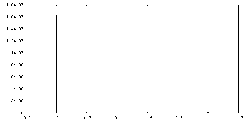
| 最終 再構成 | 解像度のタイプ: BY AUTHOR / 解像度: 12.5 Å / 解像度の算出法: FSC 0.143 CUT-OFF / 使用した粒子像数: 9305 |
|---|---|
| 初期 角度割当 | タイプ: ANGULAR RECONSTITUTION |
| 最終 角度割当 | タイプ: ANGULAR RECONSTITUTION |
| FSC曲線 (解像度の算出) | 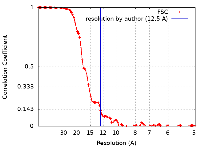 |
