ムービー
ムービー コントローラー


+ データを開く

- 基本情報
基本情報
| 登録情報 |  | ||||||||||||
|---|---|---|---|---|---|---|---|---|---|---|---|---|---|
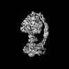
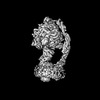
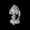
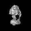
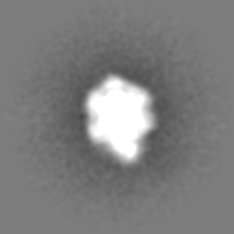
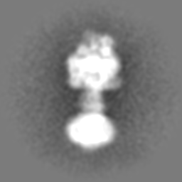
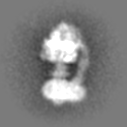
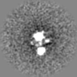
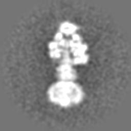
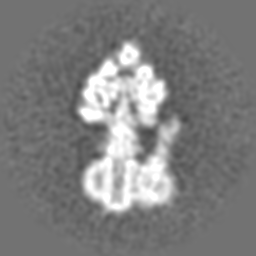
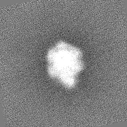
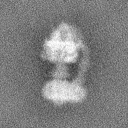
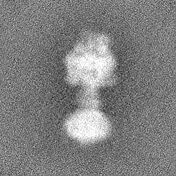
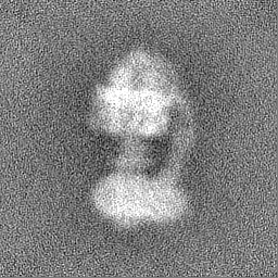
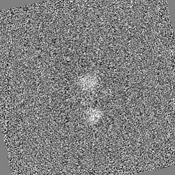
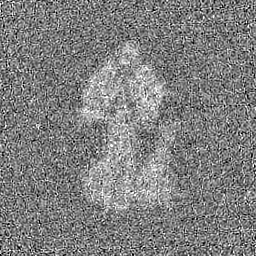
| タイトル | Yeast ATP synthase with 10 mM ATP showing strained peripheral stalk determined by a screening microscope | ||||||||||||
 マップデータ マップデータ | |||||||||||||
 試料 試料 |
| ||||||||||||
 キーワード キーワード | F1-ATPase / ATP Synthase / Hydrolase / Nanomotor / Complex | ||||||||||||
| 生物種 |  | ||||||||||||
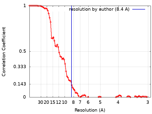
| 手法 | 単粒子再構成法 / クライオ電子顕微鏡法 / 解像度: 8.4 Å | ||||||||||||
 データ登録者 データ登録者 | Guo H / Rubinstein JL | ||||||||||||
| 資金援助 |  カナダ, 3件 カナダ, 3件
| ||||||||||||
 引用 引用 | ジャーナル: Nat Commun / 年: 2022 タイトル: Structure of ATP synthase under strain during catalysis. 著者: Hui Guo / John L Rubinstein / 要旨: ATP synthases are macromolecular machines consisting of an ATP-hydrolysis-driven F motor and a proton-translocation-driven F motor. The F and F motors oppose each other's action on a shared rotor ...ATP synthases are macromolecular machines consisting of an ATP-hydrolysis-driven F motor and a proton-translocation-driven F motor. The F and F motors oppose each other's action on a shared rotor subcomplex and are held stationary relative to each other by a peripheral stalk. Structures of resting mitochondrial ATP synthases revealed a left-handed curvature of the peripheral stalk even though rotation of the rotor, driven by either ATP hydrolysis in F or proton translocation through F, would apply a right-handed bending force to the stalk. We used cryoEM to image yeast mitochondrial ATP synthase under strain during ATP-hydrolysis-driven rotary catalysis, revealing a large deformation of the peripheral stalk. The structures show how the peripheral stalk opposes the bending force and suggests that during ATP synthesis proton translocation causes accumulation of strain in the stalk, which relaxes by driving the relative rotation of the rotor through six sub-steps within F, leading to catalysis. #1: ジャーナル: Biorxiv / 年: 2022タイトル: Structure of ATP synthase under strain during catalysis 著者: Guo H / Rubinstein JL | ||||||||||||
| 履歴 |
|
- 構造の表示
構造の表示
| 添付画像 |
|---|

- ダウンロードとリンク
-EMDBアーカイブ
| マップデータ | emd_25953.map.gz | 58.5 MB |  EMDBマップデータ形式 EMDBマップデータ形式 | |
|---|---|---|---|---|
| ヘッダ (付随情報) | emd-25953-v30.xmlemd-25953.xml | 18.7 KB 18.7 KB | 表示 表示 | EMDBヘッダ |
| FSC (解像度算出) | emd_25953_fsc.xml | 9 KB | 表示 | FSCデータファイル |
| 画像 | 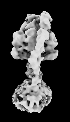 emd_25953.png emd_25953.png | 43.1 KB | ||
| マスクデータ | emd_25953_msk_1.map | 64 MB | マスクマップ | |
| Filedesc metadata | emd-25953.cif.gz | 4.4 KB | ||
| その他 | emd_25953_additional_1.map.gzemd_25953_half_map_1.map.gzemd_25953_half_map_2.map.gz | 37.9 MB 57.6 MB 57.6 MB | ||
| アーカイブディレクトリ |  http://ftp.pdbj.org/pub/emdb/structures/EMD-25953ftp://ftp.pdbj.org/pub/emdb/structures/EMD-25953 http://ftp.pdbj.org/pub/emdb/structures/EMD-25953ftp://ftp.pdbj.org/pub/emdb/structures/EMD-25953 | HTTPS FTP |
-検証レポート
| 文書・要旨 | emd_25953_validation.pdf.gz | 1.2 MB | 表示 | EMDB検証レポート |
|---|---|---|---|---|
| 文書・詳細版 | emd_25953_full_validation.pdf.gz | 1.2 MB | 表示 | |
| XML形式データ | emd_25953_validation.xml.gz | 16.7 KB | 表示 | |
| CIF形式データ | emd_25953_validation.cif.gz | 21.2 KB | 表示 | |
| アーカイブディレクトリ | https://ftp.pdbj.org/pub/emdb/validation_reports/EMD-25953ftp://ftp.pdbj.org/pub/emdb/validation_reports/EMD-25953 | HTTPS FTP |
-関連構造データ
| 関連構造データ |  7tjsC  7tjtC  7tjuC  7tjvC  7tjwC  7tjxC  7tjyC 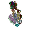 7tjzC 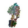 7tk0C  7tk1C 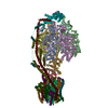 7tk2C 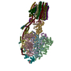 7tk3C 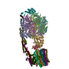 7tk4C 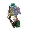 7tk5C 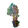 7tk6C 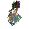 7tk7C 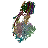 7tk8C 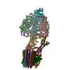 7tk9C 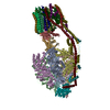 7tkaC  7tkbC 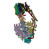 7tkcC 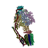 7tkdC 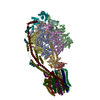 7tkeC 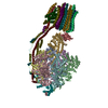 7tkfC 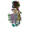 7tkgC 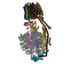 7tkhC 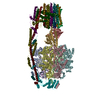 7tkiC 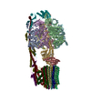 7tkjC 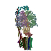 7tkkC 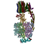 7tklC 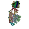 7tkmC 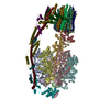 7tknC 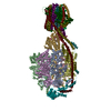 7tkoC 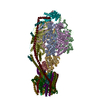 7tkpC  7tkqC  7tkrC 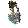 7tksC C: 同じ文献を引用 ( |
|---|


-リンク
| EMDBのページ | EMDB (EBI/PDBe) / EMDataResource |
|---|
-マップ
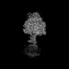
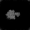
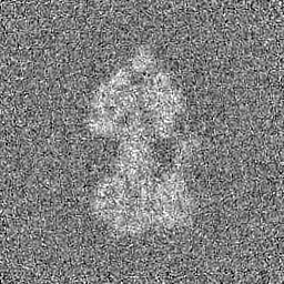
| ファイル | ダウンロード / ファイル: emd_25953.map.gz / 形式: CCP4 / 大きさ: 64 MB / タイプ: IMAGE STORED AS FLOATING POINT NUMBER (4 BYTES) | ||||||||||||||||||||||||||||||||||||
|---|---|---|---|---|---|---|---|---|---|---|---|---|---|---|---|---|---|---|---|---|---|---|---|---|---|---|---|---|---|---|---|---|---|---|---|---|---|
| 投影像・断面図 | 画像のコントロール
画像は Spider により作成 | ||||||||||||||||||||||||||||||||||||
| ボクセルのサイズ | X=Y=Z: 1.3475 Å | ||||||||||||||||||||||||||||||||||||
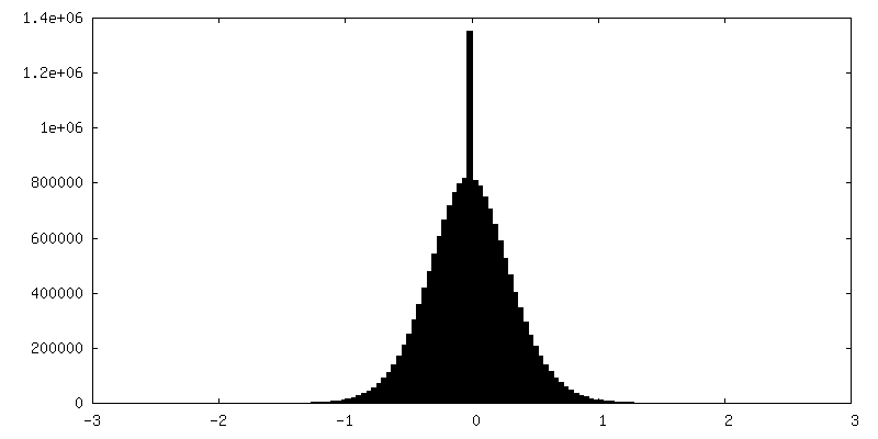
| 密度 |
| ||||||||||||||||||||||||||||||||||||
| 対称性 | 空間群: 1 | ||||||||||||||||||||||||||||||||||||
| 詳細 | EMDB XML:
|
 Z (Sec.)
 Y (Row.)
 X (Col.)


-添付データ
-マスク #1
| ファイル | emd_25953_msk_1.map | ||||||||||||
|---|---|---|---|---|---|---|---|---|---|---|---|---|---|
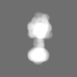
| 投影像・断面図 |
| ||||||||||||
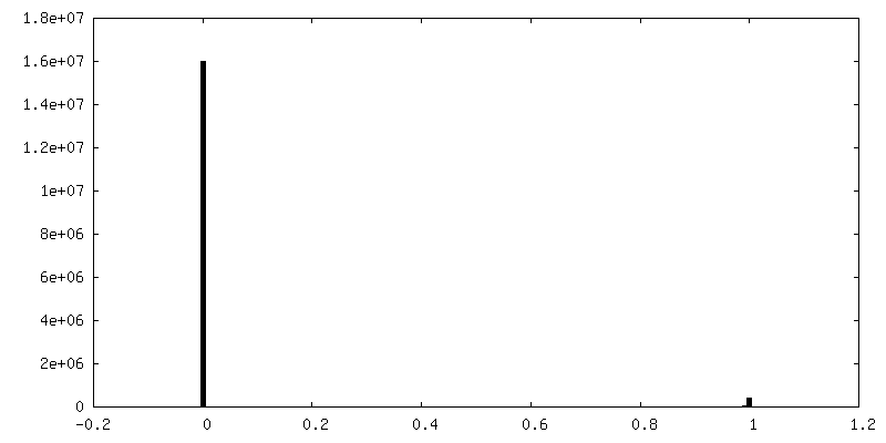
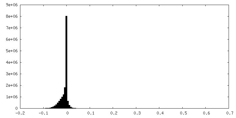
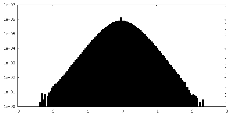
| 密度ヒストグラム |


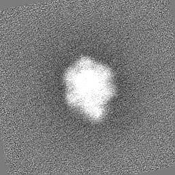
-追加マップ: Unsharpened map.
| ファイル | emd_25953_additional_1.map | ||||||||||||
|---|---|---|---|---|---|---|---|---|---|---|---|---|---|
| 注釈 | Unsharpened map. | ||||||||||||
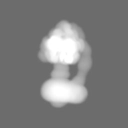
| 投影像・断面図 |
| ||||||||||||
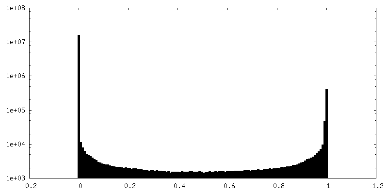
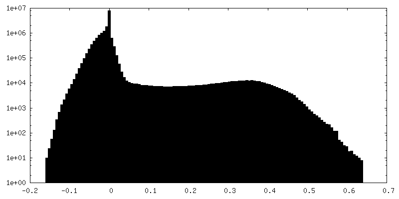
| 密度ヒストグラム |
-ハーフマップ: #1
| ファイル | emd_25953_half_map_1.map | ||||||||||||
|---|---|---|---|---|---|---|---|---|---|---|---|---|---|
| 投影像・断面図 |
| ||||||||||||
| 密度ヒストグラム |

-ハーフマップ: #2
| ファイル | emd_25953_half_map_2.map | ||||||||||||
|---|---|---|---|---|---|---|---|---|---|---|---|---|---|
| 投影像・断面図 |
| ||||||||||||
| 密度ヒストグラム |

- 試料の構成要素
-全体 : Yeast ATP synthase with 10 mM ATP showing strained peripheral sta...
| 全体 | 名称: Yeast ATP synthase with 10 mM ATP showing strained peripheral stalk determined by a screening microscope |
|---|---|
| 要素 |
|
-超分子 #1: Yeast ATP synthase with 10 mM ATP showing strained peripheral sta...
| 超分子 | 名称: Yeast ATP synthase with 10 mM ATP showing strained peripheral stalk determined by a screening microscope タイプ: complex / ID: 1 / 親要素: 0 |
|---|---|
| 由来(天然) | 生物種: |
| 分子量 | 理論値: 610 KDa |
-実験情報
-構造解析
| 手法 | クライオ電子顕微鏡法 |
|---|---|
 解析 解析 | 単粒子再構成法 |
| 試料の集合状態 | particle |
-試料調製
| 濃度 | 15 mg/mL |
|---|---|
| 緩衝液 | pH: 7.4 |
| グリッド | モデル: Homemade / 材質: COPPER/RHODIUM / メッシュ: 300 / 支持フィルム - 材質: GOLD / 支持フィルム - トポロジー: HOLEY / 支持フィルム - Film thickness: 35 / 前処理 - タイプ: GLOW DISCHARGE / 前処理 - 時間: 120 sec. |
| 凍結 | 凍結剤: ETHANE-PROPANE / チャンバー内湿度: 100 % / チャンバー内温度: 277 K / 装置: LEICA EM GP |
- 電子顕微鏡法
| 顕微鏡 | FEI TECNAI F20 |
|---|---|
| 撮影 | フィルム・検出器のモデル: GATAN K2 SUMMIT (4k x 4k) 検出モード: COUNTING / デジタル化 - サイズ - 横: 3710 pixel / デジタル化 - サイズ - 縦: 3838 pixel / デジタル化 - 画像ごとのフレーム数: 1-30 / 撮影したグリッド数: 1 / 実像数: 200 / 平均露光時間: 15.0 sec. / 平均電子線量: 36.0 e/Å2 |
| 電子線 | 加速電圧: 200 kV / 電子線源:  FIELD EMISSION GUN FIELD EMISSION GUN |
| 電子光学系 | C2レンズ絞り径: 30.0 µm / 倍率(補正後): 34483 / 照射モード: FLOOD BEAM / 撮影モード: BRIGHT FIELD / Cs: 2.0 mm / 最大 デフォーカス(公称値): 0.0025 µm / 最小 デフォーカス(公称値): 0.001 µm / 倍率(公称値): 25000 |
| 試料ステージ | 試料ホルダーモデル: GATAN 626 SINGLE TILT LIQUID NITROGEN CRYO TRANSFER HOLDER ホルダー冷却材: NITROGEN |
| 実験機器 |  モデル: Tecnai F20 / 画像提供: FEI Company |