Movie
Movie Controller

[English] 日本語
 Yorodumi
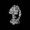
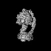
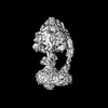
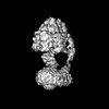
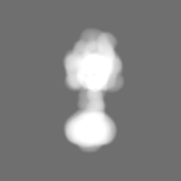
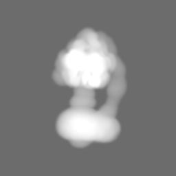
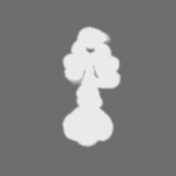
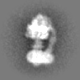
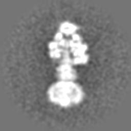
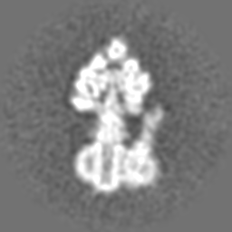
- EMDB-25953: Yeast ATP synthase with 10 mM ATP showing strained peripheral sta... -
+ Open data

- Basic information
| Entry |  | ||||||||||||
|---|---|---|---|---|---|---|---|---|---|---|---|---|---|
| Title | Yeast ATP synthase with 10 mM ATP showing strained peripheral stalk determined by a screening microscope | ||||||||||||
 Map data Map data | |||||||||||||
 Sample Sample |
| ||||||||||||
 Keywords Keywords | F1-ATPase / ATP Synthase / Hydrolase / Nanomotor / Complex | ||||||||||||
| Biological species |  | ||||||||||||
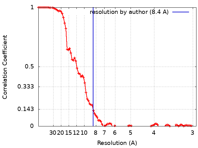
| Method | single particle reconstruction / cryo EM / Resolution: 8.4 Å | ||||||||||||
 Authors Authors | Guo H / Rubinstein JL | ||||||||||||
| Funding support |  Canada, 3 items Canada, 3 items
| ||||||||||||
 Citation Citation | Journal: Nat Commun / Year: 2022 Title: Structure of ATP synthase under strain during catalysis. Authors: Hui Guo / John L Rubinstein / Abstract: ATP synthases are macromolecular machines consisting of an ATP-hydrolysis-driven F motor and a proton-translocation-driven F motor. The F and F motors oppose each other's action on a shared rotor ...ATP synthases are macromolecular machines consisting of an ATP-hydrolysis-driven F motor and a proton-translocation-driven F motor. The F and F motors oppose each other's action on a shared rotor subcomplex and are held stationary relative to each other by a peripheral stalk. Structures of resting mitochondrial ATP synthases revealed a left-handed curvature of the peripheral stalk even though rotation of the rotor, driven by either ATP hydrolysis in F or proton translocation through F, would apply a right-handed bending force to the stalk. We used cryoEM to image yeast mitochondrial ATP synthase under strain during ATP-hydrolysis-driven rotary catalysis, revealing a large deformation of the peripheral stalk. The structures show how the peripheral stalk opposes the bending force and suggests that during ATP synthesis proton translocation causes accumulation of strain in the stalk, which relaxes by driving the relative rotation of the rotor through six sub-steps within F, leading to catalysis. #1: Journal: Biorxiv / Year: 2022Title: Structure of ATP synthase under strain during catalysis Authors: Guo H / Rubinstein JL | ||||||||||||
| History |
|
- Structure visualization
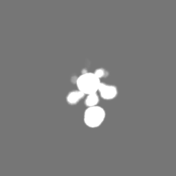
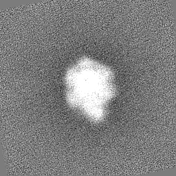
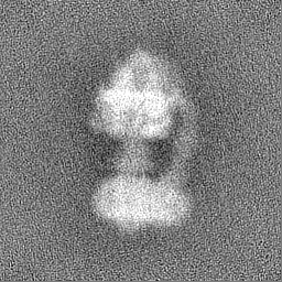
| Supplemental images |
|---|

- Downloads & links
-EMDB archive
| Map data | emd_25953.map.gz | 58.5 MB |  EMDB map data format EMDB map data format | |
|---|---|---|---|---|
| Header (meta data) | emd-25953-v30.xmlemd-25953.xml | 18.7 KB 18.7 KB | Display Display | EMDB header |
| FSC (resolution estimation) | emd_25953_fsc.xml | 9 KB | Display | FSC data file |
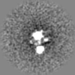
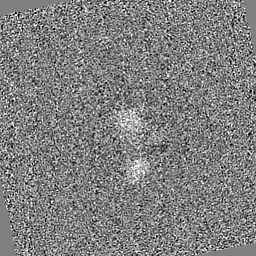
| Images | 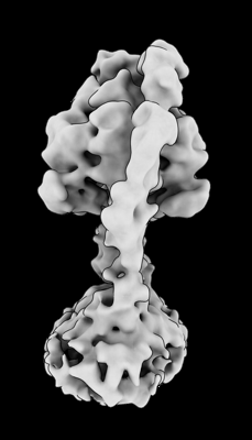 emd_25953.png emd_25953.png | 43.1 KB | ||
| Masks | emd_25953_msk_1.map | 64 MB | Mask map | |
| Filedesc metadata | emd-25953.cif.gz | 4.4 KB | ||
| Others | emd_25953_additional_1.map.gzemd_25953_half_map_1.map.gzemd_25953_half_map_2.map.gz | 37.9 MB 57.6 MB 57.6 MB | ||
| Archive directory |  http://ftp.pdbj.org/pub/emdb/structures/EMD-25953ftp://ftp.pdbj.org/pub/emdb/structures/EMD-25953 http://ftp.pdbj.org/pub/emdb/structures/EMD-25953ftp://ftp.pdbj.org/pub/emdb/structures/EMD-25953 | HTTPS FTP |
-Related structure data
| Related structure data |  7tjsC  7tjtC 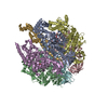 7tjuC 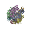 7tjvC 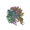 7tjwC 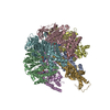 7tjxC  7tjyC 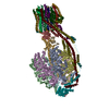 7tjzC 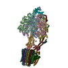 7tk0C  7tk1C 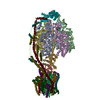 7tk2C 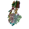 7tk3C 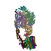 7tk4C 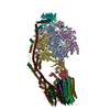 7tk5C 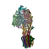 7tk6C 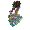 7tk7C 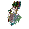 7tk8C 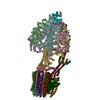 7tk9C 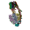 7tkaC  7tkbC 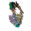 7tkcC 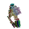 7tkdC 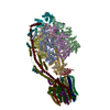 7tkeC 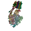 7tkfC 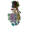 7tkgC 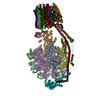 7tkhC 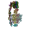 7tkiC 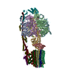 7tkjC 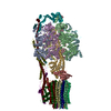 7tkkC 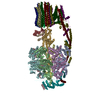 7tklC 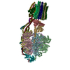 7tkmC 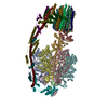 7tknC 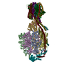 7tkoC 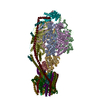 7tkpC  7tkqC  7tkrC 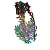 7tksC C: citing same article ( |
|---|


-Links
| EMDB pages | EMDB (EBI/PDBe) / EMDataResource |
|---|
-Map
| File | Download / File: emd_25953.map.gz / Format: CCP4 / Size: 64 MB / Type: IMAGE STORED AS FLOATING POINT NUMBER (4 BYTES) | ||||||||||||||||||||||||||||||||||||
|---|---|---|---|---|---|---|---|---|---|---|---|---|---|---|---|---|---|---|---|---|---|---|---|---|---|---|---|---|---|---|---|---|---|---|---|---|---|
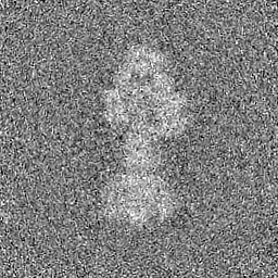
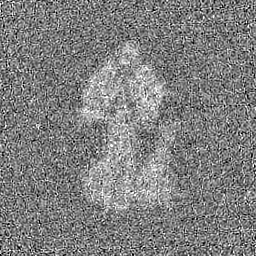
| Projections & slices | Image control
Images are generated by Spider. | ||||||||||||||||||||||||||||||||||||
| Voxel size | X=Y=Z: 1.3475 Å | ||||||||||||||||||||||||||||||||||||
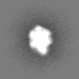
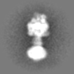
| Density |
| ||||||||||||||||||||||||||||||||||||
| Symmetry | Space group: 1 | ||||||||||||||||||||||||||||||||||||
| Details | EMDB XML:
|
 Z (Sec.)
 Y (Row.)
 X (Col.)


-Supplemental data
-Mask #1
| File | emd_25953_msk_1.map | ||||||||||||
|---|---|---|---|---|---|---|---|---|---|---|---|---|---|
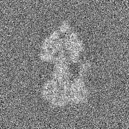
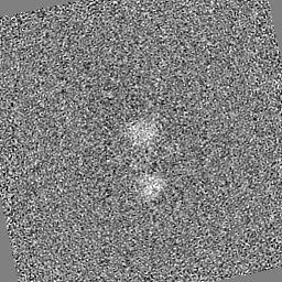
| Projections & Slices |
| ||||||||||||
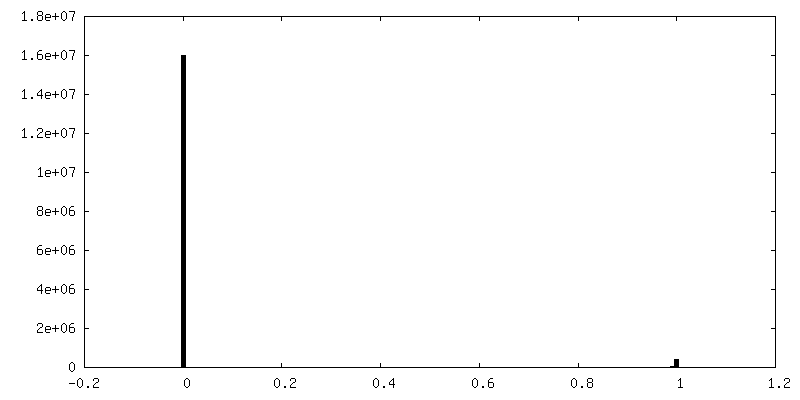
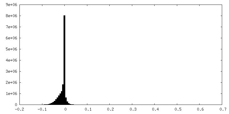
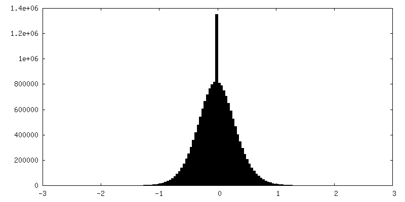
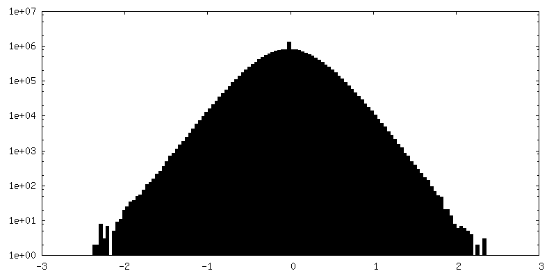
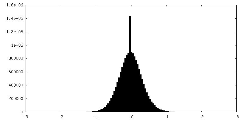
| Density Histograms |


-Additional map: Unsharpened map.
| File | emd_25953_additional_1.map | ||||||||||||
|---|---|---|---|---|---|---|---|---|---|---|---|---|---|
| Annotation | Unsharpened map. | ||||||||||||
| Projections & Slices |
| ||||||||||||
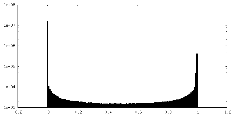
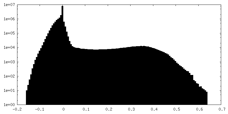
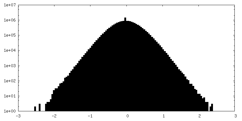
| Density Histograms |
-Half map: #1
| File | emd_25953_half_map_1.map | ||||||||||||
|---|---|---|---|---|---|---|---|---|---|---|---|---|---|
| Projections & Slices |
| ||||||||||||
| Density Histograms |

-Half map: #2
| File | emd_25953_half_map_2.map | ||||||||||||
|---|---|---|---|---|---|---|---|---|---|---|---|---|---|
| Projections & Slices |
| ||||||||||||
| Density Histograms |

- Sample components
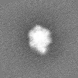
-Entire : Yeast ATP synthase with 10 mM ATP showing strained peripheral sta...
| Entire | Name: Yeast ATP synthase with 10 mM ATP showing strained peripheral stalk determined by a screening microscope |
|---|---|
| Components |
|
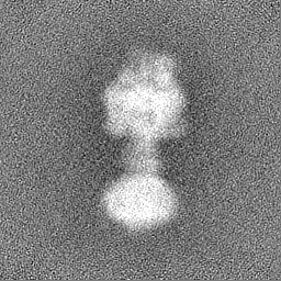
-Supramolecule #1: Yeast ATP synthase with 10 mM ATP showing strained peripheral sta...
| Supramolecule | Name: Yeast ATP synthase with 10 mM ATP showing strained peripheral stalk determined by a screening microscope type: complex / ID: 1 / Parent: 0 |
|---|---|
| Source (natural) | Organism: |
| Molecular weight | Theoretical: 610 KDa |
-Experimental details
-Structure determination
| Method | cryo EM |
|---|---|
 Processing Processing | single particle reconstruction |
| Aggregation state | particle |
-Sample preparation
| Concentration | 15 mg/mL |
|---|---|
| Buffer | pH: 7.4 |
| Grid | Model: Homemade / Material: COPPER/RHODIUM / Mesh: 300 / Support film - Material: GOLD / Support film - topology: HOLEY / Support film - Film thickness: 35 / Pretreatment - Type: GLOW DISCHARGE / Pretreatment - Time: 120 sec. |
| Vitrification | Cryogen name: ETHANE-PROPANE / Chamber humidity: 100 % / Chamber temperature: 277 K / Instrument: LEICA EM GP |
- Electron microscopy
| Microscope | FEI TECNAI F20 |
|---|---|
| Image recording | Film or detector model: GATAN K2 SUMMIT (4k x 4k) / Detector mode: COUNTING / Digitization - Dimensions - Width: 3710 pixel / Digitization - Dimensions - Height: 3838 pixel / Digitization - Frames/image: 1-30 / Number grids imaged: 1 / Number real images: 200 / Average exposure time: 15.0 sec. / Average electron dose: 36.0 e/Å2 |
| Electron beam | Acceleration voltage: 200 kV / Electron source:  FIELD EMISSION GUN FIELD EMISSION GUN |
| Electron optics | C2 aperture diameter: 30.0 µm / Calibrated magnification: 34483 / Illumination mode: FLOOD BEAM / Imaging mode: BRIGHT FIELD / Cs: 2.0 mm / Nominal defocus max: 0.0025 µm / Nominal defocus min: 0.001 µm / Nominal magnification: 25000 |
| Sample stage | Specimen holder model: GATAN 626 SINGLE TILT LIQUID NITROGEN CRYO TRANSFER HOLDER Cooling holder cryogen: NITROGEN |
| Experimental equipment |  Model: Tecnai F20 / Image courtesy: FEI Company |