[English] 日本語
Yorodumi
- EMDB-25779: Cryo-EM structure of bafilomycin A1 bound to yeast VO V-ATPase -

+
Open data


ID or keywords:

Loading...

-
Basic information

Entry
Database: EMDB / ID: EMD-25779
TitleCryo-EM structure of bafilomycin A1 bound to yeast VO V-ATPase
Map data
Sample
  • Complex: Cryo-EM structure of bafilomycin A1 bound to yeast VO V-ATPase
    • Protein or peptide: V-type proton ATPase subunit c'
    • Protein or peptide: V-type proton ATPase subunit c''
    • Protein or peptide: V0 assembly protein 1
    • Protein or peptide: V-type proton ATPase subunit e
    • Protein or peptide: V-type proton ATPase subunit c
    • Protein or peptide: Yeast V-ATPase subunit f
    • Protein or peptide: V-type proton ATPase subunit d
    • Protein or peptide: V-type proton ATPase subunit a, vacuolar isoform
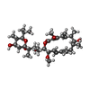
  • Ligand: (5R)-2,4-dideoxy-1-C-{(2S,3R,4S)-3-hydroxy-4-[(2R,3S,4E,6E,9R,10S,11R,12E,14Z)-10-hydroxy-3,15-dimethoxy-7,9,11,13-tetramethyl-16-oxo-1-oxacyclohexadeca-4,6,12,14-tetraen-2-yl]pentan-2-yl}-4-methyl-5-propan-2-yl-alpha-D-threo-pentopyranose
Function / homology
Function and homology information


cell wall mannoprotein biosynthetic process / ATPase-coupled ion transmembrane transporter activity / cellular response to alkaline pH / polyphosphate metabolic process / P-type proton-exporting transporter activity / Insulin receptor recycling / Transferrin endocytosis and recycling / ROS and RNS production in phagocytes / Amino acids regulate mTORC1 / Golgi lumen acidification ...cell wall mannoprotein biosynthetic process / ATPase-coupled ion transmembrane transporter activity / cellular response to alkaline pH / polyphosphate metabolic process / P-type proton-exporting transporter activity / Insulin receptor recycling / Transferrin endocytosis and recycling / ROS and RNS production in phagocytes / Amino acids regulate mTORC1 / Golgi lumen acidification / vacuolar transport / : / vacuolar proton-transporting V-type ATPase, V0 domain / endosomal lumen acidification / protein targeting to vacuole / proton-transporting V-type ATPase complex / vacuolar proton-transporting V-type ATPase complex / vacuole organization / fungal-type vacuole / vacuolar acidification / cellular hyperosmotic response / fungal-type vacuole membrane / phosphatidylinositol-3,5-bisphosphate binding / proton transmembrane transporter activity / intracellular copper ion homeostasis / RNA endonuclease activity / Neutrophil degranulation / proton-transporting ATPase activity, rotational mechanism / proton transmembrane transport / cell periphery / transmembrane transport / endocytosis / ATPase binding / protein-containing complex assembly / intracellular iron ion homeostasis / Golgi membrane / endoplasmic reticulum membrane / membrane
Similarity search - Function
Ribonuclease kappa / V-type proton ATPase subunit S1/VOA1, transmembrane domain / V0 complex accessory subunit Ac45/VOA1 transmembrane domain / ATPase, V0 complex, subunit e1/e2 / ATP synthase subunit H / ATPase, V0 complex, subunit d / V-ATPase proteolipid subunit C, eukaryotic / ATPase, V0 complex, subunit 116kDa, eukaryotic / V-ATPase proteolipid subunit / ATPase, V0 complex, c/d subunit ...Ribonuclease kappa / V-type proton ATPase subunit S1/VOA1, transmembrane domain / V0 complex accessory subunit Ac45/VOA1 transmembrane domain / ATPase, V0 complex, subunit e1/e2 / ATP synthase subunit H / ATPase, V0 complex, subunit d / V-ATPase proteolipid subunit C, eukaryotic / ATPase, V0 complex, subunit 116kDa, eukaryotic / V-ATPase proteolipid subunit / ATPase, V0 complex, c/d subunit / V-type ATPase subunit C/d / V-type ATP synthase subunit c/d subunit superfamily / V-type ATP synthase c/d subunit, domain 3 superfamily / ATP synthase (C/AC39) subunit / V-type ATPase, V0 complex, 116kDa subunit family / V-type ATPase 116kDa subunit family / V-ATPase proteolipid subunit C-like domain / F/V-ATP synthase subunit C superfamily / ATP synthase subunit C
Similarity search - Domain/homology
V-type proton ATPase subunit f / V-type proton ATPase subunit c'' / V-type proton ATPase subunit c / V-type proton ATPase subunit d / V-type proton ATPase subunit a, vacuolar isoform / V-type proton ATPase subunit c' / V0 assembly protein 1 / V-type proton ATPase subunit e
Similarity search - Component
Biological speciesSaccharomyces cerevisiae (brewer's yeast) / baker's yeast (brewer's yeast)
Methodsingle particle reconstruction / cryo EM / Resolution: 3.2 Å
AuthorsKeon KA / Rubinstein JL / Benlekbir S / Kirsch SH / Muller R
Funding support Canada, 1 items
OrganizationGrant numberCountry
Canadian Institutes of Health Research (CIHR)PJT166152 Canada
CitationJournal: ACS Chem Biol / Year: 2022
Title: Cryo-EM of the Yeast V Complex Reveals Distinct Binding Sites for Macrolide V-ATPase Inhibitors.
Authors: Kristine A Keon / Samir Benlekbir / Susanne H Kirsch / Rolf Müller / John L Rubinstein /
Abstract: Vacuolar-type adenosine triphosphatases (V-ATPases) are proton pumps found in almost all eukaryotic cells. These enzymes consist of a soluble catalytic V region that hydrolyzes ATP and a membrane- ...Vacuolar-type adenosine triphosphatases (V-ATPases) are proton pumps found in almost all eukaryotic cells. These enzymes consist of a soluble catalytic V region that hydrolyzes ATP and a membrane-embedded V region responsible for proton translocation. V-ATPase activity leads to acidification of endosomes, phagosomes, lysosomes, secretory vesicles, and the trans-Golgi network, with extracellular acidification occurring in some specialized cells. Small-molecule inhibitors of V-ATPase have played a crucial role in elucidating numerous aspects of cell biology by blocking acidification of intracellular compartments, while therapeutic use of V-ATPase inhibitors has been proposed for the treatment of cancer, osteoporosis, and some infections. Here, we determine structures of the isolated V complex from bound to two well-known macrolide inhibitors: bafilomycin A1 and archazolid A. The structures reveal different binding sites for the inhibitors on the surface of the proton-carrying c ring, with only a small amount of overlap between the two sites. Binding of both inhibitors is mediated primarily through van der Waals interactions in shallow pockets and suggests that the inhibitors block rotation of the ring. Together, these structures indicate the existence of a large chemical space available for V-ATPase inhibitors that block acidification by binding the c ring.
History
DepositionDec 21, 2021-
Header (metadata) releaseFeb 23, 2022-
Map releaseFeb 23, 2022-
UpdateMar 30, 2022-
Current statusMar 30, 2022Processing site: RCSB / Status: Released

-
Structure visualization

Movie
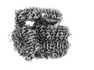
  • Surface view with section colored by density value
  • Surface level: 1
  • Imaged by UCSF Chimera
  • Download
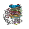
  • Surface view colored by cylindrical radius
  • Surface level: 1
  • Imaged by UCSF Chimera
  • Download
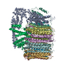
  • Surface view with fitted model
  • Atomic models: PDB-7tao
  • Surface level: 1
  • Imaged by UCSF Chimera
  • Download
Movie viewer
Structure viewerEM map:
SurfViewMolmilJmol/JSmol
Supplemental images

Downloads & links

-
Map

FileDownload / File: emd_25779.map.gz / Format: CCP4 / Size: 64 MB / Type: IMAGE STORED AS FLOATING POINT NUMBER (4 BYTES)
Voxel sizeX=Y=Z: 1.20703 Å
Density
Contour LevelBy AUTHOR: 1.0 / Movie #1: 1
Minimum - Maximum-6.0831785 - 9.427784
Average (Standard dev.)0.0014758433 (±0.23107381)
SymmetrySpace group: 1
Details

EMDB XML:

Map geometry
Axis orderXYZ
Origin000
Dimensions256256256
Spacing256256256
CellA=B=C: 308.99994 Å
α=β=γ: 90.0 °

CCP4 map header:

modeImage stored as Reals
Å/pix. X/Y/Z1.207031251.207031251.20703125
M x/y/z256256256
origin x/y/z0.0000.0000.000
length x/y/z309.000309.000309.000
α/β/γ90.00090.00090.000
MAP C/R/S123
start NC/NR/NS000
NC/NR/NS256256256
D min/max/mean-6.0839.4280.001

-
Supplemental data

-
Sample components

+
Entire : Cryo-EM structure of bafilomycin A1 bound to yeast VO V-ATPase

EntireName: Cryo-EM structure of bafilomycin A1 bound to yeast VO V-ATPase
Components
  • Complex: Cryo-EM structure of bafilomycin A1 bound to yeast VO V-ATPase
    • Protein or peptide: V-type proton ATPase subunit c'
    • Protein or peptide: V-type proton ATPase subunit c''
    • Protein or peptide: V0 assembly protein 1
    • Protein or peptide: V-type proton ATPase subunit e
    • Protein or peptide: V-type proton ATPase subunit c
    • Protein or peptide: Yeast V-ATPase subunit f
    • Protein or peptide: V-type proton ATPase subunit d
    • Protein or peptide: V-type proton ATPase subunit a, vacuolar isoform
  • Ligand: (5R)-2,4-dideoxy-1-C-{(2S,3R,4S)-3-hydroxy-4-[(2R,3S,4E,6E,9R,10S,11R,12E,14Z)-10-hydroxy-3,15-dimethoxy-7,9,11,13-tetramethyl-16-oxo-1-oxacyclohexadeca-4,6,12,14-tetraen-2-yl]pentan-2-yl}-4-methyl-5-propan-2-yl-alpha-D-threo-pentopyranose

+
Supramolecule #1: Cryo-EM structure of bafilomycin A1 bound to yeast VO V-ATPase

SupramoleculeName: Cryo-EM structure of bafilomycin A1 bound to yeast VO V-ATPase
type: complex / Chimera: Yes / ID: 1 / Parent: 0 / Macromolecule list: #1-#8
Source (natural)Organism: Saccharomyces cerevisiae (brewer's yeast)

+
Macromolecule #1: V-type proton ATPase subunit c'

MacromoleculeName: V-type proton ATPase subunit c' / type: protein_or_peptide / ID: 1 / Number of copies: 1 / Enantiomer: LEVO
Source (natural)Organism: baker's yeast (brewer's yeast)
Molecular weightTheoretical: 17.046361 KDa
SequenceString:
MSTQLASNIY APLYAPFFGF AGCAAAMVLS CLGAAIGTAK SGIGIAGIGT FKPELIMKSL IPVVMSGILA IYGLVVAVLI AGNLSPTED YTLFNGFMHL SCGLCVGFAC LSSGYAIGMV GDVGVRKYMH QPRLFVGIVL ILIFSEVLGL YGMIVALILN T RGSE

+
Macromolecule #2: V-type proton ATPase subunit c''

MacromoleculeName: V-type proton ATPase subunit c'' / type: protein_or_peptide / ID: 2 / Number of copies: 1 / Enantiomer: LEVO
Source (natural)Organism: baker's yeast (brewer's yeast)
Molecular weightTheoretical: 22.610641 KDa
SequenceString: MNKESKDDDM SLGKFSFSHF LYYLVLIVVI VYGLYKLFTG HGSDINFGKF LLRTSPYMWA NLGIALCVGL SVVGAAWGIF ITGSSMIGA GVRAPRITTK NLISIIFCEV VAIYGLIIAI VFSSKLTVAT AENMYSKSNL YTGYSLFWAG ITVGASNLIC G IAVGITGA ...String:
MNKESKDDDM SLGKFSFSHF LYYLVLIVVI VYGLYKLFTG HGSDINFGKF LLRTSPYMWA NLGIALCVGL SVVGAAWGIF ITGSSMIGA GVRAPRITTK NLISIIFCEV VAIYGLIIAI VFSSKLTVAT AENMYSKSNL YTGYSLFWAG ITVGASNLIC G IAVGITGA TAAISDAADS ALFVKILVIE IFGSILGLLG LIVGLLMAGK ASEFQ

+
Macromolecule #3: V0 assembly protein 1

MacromoleculeName: V0 assembly protein 1 / type: protein_or_peptide / ID: 3 / Number of copies: 1 / Enantiomer: LEVO
Source (natural)Organism: baker's yeast (brewer's yeast)
Molecular weightTheoretical: 29.694885 KDa
SequenceString: MVFGQLYALF IFTLSCCISK TVQADSSKES SSFISFDKES NWDTISTISS TADVISSVDS AIAVFEFDNF SLLDNLMIDE EYPFFNRFF ANDVSLTVHD DSPLNISQSL SPIMEQFTVD ELPESASDLL YEYSLDDKSI VLFKFTSDAY DLKKLDEFID S CLSFLEDK ...String:
MVFGQLYALF IFTLSCCISK TVQADSSKES SSFISFDKES NWDTISTISS TADVISSVDS AIAVFEFDNF SLLDNLMIDE EYPFFNRFF ANDVSLTVHD DSPLNISQSL SPIMEQFTVD ELPESASDLL YEYSLDDKSI VLFKFTSDAY DLKKLDEFID S CLSFLEDK SGDNLTVVIN SLGWAFEDED GDDEYATEET LSHHDNNKGK EGDDDILSSI WTEGLLMCLI VSALLLFILI VA LSWISNL DITYGALEKS TNPIKKNN

+
Macromolecule #4: V-type proton ATPase subunit e

MacromoleculeName: V-type proton ATPase subunit e / type: protein_or_peptide / ID: 4 / Number of copies: 1 / Enantiomer: LEVO
Source (natural)Organism: baker's yeast (brewer's yeast)
Molecular weightTheoretical: 8.387065 KDa
SequenceString:
MSSFYTVVGV FIVVSAMSVL FWIMAPKNNQ AVWRSTVILT LAMMFLMWAI TFLCQLHPLV APRRSDLRPE FAE

+
Macromolecule #5: V-type proton ATPase subunit c

MacromoleculeName: V-type proton ATPase subunit c / type: protein_or_peptide / ID: 5 / Number of copies: 8 / Enantiomer: LEVO
Source (natural)Organism: baker's yeast (brewer's yeast)
Molecular weightTheoretical: 16.357501 KDa
SequenceString:
MTELCPVYAP FFGAIGCASA IIFTSLGAAY GTAKSGVGIC ATCVLRPDLL FKNIVPVIMA GIIAIYGLVV SVLVCYSLGQ KQALYTGFI QLGAGLSVGL SGLAAGFAIG IVGDAGVRGS SQQPRLFVGM ILILIFAEVL GLYGLIVALL LNSRATQDVV C

+
Macromolecule #6: Yeast V-ATPase subunit f

MacromoleculeName: Yeast V-ATPase subunit f / type: protein_or_peptide / ID: 6 / Number of copies: 1 / Enantiomer: LEVO
Source (natural)Organism: baker's yeast (brewer's yeast)
Molecular weightTheoretical: 9.369934 KDa
SequenceString:
MRPVVSTGKA WCCTVLSAFG VVILSVIAHL FNTNHESFVG SINDPEDGPA VAHTVYLAAL VYLVFFVFCG FQVYLARRKP SIELR

+
Macromolecule #7: V-type proton ATPase subunit d

MacromoleculeName: V-type proton ATPase subunit d / type: protein_or_peptide / ID: 7 / Number of copies: 1 / Enantiomer: LEVO
Source (natural)Organism: baker's yeast (brewer's yeast)
Molecular weightTheoretical: 39.822484 KDa
SequenceString: MEGVYFNIDN GFIEGVVRGY RNGLLSNNQY INLTQCDTLE DLKLQLSSTD YGNFLSSVSS ESLTTSLIQE YASSKLYHEF NYIRDQSSG STRKFMDYIT YGYMIDNVAL MITGTIHDRD KGEILQRCHP LGWFDTLPTL SVATDLESLY ETVLVDTPLA P YFKNCFDT ...String:
MEGVYFNIDN GFIEGVVRGY RNGLLSNNQY INLTQCDTLE DLKLQLSSTD YGNFLSSVSS ESLTTSLIQE YASSKLYHEF NYIRDQSSG STRKFMDYIT YGYMIDNVAL MITGTIHDRD KGEILQRCHP LGWFDTLPTL SVATDLESLY ETVLVDTPLA P YFKNCFDT AEELDDMNIE IIRNKLYKAY LEDFYNFVTE EIPEPAKECM QTLLGFEADR RSINIALNSL QSSDIDPDLK SD LLPNIGK LYPLATFHLA QAQDFEGVRA ALANVYEYRG FLETGNLEDH FYQLEMELCR DAFTQQFAIS TVWAWMKSKE QEV RNITWI AECIAQNQRE RINNYISVY

+
Macromolecule #8: V-type proton ATPase subunit a, vacuolar isoform

MacromoleculeName: V-type proton ATPase subunit a, vacuolar isoform / type: protein_or_peptide / ID: 8 / Number of copies: 1 / Enantiomer: LEVO
Source (natural)Organism: baker's yeast (brewer's yeast)
Molecular weightTheoretical: 95.625484 KDa
SequenceString: MAEKEEAIFR SAEMALVQFY IPQEISRDSA YTLGQLGLVQ FRDLNSKVRA FQRTFVNEIR RLDNVERQYR YFYSLLKKHD IKLYEGDTD KYLDGSGELY VPPSGSVIDD YVRNASYLEE RLIQMEDATD QIEVQKNDLE QYRFILQSGD EFFLKGDNTD S TSYMDEDM ...String:
MAEKEEAIFR SAEMALVQFY IPQEISRDSA YTLGQLGLVQ FRDLNSKVRA FQRTFVNEIR RLDNVERQYR YFYSLLKKHD IKLYEGDTD KYLDGSGELY VPPSGSVIDD YVRNASYLEE RLIQMEDATD QIEVQKNDLE QYRFILQSGD EFFLKGDNTD S TSYMDEDM IDANGENIAA AIGASVNYVT GVIARDKVAT LEQILWRVLR GNLFFKTVEI EQPVYDVKTR EYKHKNAFIV FS HGDLIIK RIRKIAESLD ANLYDVDSSN EGRSQQLAKV NKNLSDLYTV LKTTSTTLES ELYAIAKELD SWFQDVTREK AIF EILNKS NYDTNRKILI AEGWIPRDEL ATLQARLGEM IARLGIDVPS IIQVLDTNHT PPTFHRTNKF TAGFQSICDC YGIA QYREI NAGLPTIVTF PFMFAIMFGD MGHGFLMTLA ALSLVLNEKK INKMKRGEIF DMAFTGRYII LLMGVFSMYT GFLYN DIFS KTMTIFKSGW KWPDHWKKGE SITATSVGTY PIGLDWAWHG TENALLFSNS YKMKLSILMG FIHMTYSYFF SLANHL YFN SMIDIIGNFI PGLLFMQGIF GYLSVCIVYK WAVDWVKDGK PAPGLLNMLI NMFLSPGTID DELYPHQAKV QVFLLLM AL VCIPWLLLVK PLHFKFTHKK KSHEPLPSTE ADASSEDLEA QQLISAMDAD DAEEEEVGSG SHGEDFGDIM IHQVIHTI E FCLNCVSHTA SYLRLWALSL AHAQLSSVLW TMTIQIAFGF RGFVGVFMTV ALFAMWFALT CAVLVLMEGT SAMLHSLRL HWVESMSKFF VGEGLPYEPF AFEYKDMEVA VASASSSASS

+
Macromolecule #9: (5R)-2,4-dideoxy-1-C-{(2S,3R,4S)-3-hydroxy-4-[(2R,3S,4E,6E,9R,10S...

MacromoleculeName: (5R)-2,4-dideoxy-1-C-{(2S,3R,4S)-3-hydroxy-4-[(2R,3S,4E,6E,9R,10S,11R,12E,14Z)-10-hydroxy-3,15-dimethoxy-7,9,11,13-tetramethyl-16-oxo-1-oxacyclohexadeca-4,6,12,14-tetraen-2-yl]pentan-2-yl}-4- ...Name: (5R)-2,4-dideoxy-1-C-{(2S,3R,4S)-3-hydroxy-4-[(2R,3S,4E,6E,9R,10S,11R,12E,14Z)-10-hydroxy-3,15-dimethoxy-7,9,11,13-tetramethyl-16-oxo-1-oxacyclohexadeca-4,6,12,14-tetraen-2-yl]pentan-2-yl}-4-methyl-5-propan-2-yl-alpha-D-threo-pentopyranose
type: ligand / ID: 9 / Number of copies: 9 / Formula: WEV
Molecular weightTheoretical: 622.83 Da
Chemical component information

ChemComp-WEV:
(5R)-2,4-dideoxy-1-C-{(2S,3R,4S)-3-hydroxy-4-[(2R,3S,4E,6E,9R,10S,11R,12E,14Z)-10-hydroxy-3,15-dimethoxy-7,9,11,13-tetramethyl-16-oxo-1-oxacyclohexadeca-4,6,12,14-tetraen-2-yl]pentan-2-yl}-4-methyl-5-propan-2-yl-alpha-D-threo-pentopyranose

-
Experimental details

-
Structure determination

Methodcryo EM
Processingsingle particle reconstruction
Aggregation stateparticle

-
Sample preparation

BufferpH: 7.4
VitrificationCryogen name: ETHANE

-
Electron microscopy

MicroscopeFEI TITAN KRIOS
Electron beamAcceleration voltage: 300 kV / Electron source: FIELD EMISSION GUN
Electron opticsIllumination mode: FLOOD BEAM / Imaging mode: BRIGHT FIELDBright-field microscopy / Nominal defocus max: 2.7600000000000002 µm / Nominal defocus min: 0.65 µm
Image recordingFilm or detector model: FEI FALCON IV (4k x 4k) / Average electron dose: 39.0 e/Å2
Experimental equipment
Model: Titan Krios / Image courtesy: FEI Company

-
Image processing

Startup modelType of model: PDB ENTRY
PDB model - PDB ID:
Initial angle assignmentType: OTHER
Final angle assignmentType: MAXIMUM LIKELIHOOD
Final reconstructionResolution.type: BY AUTHOR / Resolution: 3.2 Å / Resolution method: FSC 0.143 CUT-OFF / Number images used: 447983
FSC plot (resolution estimation)

+
About Yorodumi

-
News

-
Feb 9, 2022. New format data for meta-information of EMDB entries

New format data for meta-information of EMDB entries

  • Version 3 of the EMDB header file is now the official format.
  • The previous official version 1.9 will be removed from the archive.

Related info.:EMDB header

External links:wwPDB to switch to version 3 of the EMDB data model

-
Aug 12, 2020. Covid-19 info

Covid-19 info

URL: https://pdbj.org/emnavi/covid19.php

New page: Covid-19 featured information page in EM Navigator.

Related info.:Covid-19 info / Mar 5, 2020. Novel coronavirus structure data

+
Mar 5, 2020. Novel coronavirus structure data

Novel coronavirus structure data

Related info.:Yorodumi Speices / Aug 12, 2020. Covid-19 info

External links:COVID-19 featured content - PDBj / Molecule of the Month (242):Coronavirus Proteases

+
Jan 31, 2019. EMDB accession codes are about to change! (news from PDBe EMDB page)

EMDB accession codes are about to change! (news from PDBe EMDB page)

  • The allocation of 4 digits for EMDB accession codes will soon come to an end. Whilst these codes will remain in use, new EMDB accession codes will include an additional digit and will expand incrementally as the available range of codes is exhausted. The current 4-digit format prefixed with “EMD-” (i.e. EMD-XXXX) will advance to a 5-digit format (i.e. EMD-XXXXX), and so on. It is currently estimated that the 4-digit codes will be depleted around Spring 2019, at which point the 5-digit format will come into force.
  • The EM Navigator/Yorodumi systems omit the EMD- prefix.

Related info.:Q: What is EMD? / ID/Accession-code notation in Yorodumi/EM Navigator

External links:EMDB Accession Codes are Changing Soon! / Contact to PDBj

+
Jul 12, 2017. Major update of PDB

Major update of PDB

  • wwPDB released updated PDB data conforming to the new PDBx/mmCIF dictionary.
  • This is a major update changing the version number from 4 to 5, and with Remediation, in which all the entries are updated.
  • In this update, many items about electron microscopy experimental information are reorganized (e.g. em_software).
  • Now, EM Navigator and Yorodumi are based on the updated data.

External links:wwPDB Remediation / Enriched Model Files Conforming to OneDep Data Standards Now Available in the PDB FTP Archive

-
Yorodumi

Thousand views of thousand structures

  • Yorodumi is a browser for structure data from EMDB, PDB, SASBDB, etc.
  • This page is also the successor to EM Navigator detail page, and also detail information page/front-end page for Omokage search.
  • The word "yorodu" (or yorozu) is an old Japanese word meaning "ten thousand". "mi" (miru) is to see.

Related info.:EMDB / PDB / SASBDB / Comparison of 3 databanks / Yorodumi Search / Aug 31, 2016. New EM Navigator & Yorodumi / Yorodumi Papers / Jmol/JSmol / Function and homology information / Changes in new EM Navigator and Yorodumi

Read more