 ムービー
ムービー コントローラー


+ データを開く

- 基本情報
基本情報
| 登録情報 | データベース: EMDB / ID: EMD-2548 | |||||||||
|---|---|---|---|---|---|---|---|---|---|---|
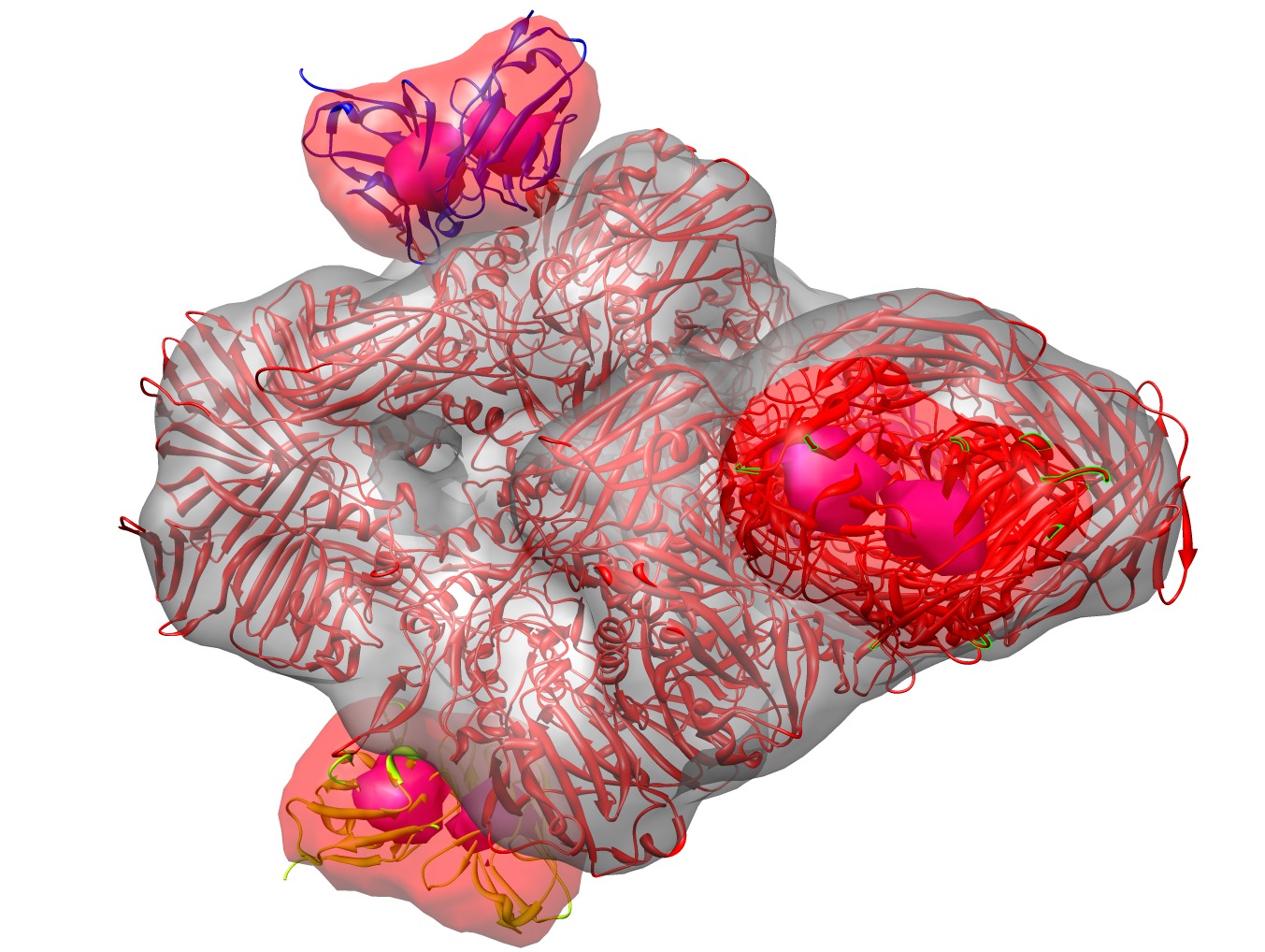
| タイトル | Single particle electron cryomicroscopy of the complex between the E.coli enzyme beta-galactosidase and the single chain Fv antibody scFv13R4. | |||||||||
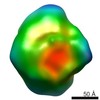
 マップデータ マップデータ | Map of the structure of the complex between beta-galactosidase and the scFv13R4 single chain antibody domain. | |||||||||
 試料 試料 |
| |||||||||
 キーワード キーワード | beta-galactosidase / antibody / scFv13R4 / single particle cryoEM | |||||||||
| 機能・相同性 |  機能・相同性情報 機能・相同性情報alkali metal ion binding / lactose catabolic process / beta-galactosidase complex / beta-galactosidase / beta-galactosidase activity / immunoglobulin mediated immune response / immunoglobulin complex / antigen binding / carbohydrate binding / adaptive immune response ...alkali metal ion binding / lactose catabolic process / beta-galactosidase complex / beta-galactosidase / beta-galactosidase activity / immunoglobulin mediated immune response / immunoglobulin complex / antigen binding / carbohydrate binding / adaptive immune response / magnesium ion binding / extracellular region / identical protein binding 類似検索 - 分子機能 | |||||||||
| 生物種 |   | |||||||||
| 手法 | 単粒子再構成法 / クライオ電子顕微鏡法 / ネガティブ染色法 / 解像度: 13.0 Å | |||||||||
 データ登録者 データ登録者 | Vinothkumar KR / McMullan G / Henderson R | |||||||||
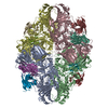
 引用 引用 | ジャーナル: Structure / 年: 2014 タイトル: Molecular mechanism of antibody-mediated activation of β-galactosidase. 著者: Kutti R Vinothkumar / Greg McMullan / Richard Henderson /  要旨: Binding of a single-chain Fv antibody to Escherichia coli β-galactosidase (β-gal) is known to stabilize the enzyme and activate several inactive point mutants, historically called antibody-mediated ...Binding of a single-chain Fv antibody to Escherichia coli β-galactosidase (β-gal) is known to stabilize the enzyme and activate several inactive point mutants, historically called antibody-mediated enzyme formation mutants. To understand the nature of this activation, we have determined by electron cryo-microscopy the structure of the complex between β-gal and the antibody scFv13R4. Our structure localizes the scFv13R4 binding site to the crevice between domains 1 and 3 in each β-gal subunit. The mutations that scFv13R4 counteracts are located between the antibody binding site and the active site of β-gal, at one end of the TIM-barrel that forms domain 3 where the substrate lactose is hydrolyzed. The mode of binding suggests how scFv stabilizes both the active site of β-gal and the tetrameric state. | |||||||||
| 履歴 |
|
- 構造の表示
構造の表示
| ムービー |
ムービービューア |
|---|---|
| 構造ビューア | EMマップ: SurfViewMolmilJmol/JSmol |
| 添付画像 |
- ダウンロードとリンク
-EMDBアーカイブ
| マップデータ | emd_2548.map.gz | 2.8 MB | EMDBマップデータ形式 | |
|---|---|---|---|---|
| ヘッダ (付随情報) | emd-2548-v30.xmlemd-2548.xml | 14.5 KB 14.5 KB | 表示 表示 | EMDBヘッダ |
| 画像 | emd_2548.tif | 1.1 MB | ||
| アーカイブディレクトリ |  http://ftp.pdbj.org/pub/emdb/structures/EMD-2548ftp://ftp.pdbj.org/pub/emdb/structures/EMD-2548 http://ftp.pdbj.org/pub/emdb/structures/EMD-2548ftp://ftp.pdbj.org/pub/emdb/structures/EMD-2548 | HTTPS FTP |
-関連構造データ


-リンク
| EMDBのページ | EMDB (EBI/PDBe) / EMDataResource |
|---|---|
| 「今月の分子」の関連する項目 |


-マップ
| ファイル | ダウンロード / ファイル: emd_2548.map.gz / 形式: CCP4 / 大きさ: 3.7 MB / タイプ: IMAGE STORED AS FLOATING POINT NUMBER (4 BYTES) | ||||||||||||||||||||||||||||||||||||||||||||||||||||||||||||||||||||
|---|---|---|---|---|---|---|---|---|---|---|---|---|---|---|---|---|---|---|---|---|---|---|---|---|---|---|---|---|---|---|---|---|---|---|---|---|---|---|---|---|---|---|---|---|---|---|---|---|---|---|---|---|---|---|---|---|---|---|---|---|---|---|---|---|---|---|---|---|---|
| 注釈 | Map of the structure of the complex between beta-galactosidase and the scFv13R4 single chain antibody domain. | ||||||||||||||||||||||||||||||||||||||||||||||||||||||||||||||||||||
| 投影像・断面図 | 画像のコントロール
画像は Spider により作成 | ||||||||||||||||||||||||||||||||||||||||||||||||||||||||||||||||||||
| ボクセルのサイズ | X=Y=Z: 3 Å | ||||||||||||||||||||||||||||||||||||||||||||||||||||||||||||||||||||
| 密度 |
| ||||||||||||||||||||||||||||||||||||||||||||||||||||||||||||||||||||
| 対称性 | 空間群: 1 | ||||||||||||||||||||||||||||||||||||||||||||||||||||||||||||||||||||
| 詳細 | EMDB XML:
CCP4マップ ヘッダ情報:
| ||||||||||||||||||||||||||||||||||||||||||||||||||||||||||||||||||||
 Z (Sec.)
 Y (Row.)
 X (Col.)


-添付データ
- 試料の構成要素
-全体 : Single chain Fv antibody domain bound to the enzyme beta-galactosidase
| 全体 | 名称: Single chain Fv antibody domain bound to the enzyme beta-galactosidase |
|---|---|
| 要素 |
|
-超分子 #1000: Single chain Fv antibody domain bound to the enzyme beta-galactosidase
| 超分子 | 名称: Single chain Fv antibody domain bound to the enzyme beta-galactosidase タイプ: sample / ID: 1000 詳細: The occupancy of the antibody domains was estimated to be 90% 集合状態: one D2 symmetry tetramer, with 4 Fv antibody domains (heavy & light chains) Number unique components: 3 |
|---|---|
| 分子量 | 理論値: 560 KDa 手法: Known stoichiometry and known molecular weights of the components, which are 450 kiloDaltons for the D2 beta-galactosidase and 28 kiloDaltons for each Fv domain. |
-分子 #1: beta-galactosidase
| 分子 | 名称: beta-galactosidase / タイプ: protein_or_peptide / ID: 1 / 詳細: Central tetrameric enzyme in complex / コピー数: 4 / 集合状態: tetramer / 組換発現: Yes |
|---|---|
| 由来(天然) | 生物種: |
| 分子量 | 理論値: 465 KDa |
| 配列 | UniProtKB: Beta-galactosidase / GO: beta-galactosidase complex / InterPro: Glycoside hydrolase family 1 |
-分子 #2: SCFV13R4 ANTIBODY FV HEAVY CHAIN
| 分子 | 名称: SCFV13R4 ANTIBODY FV HEAVY CHAIN / タイプ: protein_or_peptide / ID: 2 / コピー数: 4 / 集合状態: Tetrameric / 組換発現: Yes |
|---|---|
| 由来(天然) | 生物種: |
| 組換発現 | 生物種: |
-分子 #3: SCFV13R4 ANTIBODY FV LIGHT CHAIN
| 分子 | 名称: SCFV13R4 ANTIBODY FV LIGHT CHAIN / タイプ: protein_or_peptide / ID: 3 / コピー数: 4 / 集合状態: Tetrameric / 組換発現: Yes |
|---|---|
| 由来(天然) | 生物種: |
| 組換発現 | 生物種: |
-実験情報
-構造解析
| 手法 | ネガティブ染色法, クライオ電子顕微鏡法 |
|---|---|
 解析 解析 | 単粒子再構成法 |
| 試料の集合状態 | particle |
-試料調製
| 濃度 | 0.9 mg/mL |
|---|---|
| 緩衝液 | pH: 7.4 / 詳細: 20% phosphate buffered saline (PBS) |
| 染色 | タイプ: NEGATIVE 詳細: Sample applied to glow-discharged (30 seconds in residual air) grids, blotted and frozen rapidly in an environmental plunge-freeze apparatus (Bellare et al, J. Electr. Micros. Tech., 1988, 10, 87-111). |
| グリッド | 詳細: Qunatifoil grids with 1.2 micron holes |
| 凍結 | 凍結剤: ETHANE / チャンバー内湿度: 100 % / チャンバー内温度: 100 K / 装置: OTHER 手法: Blot for 10-20 seconds until diameter of blotted meniscus ceases to expand, before plunging. |
- 電子顕微鏡法
| 顕微鏡 | FEI POLARA 300 |
|---|---|
| 温度 | 最低: 88 K / 最高: 90 K / 平均: 89 K |
| アライメント法 | Legacy - 非点収差: Corrected at 200,000x once at start of session Legacy - Electron beam tilt params: 0 |
| 詳細 | Exposure intensity set to give 50 electrons/pixel/second at the detector. This translates into 16 electron/square_Angstrom/second at the specimen. |
| 日付 | 2012年8月2日 |
| 撮影 | カテゴリ: CCD フィルム・検出器のモデル: FEI FALCON II (4k x 4k) デジタル化 - サンプリング間隔: 14 µm / 実像数: 49 / 平均電子線量: 67 e/Å2 詳細: Every image is the average of all frames recorded by the Falcon II direct electron detector ビット/ピクセル: 16 |
| 電子線 | 加速電圧: 300 kV / 電子線源:  FIELD EMISSION GUN FIELD EMISSION GUN |
| 電子光学系 | 倍率(補正後): 81600 / 照射モード: FLOOD BEAM / 撮影モード: BRIGHT FIELD / Cs: 2.0 mm / 最大 デフォーカス(公称値): 4.027 µm / 最小 デフォーカス(公称値): 2.678 µm / 倍率(公称値): 59000 |
| 試料ステージ | 試料ホルダー: Polara cartridges / 試料ホルダーモデル: OTHER |
| 実験機器 |  モデル: Tecnai Polara / 画像提供: FEI Company |
-画像解析
| 詳細 | The map was obtained using Frealign, with a starting model consisting of a similar map from 43,000 single particle images of beta-galactosidase without antibody. |
|---|---|
| CTF補正 | 詳細: done inside FREALIGN |
| 最終 再構成 | 想定した対称性 - 点群: D2 (2回x2回 2面回転対称) アルゴリズム: OTHER / 解像度のタイプ: BY AUTHOR / 解像度: 13.0 Å / 解像度の算出法: OTHER / ソフトウェア - 名称: MRC, CTFFIND3, FREALIGN 詳細: Map was calculated from a single dataset of 2965 particles. 使用した粒子像数: 2965 |
| 最終 角度割当 | 詳細: as determined by FREALIGN. Orientations were determined using the data out to a resolution limited to 14 Angstroms |
-原子モデル構築 1
| 初期モデル | PDB ID: Chain - #0 - Chain ID: A / Chain - #1 - Chain ID: B / Chain - #2 - Chain ID: C / Chain - #3 - Chain ID: D |
|---|---|
| ソフトウェア | 名称: Chimera |
| 詳細 | Since there are three orthogonal two-fold axes, there are actually no parameters required for the fitting, except to make sure the three axes are correctly assigned (which they were at an earlier stage) and relative magnification, which was tested from 0.95 to 1.05, determined. In this case, at this resolution, a relative magnification of 1.00 was adequate. For the higher resolution map of beta-galactosidase without antibody, a 2.5% correction was necessary. In addition to 1F4A, we also fitted the coordinates of Fv antibody HYHEL-10 (anti-hen egg lysozyme) with PDB coordinates 3A6B to all four antibody domains in the map. |
| 精密化 | 空間: REAL / プロトコル: RIGID BODY FIT / 温度因子: 0 当てはまり具合の基準: FSC curve between map and model |
| 得られたモデル |  PDB-4ckd: |

