 Movie
Movie Controller

[English] 日本語
 Yorodumi
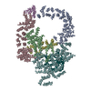
- PDB-4ckd: Model of complex between the E.coli enzyme beta-galactosidase and... -
+ Open data

- Basic information
| Entry | Database: PDB / ID: 4ckd | ||||||
|---|---|---|---|---|---|---|---|
| Title | Model of complex between the E.coli enzyme beta-galactosidase and four single chain Fv antibody domains scFv13R4. | ||||||
 Components Components |
| ||||||
 Keywords Keywords | HYDROLASE/IMMUNE SYSTEM / HYDROLASE-IMMUNE SYSTEM COMPLEX | ||||||
| Function / homology |  Function and homology information Function and homology informationalkali metal ion binding / lactose catabolic process / beta-galactosidase complex / beta-galactosidase / beta-galactosidase activity / immunoglobulin mediated immune response / immunoglobulin complex / antigen binding / carbohydrate binding / adaptive immune response ...alkali metal ion binding / lactose catabolic process / beta-galactosidase complex / beta-galactosidase / beta-galactosidase activity / immunoglobulin mediated immune response / immunoglobulin complex / antigen binding / carbohydrate binding / adaptive immune response / immune response / magnesium ion binding / extracellular region / identical protein binding / plasma membrane Similarity search - Function | ||||||
| Biological species |   | ||||||
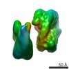
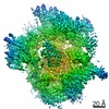
| Method | ELECTRON MICROSCOPY / single particle reconstruction / cryo EM / Resolution: 13 Å | ||||||
 Authors Authors | Vinothkumar, K.R. / McMullan, G. / Henderson, R. | ||||||
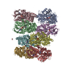
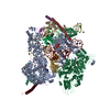
 Citation Citation | Journal: Structure / Year: 2014 Title: Molecular mechanism of antibody-mediated activation of β-galactosidase. Authors: Kutti R Vinothkumar / Greg McMullan / Richard Henderson /  Abstract: Binding of a single-chain Fv antibody to Escherichia coli β-galactosidase (β-gal) is known to stabilize the enzyme and activate several inactive point mutants, historically called antibody-mediated ...Binding of a single-chain Fv antibody to Escherichia coli β-galactosidase (β-gal) is known to stabilize the enzyme and activate several inactive point mutants, historically called antibody-mediated enzyme formation mutants. To understand the nature of this activation, we have determined by electron cryo-microscopy the structure of the complex between β-gal and the antibody scFv13R4. Our structure localizes the scFv13R4 binding site to the crevice between domains 1 and 3 in each β-gal subunit. The mutations that scFv13R4 counteracts are located between the antibody binding site and the active site of β-gal, at one end of the TIM-barrel that forms domain 3 where the substrate lactose is hydrolyzed. The mode of binding suggests how scFv stabilizes both the active site of β-gal and the tetrameric state. | ||||||
| History |
|
- Structure visualization
| Movie |
Movie viewer |
|---|---|
| Structure viewer | Molecule: MolmilJmol/JSmol |
- Downloads & links
-Download
| PDBx/mmCIF format | 4ckd.cif.gz | 970.8 KB | Display | PDBx/mmCIF format |
|---|---|---|---|---|
| PDB format | pdb4ckd.ent.gz | 788.7 KB | Display | PDB format |
| PDBx/mmJSON format | 4ckd.json.gz | Tree view | PDBx/mmJSON format | |
| Others |  Other downloads Other downloads |
-Validation report
| Arichive directory | https://data.pdbj.org/pub/pdb/validation_reports/ck/4ckdftp://data.pdbj.org/pub/pdb/validation_reports/ck/4ckd | HTTPS FTP |
|---|
-Related structure data
| Related structure data | 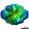 2548MC M: map data used to model this data C: citing same article ( |
|---|---|
| Similar structure data |
-Links
 PDBj
PDBj


- Assembly
| Deposited unit | 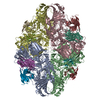
|
|---|---|
| 1 |
|
-Components
| #1: Protein | Mass: 116602.484 Da / Num. of mol.: 4 Source method: isolated from a genetically manipulated source Source: (gene. exp.) #2: Antibody | Mass: 12795.939 Da / Num. of mol.: 4 Source method: isolated from a genetically manipulated source Source: (gene. exp.) #3: Antibody | Mass: 11624.795 Da / Num. of mol.: 4 Source method: isolated from a genetically manipulated source Source: (gene. exp.) Has protein modification | Y | Sequence details | THE DEPOSITED BETA-GALACTOSIDASE SEQUENCE CONTAINS RESIDUES 4-1024 IN THE FULL GENE SEQUENCE ...THE DEPOSITED BETA-GALACTOSID | |
|---|
-Experimental details
-Experiment
| Experiment | Method: ELECTRON MICROSCOPY |
|---|---|
| EM experiment | Aggregation state: PARTICLE / 3D reconstruction method: single particle reconstruction |
- Sample preparation
| Component | Name: SINGLE CHAIN FV ANTIBODY DOMAIN BOUND TO THE ENZYME BETA-GALACTOSIDASE Type: COMPLEX / Details: 49 BEST IMAGES SELECTED OUT OF 52 RECORDED |
|---|---|
| Buffer solution | Name: 20% PHOSPHATE BUFFERED SALINE (PBS) / pH: 7.4 / Details: 20% PHOSPHATE BUFFERED SALINE (PBS) |
| Specimen | Conc.: 0.9 mg/ml / Embedding applied: NO / Shadowing applied: NO / Staining applied: NO / Vitrification applied: YES |
| Specimen support | Details: HOLEY CARBON |
| Vitrification | Cryogen name: ETHANE Details: VITRIFICATION 1 -- CRYOGEN- ETHANE, HUMIDITY- 100, TEMPERATURE- 100, INSTRUMENT- OTHER, METHOD- BLOT FOR 10-20 SECONDS UNTIL DIAMETER OF BLOTTED MENISCUS CEASES TO EXPAND, BEFORE PLUNGING. |
- Electron microscopy imaging
| Experimental equipment |  Model: Tecnai Polara / Image courtesy: FEI Company |
|---|---|
| Microscopy | Model: FEI POLARA 300 / Date: Aug 2, 2012 Details: EXPOSURE INTENSITY SET TO GIVE 50 ELECTRONS PER PIXEL PER SECOND AT THE DETECTOR. THIS TRANSLATES INTO 16 ELECTRONS PER SQUARE ANGSTROM PER SECOND AT THE SPECIMEN. |
| Electron gun | Electron source:  FIELD EMISSION GUN / Accelerating voltage: 300 kV / Illumination mode: FLOOD BEAM FIELD EMISSION GUN / Accelerating voltage: 300 kV / Illumination mode: FLOOD BEAM |
| Electron lens | Mode: BRIGHT FIELD / Nominal magnification: 59000 X / Calibrated magnification: 81600 X / Nominal defocus max: 4027 nm / Nominal defocus min: 2678 nm / Cs: 2 mm |
| Specimen holder | Temperature: 89 K |
| Image recording | Electron dose: 67 e/Å2 / Film or detector model: FEI FALCON II (4k x 4k) |
| Image scans | Num. digital images: 49 |
| Radiation wavelength | Relative weight: 1 |
- Processing
| EM software |
| ||||||||||||||||||||
|---|---|---|---|---|---|---|---|---|---|---|---|---|---|---|---|---|---|---|---|---|---|
| CTF correction | Details: DONE INSIDE FREALIGN | ||||||||||||||||||||
| Symmetry | Point symmetry: D2 (2x2 fold dihedral) | ||||||||||||||||||||
| 3D reconstruction | Method: PROJECTION MATCHING / Resolution: 13 Å / Num. of particles: 2965 / Nominal pixel size: 1.74 Å / Actual pixel size: 1.72 Å Magnification calibration: MAGNIFICATION REFINED BY MAXIMISING FSC AGAINST ATOMIC MODEL Details: FREALIGN MAP OBTAINED FROM 2965 PARTICLES USING D2 SYMMETRY THIS IS A MODEL MADE BY DOCKING TWO ATOMIC MODELS AS RIGID BODIES INTO A 13 ANGSTROM RESOLUTION DENSITY MAP. SUBMISSION BASED ON ...Details: FREALIGN MAP OBTAINED FROM 2965 PARTICLES USING D2 SYMMETRY THIS IS A MODEL MADE BY DOCKING TWO ATOMIC MODELS AS RIGID BODIES INTO A 13 ANGSTROM RESOLUTION DENSITY MAP. SUBMISSION BASED ON EXPERIMENTAL DATA FROM EMDB EMD-2548. (DEPOSITION ID: 12230). Symmetry type: POINT | ||||||||||||||||||||
| Atomic model building | Protocol: RIGID BODY FIT / Space: REAL / Target criteria: FSC CURVE BETWEEN MAP AND MODEL / Details: METHOD--RIGID BODY REFINEMENT PROTOCOL--X-RAY | ||||||||||||||||||||
| Atomic model building | PDB-ID: 1F4A Accession code: 1F4A / Source name: PDB / Type: experimental model | ||||||||||||||||||||
| Refinement | Highest resolution: 13 Å | ||||||||||||||||||||
| Refinement step | Cycle: LAST / Highest resolution: 13 Å
|

