ムービー
ムービー コントローラー


+ データを開く

- 基本情報
基本情報
| 登録情報 |  | |||||||||
|---|---|---|---|---|---|---|---|---|---|---|
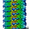
| タイトル | Composite map of ciliary C2 central pair apparatus isolated from Chlamydomonas reinhardtii | |||||||||
 マップデータ マップデータ | Ciliary C2 central pair apparatus isolated from Chlamydomonas reinhardtii | |||||||||
 試料 試料 |
| |||||||||
 キーワード キーワード | cilia / microtubule / STRUCTURAL PROTEIN | |||||||||
| 機能・相同性 |  機能・相同性情報 機能・相同性情報axonemal central pair / axonemal doublet microtubule / positive regulation of cilium-dependent cell motility / regulation of cilium beat frequency involved in ciliary motility / establishment of protein localization to organelle / cilium movement / axoneme assembly / axonemal microtubule / microtubule associated complex / motile cilium ...axonemal central pair / axonemal doublet microtubule / positive regulation of cilium-dependent cell motility / regulation of cilium beat frequency involved in ciliary motility / establishment of protein localization to organelle / cilium movement / axoneme assembly / axonemal microtubule / microtubule associated complex / motile cilium / regulation of cytoskeleton organization / axoneme / cilium assembly / microtubule-based process / structural constituent of cytoskeleton / microtubule binding / 加水分解酵素; 酸無水物に作用; GTPに作用・細胞または細胞小器官の運動に関与 / microtubule / transcription coactivator activity / hydrolase activity / ciliary basal body / cilium / GTPase activity / calcium ion binding / regulation of DNA-templated transcription / GTP binding / metal ion binding / nucleus / cytoplasm 類似検索 - 分子機能 | |||||||||
| 生物種 |   Chlamydomonas reinhardtii (クラミドモナス) Chlamydomonas reinhardtii (クラミドモナス) | |||||||||
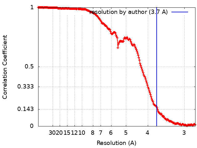
| 手法 | 単粒子再構成法 / クライオ電子顕微鏡法 / 解像度: 3.7 Å | |||||||||
 データ登録者 データ登録者 | Gui M / Wang X / Dutcher SK / Brown A / Zhang R | |||||||||
| 資金援助 |  米国, 1件 米国, 1件
| |||||||||
 引用 引用 | ジャーナル: Nat Struct Mol Biol / 年: 2022 タイトル: Ciliary central apparatus structure reveals mechanisms of microtubule patterning. 著者: Miao Gui / Xiangli Wang / Susan K Dutcher / Alan Brown / Rui Zhang / 要旨: A pair of extensively modified microtubules form the central apparatus (CA) of the axoneme of most motile cilia, where they regulate ciliary motility. The external surfaces of both CA microtubules ...A pair of extensively modified microtubules form the central apparatus (CA) of the axoneme of most motile cilia, where they regulate ciliary motility. The external surfaces of both CA microtubules are patterned asymmetrically with large protein complexes that repeat every 16 or 32 nm. The composition of these projections and the mechanisms that establish asymmetry and longitudinal periodicity are unknown. Here, by determining cryo-EM structures of the CA microtubules, we identify 48 different CA-associated proteins, which in turn reveal mechanisms for asymmetric and periodic protein binding to microtubules. We identify arc-MIPs, a novel class of microtubule inner protein, that bind laterally across protofilaments and remodel tubulin structure and lattice contacts. The binding mechanisms utilized by CA proteins may be generalizable to other microtubule-associated proteins. These structures establish a foundation to elucidate the contributions of individual CA proteins to ciliary motility and ciliopathies. | |||||||||
| 履歴 |
|
- 構造の表示
構造の表示
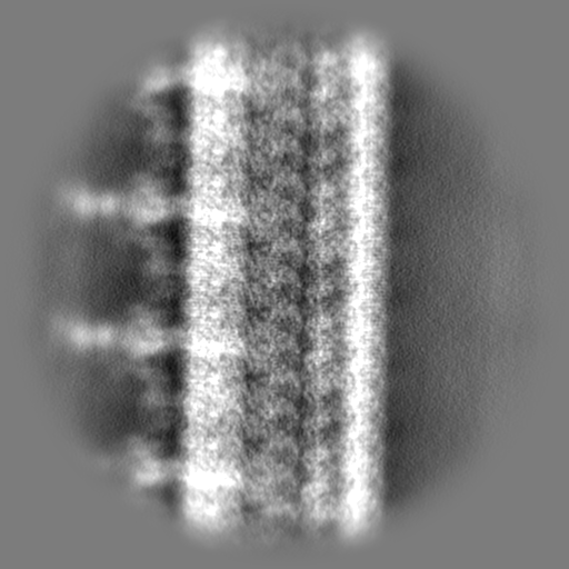
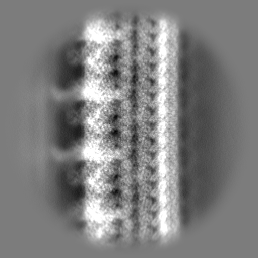
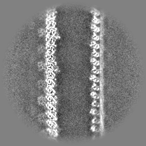
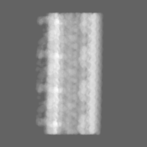
| 添付画像 |
|---|

- ダウンロードとリンク
-EMDBアーカイブ
| マップデータ | emd_25361.map.gz | 246.3 MB | EMDBマップデータ形式 | |
|---|---|---|---|---|
| ヘッダ (付随情報) | emd-25361-v30.xmlemd-25361.xml | 52.5 KB 52.5 KB | 表示 表示 | EMDBヘッダ |
| FSC (解像度算出) | emd_25361_fsc.xml | 17.8 KB | 表示 | FSCデータファイル |
| 画像 |  emd_25361.png emd_25361.png | 76 KB | ||
| マスクデータ | emd_25361_msk_1.map | 512 MB | マスクマップ | |
| Filedesc metadata | emd-25361.cif.gz | 11.9 KB | ||
| その他 | emd_25361_additional_1.map.gzemd_25361_additional_2.map.gzemd_25361_additional_3.map.gzemd_25361_additional_4.map.gzemd_25361_additional_5.map.gzemd_25361_additional_6.map.gzemd_25361_additional_7.map.gzemd_25361_additional_8.map.gzemd_25361_additional_9.map.gz | 1.9 MB 1.9 MB 1.9 MB 1.9 MB 257.5 MB 4.4 MB 1.9 MB 1.9 MB 1.9 MB | ||
| アーカイブディレクトリ |  http://ftp.pdbj.org/pub/emdb/structures/EMD-25361ftp://ftp.pdbj.org/pub/emdb/structures/EMD-25361 http://ftp.pdbj.org/pub/emdb/structures/EMD-25361ftp://ftp.pdbj.org/pub/emdb/structures/EMD-25361 | HTTPS FTP |
-検証レポート
| 文書・要旨 | emd_25361_validation.pdf.gz | 557.7 KB | 表示 | EMDB検証レポート |
|---|---|---|---|---|
| 文書・詳細版 | emd_25361_full_validation.pdf.gz | 557.3 KB | 表示 | |
| XML形式データ | emd_25361_validation.xml.gz | 16.3 KB | 表示 | |
| CIF形式データ | emd_25361_validation.cif.gz | 22.5 KB | 表示 | |
| アーカイブディレクトリ | https://ftp.pdbj.org/pub/emdb/validation_reports/EMD-25361ftp://ftp.pdbj.org/pub/emdb/validation_reports/EMD-25361 | HTTPS FTP |
-関連構造データ


-リンク
| EMDBのページ | EMDB (EBI/PDBe) / EMDataResource |
|---|---|
| 「今月の分子」の関連する項目 |


-マップ
| ファイル | ダウンロード / ファイル: emd_25361.map.gz / 形式: CCP4 / 大きさ: 512 MB / タイプ: IMAGE STORED AS FLOATING POINT NUMBER (4 BYTES) | ||||||||||||||||||||||||||||||||||||
|---|---|---|---|---|---|---|---|---|---|---|---|---|---|---|---|---|---|---|---|---|---|---|---|---|---|---|---|---|---|---|---|---|---|---|---|---|---|
| 注釈 | Ciliary C2 central pair apparatus isolated from Chlamydomonas reinhardtii | ||||||||||||||||||||||||||||||||||||
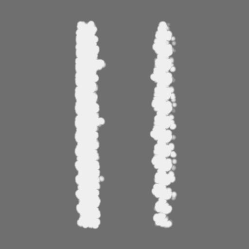
| 投影像・断面図 | 画像のコントロール
画像は Spider により作成 | ||||||||||||||||||||||||||||||||||||
| ボクセルのサイズ | X=Y=Z: 1.39 Å | ||||||||||||||||||||||||||||||||||||
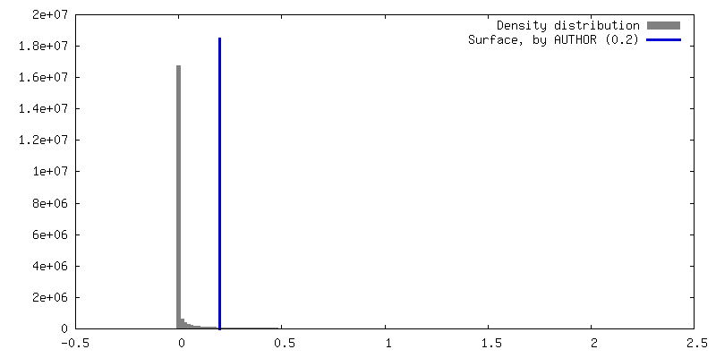
| 密度 |
| ||||||||||||||||||||||||||||||||||||
| 対称性 | 空間群: 1 | ||||||||||||||||||||||||||||||||||||
| 詳細 | EMDB XML:
|
 Z (Sec.)
 Y (Row.)
 X (Col.)


-添付データ
+マスク #1


+追加マップ: Additional map 1


+追加マップ: Additional map 2


+追加マップ: Additional map 3


+追加マップ: Additional map 4


+追加マップ: consensus refinement


+追加マップ: mask for fsc curve


+追加マップ: Additional map 7


+追加マップ: Additional map 8


+追加マップ: Additional map 9


- 試料の構成要素
+全体 : C2 central pair apparatus complex
+超分子 #1: C2 central pair apparatus complex
+分子 #1: Tubulin beta
+分子 #2: Tubulin alpha
+分子 #3: Cilia- and flagella-associated protein 20
+分子 #4: Unknown protein
+分子 #5: Unknown protein
+分子 #6: Unknown protein
+分子 #7: FAP65
+分子 #8: FAP70
+分子 #9: FAP147
+分子 #10: FAP178
+分子 #11: Flagellar WD repeat-containing protein Pf20
+分子 #12: Flagellar associated protein
+分子 #13: FAP196
+分子 #14: FAP213
+分子 #15: FAP225
+分子 #16: FAP239
+分子 #17: FAP388
+分子 #18: FAP424
+分子 #19: GUANOSINE-5'-DIPHOSPHATE

+分子 #20: GUANOSINE-5'-TRIPHOSPHATE

+分子 #21: MAGNESIUM ION
-実験情報
-構造解析
| 手法 | クライオ電子顕微鏡法 |
|---|---|
 解析 解析 | 単粒子再構成法 |
| 試料の集合状態 | filament |
-試料調製
| 緩衝液 | pH: 7.4 |
|---|---|
| グリッド | モデル: C-flat-1.2/1.3 / 材質: COPPER / 前処理 - タイプ: GLOW DISCHARGE |
| 凍結 | 凍結剤: ETHANE |
- 電子顕微鏡法
| 顕微鏡 | FEI TITAN KRIOS |
|---|---|
| 撮影 | フィルム・検出器のモデル: GATAN K2 SUMMIT (4k x 4k) 検出モード: COUNTING / 平均電子線量: 39.6 e/Å2 |
| 電子線 | 加速電圧: 300 kV / 電子線源:  FIELD EMISSION GUN FIELD EMISSION GUN |
| 電子光学系 | 照射モード: FLOOD BEAM / 撮影モード: BRIGHT FIELD |
| 試料ステージ | 試料ホルダーモデル: FEI TITAN KRIOS AUTOGRID HOLDER ホルダー冷却材: NITROGEN |
| 実験機器 |  モデル: Titan Krios / 画像提供: FEI Company |