 Movie
Movie Controller

[English] 日本語
 Yorodumi
- PDB-7som: Ciliary C2 central pair apparatus isolated from Chlamydomonas rei... -
+ Open data

- Basic information
| Entry | Database: PDB / ID: 7som | ||||||
|---|---|---|---|---|---|---|---|
| Title | Ciliary C2 central pair apparatus isolated from Chlamydomonas reinhardtii | ||||||
 Components Components |
| ||||||
 Keywords Keywords | STRUCTURAL PROTEIN / cilia / microtubule | ||||||
| Function / homology |  Function and homology information Function and homology informationaxonemal central pair / axonemal doublet microtubule / positive regulation of cilium-dependent cell motility / regulation of cilium beat frequency involved in ciliary motility / establishment of protein localization to organelle / axoneme assembly / cilium movement / axonemal microtubule / microtubule associated complex / motile cilium ...axonemal central pair / axonemal doublet microtubule / positive regulation of cilium-dependent cell motility / regulation of cilium beat frequency involved in ciliary motility / establishment of protein localization to organelle / axoneme assembly / cilium movement / axonemal microtubule / microtubule associated complex / motile cilium / regulation of cytoskeleton organization / cilium assembly / axoneme / microtubule-based process / structural constituent of cytoskeleton / microtubule binding / Hydrolases; Acting on acid anhydrides; Acting on GTP to facilitate cellular and subcellular movement / microtubule / transcription coactivator activity / ciliary basal body / hydrolase activity / GTPase activity / calcium ion binding / regulation of DNA-templated transcription / GTP binding / metal ion binding / nucleus / cytoplasm Similarity search - Function | ||||||
| Biological species |   Chlamydomonas reinhardtii (plant) Chlamydomonas reinhardtii (plant) | ||||||
| Method | ELECTRON MICROSCOPY / single particle reconstruction / cryo EM / Resolution: 3.7 Å | ||||||
 Authors Authors | Gui, M. / Wang, X. / Dutcher, S.K. / Brown, A. / Zhang, R. | ||||||
| Funding support |  United States, 1items United States, 1items
| ||||||
 Citation Citation | Journal: Nat Struct Mol Biol / Year: 2022 Title: Ciliary central apparatus structure reveals mechanisms of microtubule patterning. Authors: Miao Gui / Xiangli Wang / Susan K Dutcher / Alan Brown / Rui Zhang / Abstract: A pair of extensively modified microtubules form the central apparatus (CA) of the axoneme of most motile cilia, where they regulate ciliary motility. The external surfaces of both CA microtubules ...A pair of extensively modified microtubules form the central apparatus (CA) of the axoneme of most motile cilia, where they regulate ciliary motility. The external surfaces of both CA microtubules are patterned asymmetrically with large protein complexes that repeat every 16 or 32 nm. The composition of these projections and the mechanisms that establish asymmetry and longitudinal periodicity are unknown. Here, by determining cryo-EM structures of the CA microtubules, we identify 48 different CA-associated proteins, which in turn reveal mechanisms for asymmetric and periodic protein binding to microtubules. We identify arc-MIPs, a novel class of microtubule inner protein, that bind laterally across protofilaments and remodel tubulin structure and lattice contacts. The binding mechanisms utilized by CA proteins may be generalizable to other microtubule-associated proteins. These structures establish a foundation to elucidate the contributions of individual CA proteins to ciliary motility and ciliopathies. | ||||||
| History |
|
- Structure visualization
| Structure viewer | Molecule: MolmilJmol/JSmol |
|---|
- Downloads & links
-Download
| PDBx/mmCIF format | 7som.cif.gz | 13.2 MB | Display | PDBx/mmCIF format |
|---|---|---|---|---|
| PDB format | pdb7som.ent.gz | Display | PDB format | |
| PDBx/mmJSON format | 7som.json.gz | Tree view | PDBx/mmJSON format | |
| Others |  Other downloads Other downloads |
-Validation report
| Arichive directory | https://data.pdbj.org/pub/pdb/validation_reports/so/7somftp://data.pdbj.org/pub/pdb/validation_reports/so/7som | HTTPS FTP |
|---|
-Related structure data
| Related structure data |  25361MC  7sqcC M: map data used to model this data C: citing same article ( |
|---|---|
| Similar structure data |

-Links
 PDBj
PDBj


- Assembly
| Deposited unit | 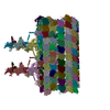
|
|---|---|
| 1 |
|
-Components
-Protein , 16 types, 198 molecules AAACAEAGAIAKBABCBEBGBIBKCCCECGCICKDCDEDGDIDKEAECEEEGEIEKFAFC...
| #1: Protein | Mass: 49665.809 Da / Num. of mol.: 74 / Source method: isolated from a natural source / Source: (natural) Chlamydomonas reinhardtii (plant) / References: UniProt: P04690#2: Protein | Mass: 49638.008 Da / Num. of mol.: 74 / Source method: isolated from a natural source / Source: (natural) Chlamydomonas reinhardtii (plant) / References: UniProt: P09204#3: Protein | Mass: 64880.324 Da / Num. of mol.: 4 / Source method: isolated from a natural source / Source: (natural) Chlamydomonas reinhardtii (plant) / References: UniProt: A8IU92#4: Protein | Mass: 22262.674 Da / Num. of mol.: 3 / Source method: isolated from a natural source / Source: (natural) Chlamydomonas reinhardtii (plant)#5: Protein | Mass: 81430.711 Da / Num. of mol.: 3 / Source method: isolated from a natural source / Source: (natural) Chlamydomonas reinhardtii (plant)#6: Protein | Mass: 57734.598 Da / Num. of mol.: 3 / Source method: isolated from a natural source / Source: (natural) Chlamydomonas reinhardtii (plant)#7: Protein | Mass: 46261.570 Da / Num. of mol.: 3 / Source method: isolated from a natural source / Source: (natural) Chlamydomonas reinhardtii (plant) / References: UniProt: A0A2K3DLJ2#8: Protein | Mass: 10089.324 Da / Num. of mol.: 3 / Source method: isolated from a natural source / Source: (natural) Chlamydomonas reinhardtii (plant) / References: UniProt: A0A2K3DKW3#9: Protein | Mass: 22193.566 Da / Num. of mol.: 3 / Source method: isolated from a natural source / Source: (natural) Chlamydomonas reinhardtii (plant) / References: UniProt: A0A2K3DUG8#10: Protein | Mass: 65929.133 Da / Num. of mol.: 7 / Source method: isolated from a natural source / Source: (natural) Chlamydomonas reinhardtii (plant) / References: UniProt: A0A2K3D8Z6#11: Protein | Mass: 10336.616 Da / Num. of mol.: 4 / Source method: isolated from a natural source / Source: (natural) Chlamydomonas reinhardtii (plant) / References: UniProt: P93107#12: Protein | Mass: 233221.547 Da / Num. of mol.: 3 / Source method: isolated from a natural source / Source: (natural) Chlamydomonas reinhardtii (plant) / References: UniProt: A8I439#13: Protein | Mass: 111116.258 Da / Num. of mol.: 4 / Source method: isolated from a natural source / Source: (natural) Chlamydomonas reinhardtii (plant) / References: UniProt: A0A2K3CQT7#14: Protein | Mass: 102141.609 Da / Num. of mol.: 2 / Source method: isolated from a natural source / Source: (natural) Chlamydomonas reinhardtii (plant) / References: UniProt: A8JB78#15: Protein | Mass: 23511.406 Da / Num. of mol.: 6 / Source method: isolated from a natural source / Source: (natural) Chlamydomonas reinhardtii (plant) / References: UniProt: A8HNF2#17: Protein | Mass: 6571.091 Da / Num. of mol.: 2 / Source method: isolated from a natural source / Source: (natural) Chlamydomonas reinhardtii (plant) / References: UniProt: A0A2K3CXB9 |
|---|
-Protein/peptide , 2 types, 2 molecules A1A3
| #16: Protein/peptide | Mass: 4103.049 Da / Num. of mol.: 1 / Source method: isolated from a natural source / Source: (natural) Chlamydomonas reinhardtii (plant) / References: UniProt: A0A2K3DV98 |
|---|---|
| #18: Protein/peptide | Mass: 4017.944 Da / Num. of mol.: 1 / Source method: isolated from a natural source / Source: (natural) Chlamydomonas reinhardtii (plant) / References: UniProt: A8HZB8 |
-Non-polymers , 3 types, 222 molecules 


| #19: Chemical | ChemComp-GDP /  Type: RNA linking / Mass: 443.201 Da / Num. of mol.: 74 / Source method: obtained synthetically / Formula: C10H15N5O11P2 / Comment: GDP, energy-carrying molecule*YM Type: RNA linking / Mass: 443.201 Da / Num. of mol.: 74 / Source method: obtained synthetically / Formula: C10H15N5O11P2 / Comment: GDP, energy-carrying molecule*YM#20: Chemical | ChemComp-GTP /  Mass: 523.180 Da / Num. of mol.: 74 / Source method: obtained synthetically / Formula: C10H16N5O14P3 / Comment: GTP, energy-carrying molecule*YM Mass: 523.180 Da / Num. of mol.: 74 / Source method: obtained synthetically / Formula: C10H16N5O14P3 / Comment: GTP, energy-carrying molecule*YM#21: Chemical | ChemComp-MG /  Mass: 24.305 Da / Num. of mol.: 74 / Source method: obtained synthetically / Formula: Mg Mass: 24.305 Da / Num. of mol.: 74 / Source method: obtained synthetically / Formula: Mg |
|---|
-Details
| Has ligand of interest | N |
|---|
-Experimental details
-Experiment
| Experiment | Method: ELECTRON MICROSCOPY |
|---|---|
| EM experiment | Aggregation state: FILAMENT / 3D reconstruction method: single particle reconstruction |
- Sample preparation
| Component | Name: C2 central pair apparatus complex / Type: COMPLEX / Entity ID: #1-#2, #13-#18, #3, #11-#12, #7-#10, #4-#6 / Source: NATURAL |
|---|---|
| Molecular weight | Experimental value: NO |
| Source (natural) | Organism: Chlamydomonas reinhardtii (plant) |
| Buffer solution | pH: 7.4 |
| Specimen | Embedding applied: NO / Shadowing applied: NO / Staining applied: NO / Vitrification applied: YES |
| Specimen support | Grid material: COPPER / Grid type: C-flat-1.2/1.3 |
| Vitrification | Cryogen name: ETHANE |
- Electron microscopy imaging
| Experimental equipment |  Model: Titan Krios / Image courtesy: FEI Company |
|---|---|
| Microscopy | Model: FEI TITAN KRIOS |
| Electron gun | Electron source:  FIELD EMISSION GUN / Accelerating voltage: 300 kV / Illumination mode: FLOOD BEAM FIELD EMISSION GUN / Accelerating voltage: 300 kV / Illumination mode: FLOOD BEAM |
| Electron lens | Mode: BRIGHT FIELD / Alignment procedure: COMA FREE |
| Specimen holder | Cryogen: NITROGEN / Specimen holder model: FEI TITAN KRIOS AUTOGRID HOLDER |
| Image recording | Electron dose: 39.6 e/Å2 / Detector mode: COUNTING / Film or detector model: GATAN K2 SUMMIT (4k x 4k) |
- Processing
| EM software |
| ||||||||||||
|---|---|---|---|---|---|---|---|---|---|---|---|---|---|
| CTF correction | Type: PHASE FLIPPING AND AMPLITUDE CORRECTION | ||||||||||||
| 3D reconstruction | Resolution: 3.7 Å / Resolution method: FSC 0.143 CUT-OFF / Num. of particles: 104806 / Symmetry type: POINT |
