 ムービー
ムービー コントローラー


+ データを開く

- 基本情報
基本情報
| 登録情報 | データベース: EMDB / ID: EMD-2531 | |||||||||
|---|---|---|---|---|---|---|---|---|---|---|
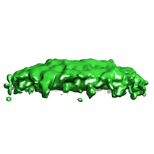
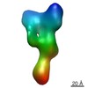
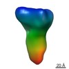
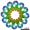
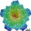
| タイトル | Tomographic subvolume average of EFF-1 fusogen on extracellular vesicles | |||||||||
 マップデータ マップデータ | Segmented membrane structure from EFF-1 coated vesicles | |||||||||
 試料 試料 |
| |||||||||
 キーワード キーワード | cell-cell fusion / extracellular fusion / membrane fusion / fusogen / pre-fusion state | |||||||||
| 生物種 |  Mesocricetus auratus (ネズミ) Mesocricetus auratus (ネズミ) | |||||||||
| 手法 | サブトモグラム平均法 / クライオ電子顕微鏡法 | |||||||||
 データ登録者 データ登録者 | Zeev-Ben-Mordehai T / Vasishtan D / Siebert CA / Grunewald K | |||||||||
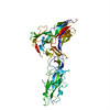
 引用 引用 | ジャーナル: Nat Commun / 年: 2014 タイトル: The full-length cell-cell fusogen EFF-1 is monomeric and upright on the membrane. 著者: Tzviya Zeev-Ben-Mordehai / Daven Vasishtan / C Alistair Siebert / Kay Grünewald /  要旨: Fusogens are membrane proteins that remodel lipid bilayers to facilitate membrane merging. Although several fusogen ectodomain structures have been solved, structural information on full-length, ...Fusogens are membrane proteins that remodel lipid bilayers to facilitate membrane merging. Although several fusogen ectodomain structures have been solved, structural information on full-length, natively membrane-anchored fusogens is scarce. Here we present the electron cryo microscopy three-dimensional reconstruction of the Caenorhabditis elegans epithelial fusion failure 1 (EFF-1) protein natively anchored in cell-derived membrane vesicles. This reveals a membrane protruding, asymmetric, elongated monomer. Flexible fitting of a protomer of the EFF-1 crystal structure, which is homologous to viral class-II fusion proteins, shows that EFF-1 has a hairpin monomeric conformation before fusion. These structural insights, when combined with our observations of membrane-merging intermediates between vesicles, enable us to propose a model for EFF-1 mediated fusion. This process, involving identical proteins on both membranes to be fused, follows a mechanism that shares features of SNARE-mediated fusion while using the structural building blocks of the unilaterally acting class-II viral fusion proteins. | |||||||||
| 履歴 |
|
- 構造の表示
構造の表示
| ムービー |
 ムービービューア ムービービューア |
|---|---|
| 構造ビューア | EMマップ: SurfViewMolmilJmol/JSmol |
| 添付画像 |
- ダウンロードとリンク
-EMDBアーカイブ
| マップデータ | emd_2531.map.gz | 224 KB | EMDBマップデータ形式 | |
|---|---|---|---|---|
| ヘッダ (付随情報) | emd-2531-v30.xmlemd-2531.xml | 9.5 KB 9.5 KB | 表示 表示 | EMDBヘッダ |
| 画像 |  EMD-2531.png EMD-2531.png | 70.6 KB | ||
| アーカイブディレクトリ |  http://ftp.pdbj.org/pub/emdb/structures/EMD-2531ftp://ftp.pdbj.org/pub/emdb/structures/EMD-2531 http://ftp.pdbj.org/pub/emdb/structures/EMD-2531ftp://ftp.pdbj.org/pub/emdb/structures/EMD-2531 | HTTPS FTP |
-検証レポート
| 文書・要旨 | emd_2531_validation.pdf.gz | 195.2 KB | 表示 | EMDB検証レポート |
|---|---|---|---|---|
| 文書・詳細版 | emd_2531_full_validation.pdf.gz | 194.3 KB | 表示 | |
| XML形式データ | emd_2531_validation.xml.gz | 4.3 KB | 表示 | |
| アーカイブディレクトリ | https://ftp.pdbj.org/pub/emdb/validation_reports/EMD-2531ftp://ftp.pdbj.org/pub/emdb/validation_reports/EMD-2531 | HTTPS FTP |
-関連構造データ

-リンク
| EMDBのページ | EMDB (EBI/PDBe) / EMDataResource |
|---|
-マップ
| ファイル | ダウンロード / ファイル: emd_2531.map.gz / 形式: CCP4 / 大きさ: 1.1 MB / タイプ: IMAGE STORED AS FLOATING POINT NUMBER (4 BYTES) | ||||||||||||||||||||||||||||||||||||||||||||||||||||||||||||
|---|---|---|---|---|---|---|---|---|---|---|---|---|---|---|---|---|---|---|---|---|---|---|---|---|---|---|---|---|---|---|---|---|---|---|---|---|---|---|---|---|---|---|---|---|---|---|---|---|---|---|---|---|---|---|---|---|---|---|---|---|---|
| 注釈 | Segmented membrane structure from EFF-1 coated vesicles | ||||||||||||||||||||||||||||||||||||||||||||||||||||||||||||
| 投影像・断面図 | 画像のコントロール
画像は Spider により作成 これらの図は立方格子座標系で作成されたものです | ||||||||||||||||||||||||||||||||||||||||||||||||||||||||||||
| ボクセルのサイズ | X=Y=Z: 3.8 Å | ||||||||||||||||||||||||||||||||||||||||||||||||||||||||||||
| 密度 |
| ||||||||||||||||||||||||||||||||||||||||||||||||||||||||||||
| 対称性 | 空間群: 1 | ||||||||||||||||||||||||||||||||||||||||||||||||||||||||||||
| 詳細 | EMDB XML:
CCP4マップ ヘッダ情報:
| ||||||||||||||||||||||||||||||||||||||||||||||||||||||||||||
 Z (Sec.)
 Y (Row.)
 X (Col.)


-添付データ
- 試料の構成要素
-全体 : Segmented membrane from epithelial fusion failure 1 (EFF-1) Isofo...
| 全体 | 名称: Segmented membrane from epithelial fusion failure 1 (EFF-1) Isoform A on extracellular vesicles |
|---|---|
| 要素 |
|
-超分子 #1000: Segmented membrane from epithelial fusion failure 1 (EFF-1) Isofo...
| 超分子 | 名称: Segmented membrane from epithelial fusion failure 1 (EFF-1) Isoform A on extracellular vesicles タイプ: sample / ID: 1000 / Number unique components: 1 |
|---|
-超分子 #1: Cell-derived vesicle membrane
| 超分子 | 名称: Cell-derived vesicle membrane / タイプ: organelle_or_cellular_component / ID: 1 詳細: Membrane structure of EFF-1 coated extracellular vesicles 組換発現: No |
|---|---|
| 由来(天然) | 生物種: Mesocricetus auratus (ネズミ) / 株: clone 13 / 別称: Golden Hamster / 組織: Kidney / 細胞: Fibroblast BHK 21 / 細胞中の位置: Plasma membrane |
-実験情報
-構造解析
| 手法 | クライオ電子顕微鏡法 |
|---|---|
 解析 解析 | サブトモグラム平均法 |
| 試料の集合状態 | particle |
-試料調製
| 緩衝液 | pH: 7.4 / 詳細: 25mM HEPES, 130mM NaCl |
|---|---|
| グリッド | 詳細: Holey carbon on top of 200 mesh gold grid. |
| 凍結 | 凍結剤: ETHANE-PROPANE MIXTURE / チャンバー内温度: 77 K / 装置: HOMEMADE PLUNGER / 手法: Blot for 3 seconds before plunging |
- 電子顕微鏡法
| 顕微鏡 | FEI POLARA 300 |
|---|---|
| 温度 | 最低: 80 K / 最高: 100 K / 平均: 85 K |
| アライメント法 | Legacy - 非点収差: Objective lens astigmatism was corrected at 115,000 times magnification Legacy - Electron beam tilt params: +1 to -1 mradians |
| 特殊光学系 | エネルギーフィルター - 名称: Gatan Quantum 964 エネルギーフィルター - エネルギー下限: 0.0 eV エネルギーフィルター - エネルギー上限: 20.0 eV |
| 日付 | 2013年3月3日 |
| 撮影 | カテゴリ: CCD フィルム・検出器のモデル: GATAN ULTRASCAN 4000 (4k x 4k) デジタル化 - サンプリング間隔: 30 µm / 実像数: 78 / 平均電子線量: 60 e/Å2 / 詳細: 2 tomograms each created from 39 projection images / ビット/ピクセル: 16 |
| 電子線 | 加速電圧: 200 kV / 電子線源:  FIELD EMISSION GUN FIELD EMISSION GUN |
| 電子光学系 | 倍率(補正後): 78950 / 照射モード: FLOOD BEAM / 撮影モード: BRIGHT FIELD / Cs: 2 mm / 最大 デフォーカス(公称値): 2.0 µm / 最小 デフォーカス(公称値): 2.0 µm / 倍率(公称値): 95000 |
| 試料ステージ | 試料ホルダー: Liquid nitrogen cooled / 試料ホルダーモデル: GATAN HELIUM / Tilt series - Axis1 - Min angle: -60 ° / Tilt series - Axis1 - Max angle: 60 ° |
| 実験機器 |  モデル: Tecnai Polara / 画像提供: FEI Company |
-画像解析
| 詳細 | Subtomograms were selected using a semi-automated picking algorithm |
|---|---|
| 最終 再構成 | 想定した対称性 - 点群: C1 (非対称) / アルゴリズム: OTHER / ソフトウェア - 名称: PEET / 使用したサブトモグラム数: 1973 |
| 最終 3次元分類 | クラス数: 1 |
