 Movie
Movie Controller


+ Open data

- Basic information
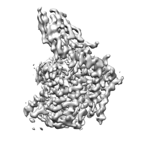
| Entry | Database: EMDB / ID: EMD-25195 | |||||||||
|---|---|---|---|---|---|---|---|---|---|---|
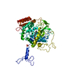
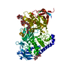
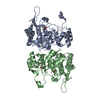
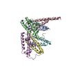
| Title | CryoEM structure of SMCT1 | |||||||||
 Map data Map data | sodium monocarboxylate cotransporter | |||||||||
 Sample Sample |
| |||||||||
| Function / homology |  Function and homology information Function and homology informationacetate transport / propionate transmembrane transporter activity / propanoate transmembrane transport / passive transmembrane transporter activity / monocarboxylate:sodium symporter activity / nicotinate transport / : / : / lactate transport / organic acid:sodium symporter activity ...acetate transport / propionate transmembrane transporter activity / propanoate transmembrane transport / passive transmembrane transporter activity / monocarboxylate:sodium symporter activity / nicotinate transport / : / : / lactate transport / organic acid:sodium symporter activity / Organic anion transport by SLC5/17/25 transporters / pyruvate transport / lactate transmembrane transporter activity / nitrate transmembrane transport / monocarboxylic acid transmembrane transporter activity / iodide transport / GINS complex / chloride transport / sodium ion transport / monoatomic ion transport / apical plasma membrane / apoptotic process / extracellular exosome / plasma membrane Similarity search - Function | |||||||||
| Biological species |  Homo sapiens (human) / synthetic construct (others) Homo sapiens (human) / synthetic construct (others) | |||||||||
| Method | single particle reconstruction / cryo EM / Resolution: 3.5 Å | |||||||||
 Authors Authors | Qu Q / Han L / Panova O / Feng L / Skiniotis G | |||||||||
| Funding support | 1 items
| |||||||||
 Citation Citation | Journal: Nature / Year: 2022 Title: Structure and mechanism of the SGLT family of glucose transporters. Authors: Lei Han / Qianhui Qu / Deniz Aydin / Ouliana Panova / Michael J Robertson / Yan Xu / Ron O Dror / Georgios Skiniotis / Liang Feng /   Abstract: Glucose is a primary energy source in living cells. The discovery in 1960s that a sodium gradient powers the active uptake of glucose in the intestine heralded the concept of a secondary active ...Glucose is a primary energy source in living cells. The discovery in 1960s that a sodium gradient powers the active uptake of glucose in the intestine heralded the concept of a secondary active transporter that can catalyse the movement of a substrate against an electrochemical gradient by harnessing energy from another coupled substrate. Subsequently, coupled Na/glucose transport was found to be mediated by sodium-glucose cotransporters (SGLTs). SGLTs are responsible for active glucose and galactose absorption in the intestine and for glucose reabsorption in the kidney, and are targeted by multiple drugs to treat diabetes. Several members within the SGLT family transport key metabolites other than glucose. Here we report cryo-electron microscopy structures of the prototypic human SGLT1 and a related monocarboxylate transporter SMCT1 from the same family. The structures, together with molecular dynamics simulations and functional studies, define the architecture of SGLTs, uncover the mechanism of substrate binding and selectivity, and shed light on water permeability of SGLT1. These results provide insights into the multifaceted functions of SGLTs. | |||||||||
| History |
|
- Structure visualization
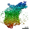
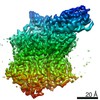
| Movie |
Movie viewer |
|---|---|
| Structure viewer | EM map: SurfViewMolmilJmol/JSmol |
| Supplemental images |
- Downloads & links
-EMDB archive
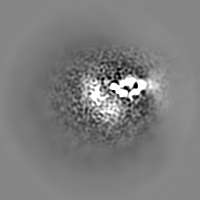
| Map data | emd_25195.map.gz | 21 MB | EMDB map data format | |
|---|---|---|---|---|
| Header (meta data) | emd-25195-v30.xmlemd-25195.xml | 11.3 KB 11.3 KB | Display Display | EMDB header |
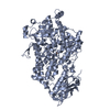
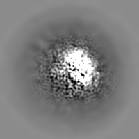
| Images |  emd_25195.png emd_25195.png | 51.5 KB | ||
| Archive directory |  http://ftp.pdbj.org/pub/emdb/structures/EMD-25195ftp://ftp.pdbj.org/pub/emdb/structures/EMD-25195 http://ftp.pdbj.org/pub/emdb/structures/EMD-25195ftp://ftp.pdbj.org/pub/emdb/structures/EMD-25195 | HTTPS FTP |
-Related structure data
| Related structure data | 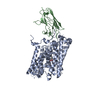 7sl9MC  7sl8C  7slaC C: citing same article ( M: atomic model generated by this map |
|---|---|
| Similar structure data |

-Links
| EMDB pages | EMDB (EBI/PDBe) / EMDataResource |
|---|
-Map
| File | Download / File: emd_25195.map.gz / Format: CCP4 / Size: 30.5 MB / Type: IMAGE STORED AS FLOATING POINT NUMBER (4 BYTES) | ||||||||||||||||||||||||||||||||||||||||||||||||||||||||||||
|---|---|---|---|---|---|---|---|---|---|---|---|---|---|---|---|---|---|---|---|---|---|---|---|---|---|---|---|---|---|---|---|---|---|---|---|---|---|---|---|---|---|---|---|---|---|---|---|---|---|---|---|---|---|---|---|---|---|---|---|---|---|
| Annotation | sodium monocarboxylate cotransporter | ||||||||||||||||||||||||||||||||||||||||||||||||||||||||||||
| Projections & slices | Image control
Images are generated by Spider. | ||||||||||||||||||||||||||||||||||||||||||||||||||||||||||||
| Voxel size | X=Y=Z: 1.06 Å | ||||||||||||||||||||||||||||||||||||||||||||||||||||||||||||
| Density |
| ||||||||||||||||||||||||||||||||||||||||||||||||||||||||||||
| Symmetry | Space group: 1 | ||||||||||||||||||||||||||||||||||||||||||||||||||||||||||||
| Details | EMDB XML:
CCP4 map header:
| ||||||||||||||||||||||||||||||||||||||||||||||||||||||||||||
 Z (Sec.)
 Y (Row.)
 X (Col.)


-Supplemental data
- Sample components
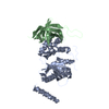
-Entire : SMCT1 and nanobody complex
| Entire | Name: SMCT1 and nanobody complex |
|---|---|
| Components |
|
-Supramolecule #1: SMCT1 and nanobody complex
| Supramolecule | Name: SMCT1 and nanobody complex / type: complex / ID: 1 / Parent: 0 / Macromolecule list: #1-#2 |
|---|---|
| Source (natural) | Organism: Homo sapiens (human) |
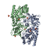
-Macromolecule #1: Sodium-coupled monocarboxylate transporter 1
| Macromolecule | Name: Sodium-coupled monocarboxylate transporter 1 / type: protein_or_peptide / ID: 1 / Number of copies: 1 / Enantiomer: LEVO |
|---|---|
| Source (natural) | Organism: Homo sapiens (human) |
| Molecular weight | Theoretical: 67.636219 KDa |
| Recombinant expression | Organism: Homo sapiens (human) |
| Sequence | String: MDTPRGIGTF VVWDYVVFAG MLVISAAIGI YYAFAGGGQQ TSKDFLMGGR RMTAVPVALS LTASFMSAVT VLGTPSEVYR FGAIFSIFA FTYFFVVVIS AEVFLPVFYK LGITSTYEYL ELRFNKCVRL CGTVLFIVQT ILYTGIVIYA PALALNQVTG F DLWGAVVA ...String: MDTPRGIGTF VVWDYVVFAG MLVISAAIGI YYAFAGGGQQ TSKDFLMGGR RMTAVPVALS LTASFMSAVT VLGTPSEVYR FGAIFSIFA FTYFFVVVIS AEVFLPVFYK LGITSTYEYL ELRFNKCVRL CGTVLFIVQT ILYTGIVIYA PALALNQVTG F DLWGAVVA TGVVCTFYCT LGGLKAVIWT DVFQVGIMVA GFASVIIQAV VMQGGISTIL NDAYDGGRLN FWNFNPNPLQ RH TFWTIII GGTFTWTSIY GVNQSQVQRY ISCKSRFQAK LSLYINLVGL WAILTCSVFC GLALYSRYHD CDPWTAKKVS APD QLMPYL VLDILQDYPG LPGLFVACAY SGTLSTVSSS INALAAVTVE DLIKPYFRSL SERSLSWISQ GMSVVYGALC IGMA ALASL MGALLQAALS VFGMVGGPLM GLFALGILVP FANSIGALVG LMAGFAISLW VGIGAQIYPP LPERTLPLHL DIQGC NSTY NETNLMTTTE MPFTTSVFQI YNVQRTPLMD NWYSLSYLYF STVGTLVTLL VGILVSLSTG GRKQNLDPRY ILTKED FLS NFDIFKKKKH VLSYKSHPVE DGGTDNPAFN HIELNSDQSG KSNGTRLSNS LEVLFQ |
-Macromolecule #2: nanobody Nb2
| Macromolecule | Name: nanobody Nb2 / type: protein_or_peptide / ID: 2 / Number of copies: 1 / Enantiomer: LEVO |
|---|---|
| Source (natural) | Organism: synthetic construct (others) |
| Molecular weight | Theoretical: 14.441961 KDa |
| Recombinant expression | Organism:  |
| Sequence | String: MAQVQLQESG GGLVQAGGSL RLSCAASGNI STRAGMGWYR QAPGKEREFV ASINWGAITN YADSVKGRFT ISRDNAKNTV YLQMNSLKP EDTAVYYCAV EYKYGPQRSD TYYYWGQGTQ VTVSSHHHHH H |
-Macromolecule #3: butanoic acid
| Macromolecule | Name: butanoic acid / type: ligand / ID: 3 / Number of copies: 1 / Formula: BUA |
|---|---|
| Molecular weight | Theoretical: 88.105 Da |
| Chemical component information |  ChemComp-BUA: |
-Experimental details
-Structure determination
| Method | cryo EM |
|---|---|
 Processing Processing | single particle reconstruction |
| Aggregation state | particle |
-Sample preparation
| Buffer | pH: 7 |
|---|---|
| Vitrification | Cryogen name: ETHANE |
- Electron microscopy
| Microscope | FEI TITAN KRIOS |
|---|---|
| Image recording | Film or detector model: GATAN K2 SUMMIT (4k x 4k) / Detector mode: COUNTING / Average electron dose: 56.0 e/Å2 |
| Electron beam | Acceleration voltage: 300 kV / Electron source:  FIELD EMISSION GUN FIELD EMISSION GUN |
| Electron optics | Illumination mode: FLOOD BEAM / Imaging mode: BRIGHT FIELD |
| Experimental equipment |  Model: Titan Krios / Image courtesy: FEI Company |
-Image processing
| Final reconstruction | Resolution.type: BY AUTHOR / Resolution: 3.5 Å / Resolution method: FSC 0.143 CUT-OFF / Number images used: 173859 |
|---|---|
| Initial angle assignment | Type: MAXIMUM LIKELIHOOD |
| Final angle assignment | Type: MAXIMUM LIKELIHOOD |
