+ Open data
- Basic information
| Entry | Database: EMDB / ID: EMD-25180 | |||||||||
|---|---|---|---|---|---|---|---|---|---|---|
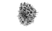
| Title | SMARCAD1-nucleosome map 1 | |||||||||
 Map data Map data | ||||||||||
 Sample Sample |
| |||||||||
| Biological species |  Homo sapiens (human) / synthetic construct (others) Homo sapiens (human) / synthetic construct (others) | |||||||||
| Method | single particle reconstruction / cryo EM / Resolution: 6.49 Å | |||||||||
 Authors Authors | Markert J / Luger K | |||||||||
| Funding support |  United States, 1 items United States, 1 items
| |||||||||
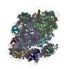
 Citation Citation |  Journal: Sci Adv / Year: 2021 Journal: Sci Adv / Year: 2021Title: SMARCAD1 is an ATP-dependent histone octamer exchange factor with de novo nucleosome assembly activity. Authors: Jonathan Markert / Keda Zhou / Karolin Luger /  Abstract: The adenosine 5′-triphosphate (ATP)–dependent chromatin remodeler SMARCAD1 acts on nucleosomes during DNA replication, repair, and transcription, but despite its implication in disease, ...The adenosine 5′-triphosphate (ATP)–dependent chromatin remodeler SMARCAD1 acts on nucleosomes during DNA replication, repair, and transcription, but despite its implication in disease, information on its function and biochemical activities is scarce. Chromatin remodelers use the energy of ATP hydrolysis to slide nucleosomes, evict histones, or exchange histone variants. Here, we show that SMARCAD1 transfers the entire histone octamer from one DNA segment to another in an ATP-dependent manner but is also capable of de novo nucleosome assembly from histone octamer because of its ability to simultaneously bind all histones. We present a low-resolution cryo–electron microscopy structure of SMARCAD1 in complex with a nucleosome and show that the adenosine triphosphatase domains engage their substrate unlike any other chromatin remodeler. Our biochemical and structural data provide mechanistic insights into SMARCAD1-induced nucleosome disassembly and reassembly. | |||||||||
| History |
|
- Structure visualization
| Movie |
 Movie viewer Movie viewer |
|---|---|
| Structure viewer | EM map:  SurfView SurfView Molmil Molmil Jmol/JSmol Jmol/JSmol |
| Supplemental images |
- Downloads & links
-EMDB archive
| Map data |  emd_25180.map.gz emd_25180.map.gz | 49.1 MB |  EMDB map data format EMDB map data format | |
|---|---|---|---|---|
| Header (meta data) |  emd-25180-v30.xml emd-25180-v30.xml emd-25180.xml emd-25180.xml | 8.1 KB 8.1 KB | Display Display |  EMDB header EMDB header |
| Images |  emd_25180.png emd_25180.png | 33.9 KB | ||
| Archive directory |  http://ftp.pdbj.org/pub/emdb/structures/EMD-25180 http://ftp.pdbj.org/pub/emdb/structures/EMD-25180 ftp://ftp.pdbj.org/pub/emdb/structures/EMD-25180 ftp://ftp.pdbj.org/pub/emdb/structures/EMD-25180 | HTTPS FTP |
-Related structure data
| Related structure data | C: citing same article ( |
|---|---|
| Similar structure data | |
| EM raw data |  EMPIAR-10939 (Title: Cryo electron microscopy SMARCAD1 with nucleosome in ADP-BeF3 bound state EMPIAR-10939 (Title: Cryo electron microscopy SMARCAD1 with nucleosome in ADP-BeF3 bound stateData size: 6.2 TB Data #1: Unaligned multi-frame micrographs of SMARCAD1-nucleosome with ADP-BeF3 [micrographs - multiframe]) |
- Links
Links
| EMDB pages |  EMDB (EBI/PDBe) / EMDB (EBI/PDBe) /  EMDataResource EMDataResource |
|---|
- Map
Map
| File |  Download / File: emd_25180.map.gz / Format: CCP4 / Size: 64 MB / Type: IMAGE STORED AS FLOATING POINT NUMBER (4 BYTES) Download / File: emd_25180.map.gz / Format: CCP4 / Size: 64 MB / Type: IMAGE STORED AS FLOATING POINT NUMBER (4 BYTES) | ||||||||||||||||||||||||||||||||||||||||||||||||||||||||||||
|---|---|---|---|---|---|---|---|---|---|---|---|---|---|---|---|---|---|---|---|---|---|---|---|---|---|---|---|---|---|---|---|---|---|---|---|---|---|---|---|---|---|---|---|---|---|---|---|---|---|---|---|---|---|---|---|---|---|---|---|---|---|
| Projections & slices | Image control
Images are generated by Spider. | ||||||||||||||||||||||||||||||||||||||||||||||||||||||||||||
| Voxel size | X=Y=Z: 1.065 Å | ||||||||||||||||||||||||||||||||||||||||||||||||||||||||||||
| Density |
| ||||||||||||||||||||||||||||||||||||||||||||||||||||||||||||
| Symmetry | Space group: 1 | ||||||||||||||||||||||||||||||||||||||||||||||||||||||||||||
| Details | EMDB XML:
CCP4 map header:
| ||||||||||||||||||||||||||||||||||||||||||||||||||||||||||||
-Supplemental data
- Sample components
-Entire : SMARCAD1-nucleosome
| Entire | Name: SMARCAD1-nucleosome |
|---|---|
| Components |
|
-Supramolecule #1: SMARCAD1-nucleosome
| Supramolecule | Name: SMARCAD1-nucleosome / type: complex / ID: 1 / Parent: 0 |
|---|
-Supramolecule #2: SMARCAD1
| Supramolecule | Name: SMARCAD1 / type: complex / ID: 2 / Parent: 1 |
|---|---|
| Source (natural) | Organism:  Homo sapiens (human) Homo sapiens (human) |
| Recombinant expression | Organism:  |
-Supramolecule #3: nucleosome histones
| Supramolecule | Name: nucleosome histones / type: complex / ID: 3 / Parent: 1 |
|---|---|
| Source (natural) | Organism:  Homo sapiens (human) Homo sapiens (human) |
| Recombinant expression | Organism:  |
-Supramolecule #4: nucleosome DNA
| Supramolecule | Name: nucleosome DNA / type: complex / ID: 4 / Parent: 1 |
|---|---|
| Source (natural) | Organism: synthetic construct (others) |
-Experimental details
-Structure determination
| Method | cryo EM |
|---|---|
 Processing Processing | single particle reconstruction |
| Aggregation state | particle |
- Sample preparation
| Buffer | pH: 7.5 |
|---|---|
| Vitrification | Cryogen name: ETHANE |
- Electron microscopy
| Microscope | FEI TITAN KRIOS |
|---|---|
| Image recording | Film or detector model: GATAN K3 (6k x 4k) / Average electron dose: 1.0 e/Å2 |
| Electron beam | Acceleration voltage: 300 kV / Electron source:  FIELD EMISSION GUN FIELD EMISSION GUN |
| Electron optics | Illumination mode: FLOOD BEAM / Imaging mode: BRIGHT FIELD |
| Experimental equipment |  Model: Titan Krios / Image courtesy: FEI Company |
- Image processing
| Final reconstruction | Resolution.type: BY AUTHOR / Resolution: 6.49 Å / Resolution method: FSC 0.143 CUT-OFF / Number images used: 20135 |
|---|---|
| Initial angle assignment | Type: ANGULAR RECONSTITUTION |
| Final angle assignment | Type: ANGULAR RECONSTITUTION |
 Movie
Movie Controller


 Z (Sec.)
 Y (Row.)
 X (Col.)


