Journal: Structure / Year: 2014 Title: Structural interactions between inhibitor and substrate docking sites give insight into mechanisms of human PS1 complexes. Authors: Yi Li / Stephen Hsueh-Jeng Lu / Ching-Ju Tsai / Christopher Bohm / Seema Qamar / Roger B Dodd / William Meadows / Amy Jeon / Adam McLeod / Fusheng Chen / Muriel Arimon / Oksana Berezovska / ...Authors: Yi Li / Stephen Hsueh-Jeng Lu / Ching-Ju Tsai / Christopher Bohm / Seema Qamar / Roger B Dodd / William Meadows / Amy Jeon / Adam McLeod / Fusheng Chen / Muriel Arimon / Oksana Berezovska / Bradley T Hyman / Taisuke Tomita / Takeshi Iwatsubo / Christopher M Johnson / Lindsay A Farrer / Gerold Schmitt-Ulms / Paul E Fraser / Peter H St George-Hyslop / Abstract: Presenilin-mediated endoproteolysis of transmembrane proteins plays a key role in physiological signaling and in the pathogenesis of Alzheimer disease and some cancers. Numerous inhibitors have been ...Presenilin-mediated endoproteolysis of transmembrane proteins plays a key role in physiological signaling and in the pathogenesis of Alzheimer disease and some cancers. Numerous inhibitors have been found via library screens, but their structural mechanisms remain unknown. We used several biophysical techniques to investigate the structure of human presenilin complexes and the effects of peptidomimetic γ-secretase inhibitors. The complexes are bilobed. The head contains nicastrin ectodomain. The membrane-embedded base has a central channel and a lateral cleft, which may represent the initial substrate docking site. Inhibitor binding induces widespread structural changes, including rotation of the head and closure of the lateral cleft. These changes block substrate access to the catalytic pocket and inhibit the enzyme. Intriguingly, peptide substrate docking has reciprocal effects on the inhibitor binding site. Similar reciprocal shifts may underlie the mechanisms of other inhibitors and of the "lateral gate" through which substrates access to the catalytic site.
History
Deposition
Sep 23, 2013
-
Header (metadata) release
Nov 13, 2013
-
Map release
Nov 20, 2013
-
Update
Mar 2, 2016
-
Current status
Mar 2, 2016
Processing site: PDBe / Status: Released
-
Structure visualization
Movie
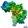
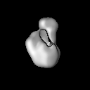
Surface view with section colored by density value
Entire : Compound E-bound human Presenilin 1 (PS1) complex
Entire
Name: Compound E-bound human Presenilin 1 (PS1) complex
Components
Sample: Compound E-bound human Presenilin 1 (PS1) complex
Protein or peptide: Presenilin-1
Protein or peptide: Nicastrin
Protein or peptide: Gamma-secretase subunit APH-1A
Protein or peptide: Gamma-secretase subunit PEN-2
Ligand: E ((S,S)- 2-[2-(3,5-Difluorophenyl)-acetylamino]-N-(1-methyl-2-oxo-5-phenyl-2,3-dihydro-1H-benzo[e][1,4]diazepin-3-yl)-propionamide)
-
Supramolecule #1000: Compound E-bound human Presenilin 1 (PS1) complex
Supramolecule
Name: Compound E-bound human Presenilin 1 (PS1) complex / type: sample / ID: 1000 / Details: The sample was monodisperse / Oligomeric state: 1:1:1:1 / Number unique components: 5
Molecular weight
Theoretical: 200 KDa
-
Macromolecule #1: Presenilin-1
Macromolecule
Name: Presenilin-1 / type: protein_or_peptide / ID: 1 / Name.synonym: Protein S182 Details: N-terminus as tagged with TAP tag composed of Protein G and Streptavidin binding peptide tags separated by tobacco etch virus protease cleavage site Number of copies: 1 / Recombinant expression: Yes
Source (natural)
Organism: Homo sapiens (human) / synonym: Human
Recombinant expression
Organism: Homo sapiens (human) / Recombinant cell: HEK293
Sequence
UniProtKB: Presenilin-1
-
Macromolecule #2: Nicastrin
Macromolecule
Name: Nicastrin / type: protein_or_peptide / ID: 2 / Name.synonym: KIAA0253 / Number of copies: 1 / Recombinant expression: No
Source (natural)
Organism: Homo sapiens (human) / synonym: Human / Cell: HEK293
Sequence
UniProtKB: Nicastrin
-
Macromolecule #3: Gamma-secretase subunit APH-1A
Macromolecule
Name: Gamma-secretase subunit APH-1A / type: protein_or_peptide / ID: 3 / Name.synonym: Aph-1alpha / Number of copies: 1 / Recombinant expression: No
Source (natural)
Organism: Homo sapiens (human) / synonym: Human / Cell: HEK293
Sequence
UniProtKB: Gamma-secretase subunit APH-1A
-
Macromolecule #4: Gamma-secretase subunit PEN-2
Macromolecule
Name: Gamma-secretase subunit PEN-2 / type: protein_or_peptide / ID: 4 / Name.synonym: Presenilin enhancer protein 2 / Number of copies: 1 / Recombinant expression: No
Source (natural)
Organism: Homo sapiens (human) / synonym: Human / Cell: HEK293
Sequence
UniProtKB: Gamma-secretase subunit PEN-2
-
Macromolecule #5: E ((S,S)- 2-[2-(3,5-Difluorophenyl)-acetylamino]-N-(1-methyl-2-ox...
Macromolecule
Name: E ((S,S)- 2-[2-(3,5-Difluorophenyl)-acetylamino]-N-(1-methyl-2-oxo-5-phenyl-2,3-dihydro-1H-benzo[e][1,4]diazepin-3-yl)-propionamide) type: ligand / ID: 5 / Name.synonym: gamma-Secretase Inhibitor XXI / Number of copies: 1 / Recombinant expression: No
Source (natural)
Organism: synthetic construct (others)
-
Experimental details
-
Structure determination
Method
negative staining
Processing
single particle reconstruction
Aggregation state
particle
-
Sample preparation
Concentration
0.02 mg/mL
Buffer
pH: 7.4 Details: 50 mM Tris-HCl, 150 mM NaCl, 2 mM EDTA, 5 mM MgCl2, 5 mM CaCl2
Staining
Type: NEGATIVE Details: Grids with adsorbed protein floated on 1% w/v uranyl acetate for 2-10 seconds
Grid
Details: Carbon-coated 400-mesh copper grids were glow discharged in air at 600-700 V for 30-60 seconds on an Edward S150B sputter coater.
Vitrification
Cryogen name: NONE / Instrument: OTHER
-
Electron microscopy
Microscope
FEI TECNAI 12
Date
Sep 19, 2009
Image recording
Category: CCD / Film or detector model: TVIPS TEMCAM-F224 (2k x 2k) / Number real images: 300
Electron beam
Acceleration voltage: 120 kV / Electron source: TUNGSTEN HAIRPIN
Electron optics
Illumination mode: FLOOD BEAM / Imaging mode: BRIGHT FIELD / Cs: 2 mm / Nominal defocus max: 1.0 µm
Sample stage
Specimen holder model: OTHER
-
Image processing
Details
The particles were selected using EMAN2
Final reconstruction
Applied symmetry - Point group: C1 (asymmetric) / Algorithm: OTHER / Resolution.type: BY AUTHOR / Resolution: 17.4 Å / Resolution method: OTHER / Software - Name: EMAN2, RELION / Number images used: 10651
+
About Yorodumi
-
News
-
Feb 9, 2022. New format data for meta-information of EMDB entries
New format data for meta-information of EMDB entries
Version 3 of the EMDB header file is now the official format.
The previous official version 1.9 will be removed from the archive.
In the structure databanks used in Yorodumi, some data are registered as the other names, "COVID-19 virus" and "2019-nCoV". Here are the details of the virus and the list of structure data.
Jan 31, 2019. EMDB accession codes are about to change! (news from PDBe EMDB page)
EMDB accession codes are about to change! (news from PDBe EMDB page)
The allocation of 4 digits for EMDB accession codes will soon come to an end. Whilst these codes will remain in use, new EMDB accession codes will include an additional digit and will expand incrementally as the available range of codes is exhausted. The current 4-digit format prefixed with “EMD-” (i.e. EMD-XXXX) will advance to a 5-digit format (i.e. EMD-XXXXX), and so on. It is currently estimated that the 4-digit codes will be depleted around Spring 2019, at which point the 5-digit format will come into force.
The EM Navigator/Yorodumi systems omit the EMD- prefix.
Related info.:Q: What is EMD? / ID/Accession-code notation in Yorodumi/EM Navigator
Yorodumi is a browser for structure data from EMDB, PDB, SASBDB, etc.
This page is also the successor to EM Navigator detail page, and also detail information page/front-end page for Omokage search.
The word "yorodu" (or yorozu) is an old Japanese word meaning "ten thousand". "mi" (miru) is to see.
Related info.:EMDB / PDB / SASBDB / Comparison of 3 databanks / Yorodumi Search / Aug 31, 2016. New EM Navigator & Yorodumi / Yorodumi Papers / Jmol/JSmol / Function and homology information / Changes in new EM Navigator and Yorodumi
 Movie
Movie Controller

 Yorodumi
 Open data
 Basic information
 Map data
 Sample
 Function and homology information
 Homo sapiens (human) / synthetic construct (others)
 Authors
 Citation


 Structure visualization
 UCSF Chimera
 Downloads & links
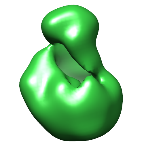 EMD-2478.png
 http://ftp.pdbj.org/pub/emdb/structures/EMD-2478


 Z (Sec.)
 Y (Row.)
 X (Col.)


 Sample components
 Processing
 Electron microscopy