 Movie
Movie Controller

[English] 日本語
 Yorodumi
- EMDB-2411: Hsc70-induced Changes in Clathrin-Auxilin Cage Structure Suggest ... -
+ Open data

- Basic information
| Entry | Database: EMDB / ID: EMD-2411 | |||||||||
|---|---|---|---|---|---|---|---|---|---|---|
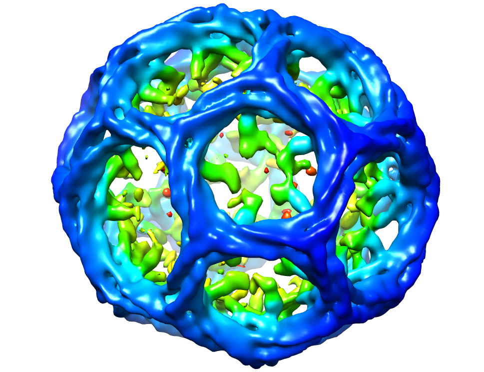
| Title | Hsc70-induced Changes in Clathrin-Auxilin Cage Structure Suggest a Role for Clathrin Light Chains in Cage Disassembly | |||||||||
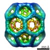
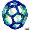
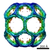
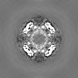
 Map data Map data | Map of Clathrin-Auxilin complex. Sharpened to match a Fourier amplitude profile derived from a model of the hexagonal barrel (the biological unit) created from the crystal structure atomic model pdb id1XI5. Fourier filtered to 30 Angstroms. | |||||||||
 Sample Sample |
| |||||||||
 Keywords Keywords | Endocytosis / coated vesicles / clathrin / auxilin | |||||||||
| Biological species |  | |||||||||
| Method | single particle reconstruction / cryo EM / Resolution: 31.0 Å | |||||||||
 Authors Authors | Young A / Stoilova-McPhie S / Rothnie A / Vallis Y / Harvey-Smith P / Ranson N / Kent H / Brodsky FM / Pearse BM / Roseman A / Smith CJ | |||||||||
 Citation Citation | Journal: J Mol Biol / Year: 2004 Title: Location of auxilin within a clathrin cage. Authors: Corinne J Smith / Timothy R Dafforn / Helen Kent / Catherine A Sims / Kavita Khubchandani-Aswani / Lin Zhang / Helen R Saibil / Barbara M F Pearse /  Abstract: The Dna J homologue, auxilin, acts as a co-chaperone for Hsc70 in the uncoating of clathrin-coated vesicles during endocytosis. Biochemical studies have aided understanding of the uncoating mechanism ...The Dna J homologue, auxilin, acts as a co-chaperone for Hsc70 in the uncoating of clathrin-coated vesicles during endocytosis. Biochemical studies have aided understanding of the uncoating mechanism but until now there was no structural information on how auxilin interacts with the clathrin cage. Here we have determined the three-dimensional structure of a complex of auxilin with clathrin cages by cryo-electron microscopy and single particle analysis. We show that auxilin forms a discrete shell of density on the inside of the clathrin cage. Peptide competition assays confirm that a candidate clathrin box motif in auxilin, LLGLE, can bind to a clathrin construct containing the beta-propeller domain and also displace the well-characterised LLNLD clathrin box motif derived from the beta-adaptin hinge region. The means by which auxilin could both aid clathrin coat assembly and displace clathrin from AP2 during uncoating is discussed. | |||||||||
| History |
|
- Structure visualization
| Movie |
 Movie viewer Movie viewer |
|---|---|
| Structure viewer | EM map: SurfViewMolmilJmol/JSmol |
| Supplemental images |
- Downloads & links
-EMDB archive
| Map data | emd_2411.map.gz | 58.8 MB | EMDB map data format | |
|---|---|---|---|---|
| Header (meta data) | emd-2411-v30.xmlemd-2411.xml | 10.2 KB 10.2 KB | Display Display | EMDB header |
| Images |  emd_2411.png emd_2411.png | 530.7 KB | ||
| Archive directory |  http://ftp.pdbj.org/pub/emdb/structures/EMD-2411ftp://ftp.pdbj.org/pub/emdb/structures/EMD-2411 http://ftp.pdbj.org/pub/emdb/structures/EMD-2411ftp://ftp.pdbj.org/pub/emdb/structures/EMD-2411 | HTTPS FTP |
-Related structure data

-Links
| EMDB pages | EMDB (EBI/PDBe) / EMDataResource |
|---|
-Map
| File | Download / File: emd_2411.map.gz / Format: CCP4 / Size: 62.5 MB / Type: IMAGE STORED AS FLOATING POINT NUMBER (4 BYTES) | ||||||||||||||||||||||||||||||||||||||||||||||||||||||||||||||||||||
|---|---|---|---|---|---|---|---|---|---|---|---|---|---|---|---|---|---|---|---|---|---|---|---|---|---|---|---|---|---|---|---|---|---|---|---|---|---|---|---|---|---|---|---|---|---|---|---|---|---|---|---|---|---|---|---|---|---|---|---|---|---|---|---|---|---|---|---|---|---|
| Annotation | Map of Clathrin-Auxilin complex. Sharpened to match a Fourier amplitude profile derived from a model of the hexagonal barrel (the biological unit) created from the crystal structure atomic model pdb id1XI5. Fourier filtered to 30 Angstroms. | ||||||||||||||||||||||||||||||||||||||||||||||||||||||||||||||||||||
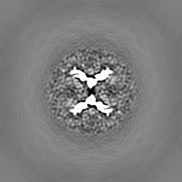
| Projections & slices | Image control
Images are generated by Spider. | ||||||||||||||||||||||||||||||||||||||||||||||||||||||||||||||||||||
| Voxel size | X=Y=Z: 6.4 Å | ||||||||||||||||||||||||||||||||||||||||||||||||||||||||||||||||||||
| Density |
| ||||||||||||||||||||||||||||||||||||||||||||||||||||||||||||||||||||
| Symmetry | Space group: 1 | ||||||||||||||||||||||||||||||||||||||||||||||||||||||||||||||||||||
| Details | EMDB XML:
CCP4 map header:
| ||||||||||||||||||||||||||||||||||||||||||||||||||||||||||||||||||||
 Z (Sec.)
 Y (Row.)
 X (Col.)


-Supplemental data
- Sample components
-Entire : Clathrin bound to auxilin
| Entire | Name: Clathrin bound to auxilin |
|---|---|
| Components |
|
-Supramolecule #1000: Clathrin bound to auxilin
| Supramolecule | Name: Clathrin bound to auxilin / type: sample / ID: 1000 / Number unique components: 2 |
|---|
-Macromolecule #1: Clathrin
| Macromolecule | Name: Clathrin / type: protein_or_peptide / ID: 1 / Recombinant expression: No |
|---|---|
| Source (natural) | Organism: |
-Macromolecule #2: Auxilin
| Macromolecule | Name: Auxilin / type: protein_or_peptide / ID: 2 / Recombinant expression: No |
|---|---|
| Source (natural) | Organism: |
-Experimental details
-Structure determination
| Method | cryo EM |
|---|---|
 Processing Processing | single particle reconstruction |
| Aggregation state | particle |
-Sample preparation
| Buffer | pH: 6 Details: 20 mM Mes (pH 6.0), 2 mM magnesium acetate, 25 mM KCl, 10 mM (NH4)2SO4, 1 mM DTT |
|---|---|
| Vitrification | Cryogen name: ETHANE / Instrument: HOMEMADE PLUNGER |
- Electron microscopy #1
| Microscopy ID | 1 |
|---|---|
| Microscope | FEI/PHILIPS CM200FEG |
| Date | Feb 2, 2000 |
| Image recording | Digitization - Scanner: ZEISS SCAI |
| Electron beam | Acceleration voltage: 200 kV / Electron source:  FIELD EMISSION GUN FIELD EMISSION GUN |
| Electron optics | Illumination mode: FLOOD BEAM / Imaging mode: BRIGHT FIELD |
| Sample stage | Specimen holder model: GATAN LIQUID NITROGEN |
-Electron microscopy #2
| Microscopy ID | 2 |
|---|---|
| Microscope | FEI/PHILIPS CM200FEG |
| Date | Feb 23, 2000 |
| Image recording | Digitization - Scanner: ZEISS SCAI |
| Electron beam | Acceleration voltage: 200 kV / Electron source: FIELD EMISSION GUN |
| Electron optics | Illumination mode: FLOOD BEAM / Imaging mode: BRIGHT FIELD |
| Sample stage | Specimen holder model: GATAN LIQUID NITROGEN |
-Image processing
| CTF correction | Details: Whole micrographs |
|---|---|
| Final reconstruction | Applied symmetry - Point group: D6 (2x6 fold dihedral) / Algorithm: OTHER / Resolution.type: BY AUTHOR / Resolution: 31.0 Å / Resolution method: FSC 0.143 CUT-OFF / Software - Name: MRC, SPIDER, FREALIGN Details: Sharpened to match a Fourier amplitude profile derived from a model of the hexagonal barrel (the biological unit) created from the crystal structure atomic model pdb id1XI5. Fourier filtered to 30 Angstroms. Number images used: 1745 |
