 ムービー
ムービー コントローラー


+ データを開く

- 基本情報
基本情報
| 登録情報 | データベース: EMDB / ID: EMD-2368 | |||||||||
|---|---|---|---|---|---|---|---|---|---|---|
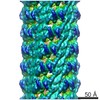
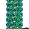
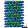
| タイトル | Negative-staining electron microscopy structure of the Agrobacterium tumefaciens T-complex, composed of the protein VirE2 coating single-stranded DNA. | |||||||||
 マップデータ マップデータ | Negative-staining EM reconstruction of the Agrobacterium T-complex | |||||||||
 試料 試料 |
| |||||||||
 キーワード キーワード | T-complex / agrobacterium / helical reconstruction / negative staining | |||||||||
| 生物種 |  Agrobacterium fabrum str. C58 (バクテリア) Agrobacterium fabrum str. C58 (バクテリア) | |||||||||
| 手法 | らせん対称体再構成法 / ネガティブ染色法 / 解像度: 25.0 Å | |||||||||
 データ登録者 データ登録者 | Abu-Arish A / Frenkiel-Krispin D / Fricke T / Tzfira T / Citovsky V / Wolf SG / Elbaum M | |||||||||
 引用 引用 | ジャーナル: J Biol Chem / 年: 2004 タイトル: Three-dimensional reconstruction of Agrobacterium VirE2 protein with single-stranded DNA. 著者: Asmahan Abu-Arish / Daphna Frenkiel-Krispin / Tobin Fricke / Tzvi Tzfira / Vitaly Citovsky / Sharon Grayer Wolf / Michael Elbaum /  要旨: Agrobacterium tumefaciens infects plant cells by a unique mechanism involving an interkingdom genetic transfer. A single-stranded DNA substrate is transported across the two cell walls along with the ...Agrobacterium tumefaciens infects plant cells by a unique mechanism involving an interkingdom genetic transfer. A single-stranded DNA substrate is transported across the two cell walls along with the bacterial virulence proteins VirD2 and VirE2. A single VirD2 molecule covalently binds to the 5'-end of the single-stranded DNA, while the VirE2 protein binds stoichiometrically along the length of the DNA, without sequence specificity. An earlier transmission/scanning transmission electron microscopy study indicated a solenoidal ("telephone coil") organization of the VirE2-DNA complex. Here we report a three-dimensional reconstruction of this complex using electron microscopy and single-particle image-processing methods. We find a hollow helical structure of 15.7-nm outer diameter, with a helical rise of 51.5 nm and 4.25 VirE2 proteins/turn. The inner face of the protein units contains a continuous wall and an inward protruding shelf. These structures appear to accommodate the DNA binding. Such a quaternary arrangement naturally sequesters the DNA from cytoplasmic nucleases and suggests a mechanism for its nuclear import by decoration with host cell factors. Coexisting with the helices, we also found VirE2 tetrameric ring structures. A two-dimensional average of the latter confirms the major features of the three-dimensional reconstruction. | |||||||||
| 履歴 |
|
- 構造の表示
構造の表示
| ムービー |
 ムービービューア ムービービューア |
|---|---|
| 構造ビューア | EMマップ: SurfViewMolmilJmol/JSmol |
| 添付画像 |

- ダウンロードとリンク
-EMDBアーカイブ
| マップデータ | emd_2368.map.gz | 1.5 MB | EMDBマップデータ形式 | |
|---|---|---|---|---|
| ヘッダ (付随情報) | emd-2368-v30.xmlemd-2368.xml | 9.3 KB 9.3 KB | 表示 表示 | EMDBヘッダ |
| 画像 | 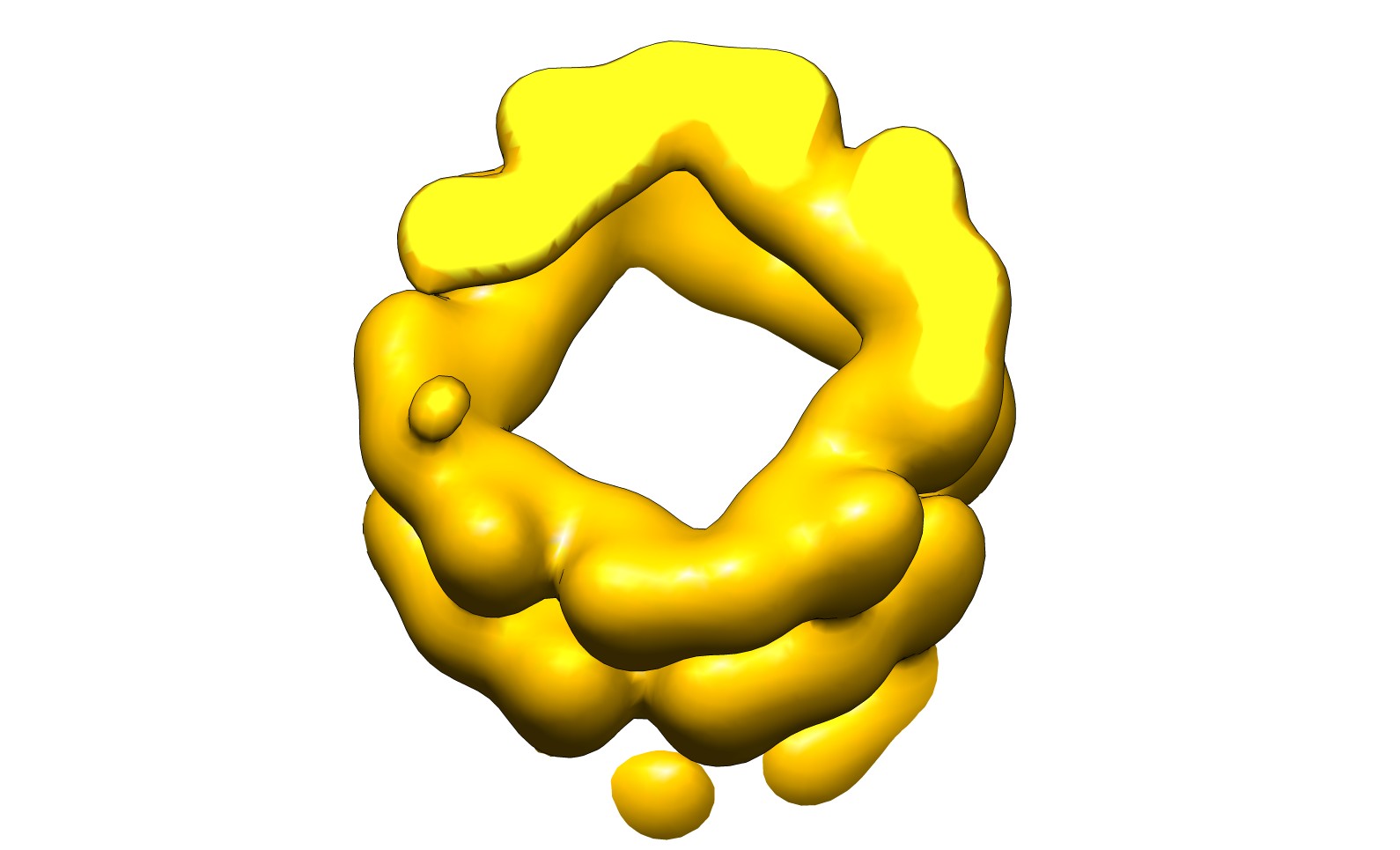 emd_2368.jpg emd_2368.jpg | 97.2 KB | ||
| アーカイブディレクトリ |  http://ftp.pdbj.org/pub/emdb/structures/EMD-2368ftp://ftp.pdbj.org/pub/emdb/structures/EMD-2368 http://ftp.pdbj.org/pub/emdb/structures/EMD-2368ftp://ftp.pdbj.org/pub/emdb/structures/EMD-2368 | HTTPS FTP |
-検証レポート
| 文書・要旨 | emd_2368_validation.pdf.gz | 223.7 KB | 表示 | EMDB検証レポート |
|---|---|---|---|---|
| 文書・詳細版 | emd_2368_full_validation.pdf.gz | 222.8 KB | 表示 | |
| XML形式データ | emd_2368_validation.xml.gz | 4.7 KB | 表示 | |
| アーカイブディレクトリ | https://ftp.pdbj.org/pub/emdb/validation_reports/EMD-2368ftp://ftp.pdbj.org/pub/emdb/validation_reports/EMD-2368 | HTTPS FTP |
-関連構造データ
| 類似構造データ |
|---|

-リンク
| EMDBのページ | EMDB (EBI/PDBe) / EMDataResource |
|---|
-マップ
| ファイル | ダウンロード / ファイル: emd_2368.map.gz / 形式: CCP4 / 大きさ: 1.9 MB / タイプ: IMAGE STORED AS FLOATING POINT NUMBER (4 BYTES) | ||||||||||||||||||||||||||||||||||||||||||||||||||||||||||||||||||||
|---|---|---|---|---|---|---|---|---|---|---|---|---|---|---|---|---|---|---|---|---|---|---|---|---|---|---|---|---|---|---|---|---|---|---|---|---|---|---|---|---|---|---|---|---|---|---|---|---|---|---|---|---|---|---|---|---|---|---|---|---|---|---|---|---|---|---|---|---|---|
| 注釈 | Negative-staining EM reconstruction of the Agrobacterium T-complex | ||||||||||||||||||||||||||||||||||||||||||||||||||||||||||||||||||||
| 投影像・断面図 | 画像のコントロール
画像は Spider により作成 | ||||||||||||||||||||||||||||||||||||||||||||||||||||||||||||||||||||
| ボクセルのサイズ | X=Y=Z: 3.2 Å | ||||||||||||||||||||||||||||||||||||||||||||||||||||||||||||||||||||
| 密度 |
| ||||||||||||||||||||||||||||||||||||||||||||||||||||||||||||||||||||
| 対称性 | 空間群: 1 | ||||||||||||||||||||||||||||||||||||||||||||||||||||||||||||||||||||
| 詳細 | EMDB XML:
CCP4マップ ヘッダ情報:
| ||||||||||||||||||||||||||||||||||||||||||||||||||||||||||||||||||||
 Z (Sec.)
 Y (Row.)
 X (Col.)


-添付データ
- 試料の構成要素
-全体 : Negative-staining recosntruction of the Agrobacterium T-complex
| 全体 | 名称: Negative-staining recosntruction of the Agrobacterium T-complex |
|---|---|
| 要素 |
|
-超分子 #1000: Negative-staining recosntruction of the Agrobacterium T-complex
| 超分子 | 名称: Negative-staining recosntruction of the Agrobacterium T-complex タイプ: sample / ID: 1000 / 詳細: Helical assembly / 集合状態: Helical / Number unique components: 2 |
|---|
-分子 #1: Agrobacterium tumefaciens VirE2 protein
| 分子 | 名称: Agrobacterium tumefaciens VirE2 protein / タイプ: protein_or_peptide / ID: 1 詳細: Negative-staining EM reconstruction of the Agrobacterium T-complex 集合状態: Helical / 組換発現: Yes |
|---|---|
| 由来(天然) | 生物種: Agrobacterium fabrum str. C58 (バクテリア) |
| 分子量 | 理論値: 58 KDa |
| 組換発現 | 生物種: |
-分子 #2: short oligomeric 26mer DNA
| 分子 | 名称: short oligomeric 26mer DNA / タイプ: dna / ID: 2 / 分類: DNA / Structure: DOUBLE HELIX / Synthetic?: Yes |
|---|---|
| 由来(天然) | 生物種: Agrobacterium fabrum str. C58 (バクテリア) |
-実験情報
-構造解析
| 手法 | ネガティブ染色法 |
|---|---|
 解析 解析 | らせん対称体再構成法 |
| 試料の集合状態 | helical array |
-試料調製
| 染色 | タイプ: NEGATIVE / 詳細: 1.5% Uranyl acetate staining |
|---|---|
| グリッド | 詳細: Carbon coated grids |
| 凍結 | 凍結剤: NONE / 装置: OTHER |
| 詳細 | Protein was mixed with M13 circular single-stranded DNA. |
- 電子顕微鏡法
| 顕微鏡 | FEI TECNAI F20 |
|---|---|
| 日付 | 2004年1月1日 |
| 撮影 | カテゴリ: CCD / フィルム・検出器のモデル: GENERIC TVIPS |
| 電子線 | 加速電圧: 200 kV / 電子線源:  FIELD EMISSION GUN FIELD EMISSION GUN |
| 電子光学系 | 照射モード: FLOOD BEAM / 撮影モード: BRIGHT FIELD / Cs: 2 mm / 最大 デフォーカス(公称値): 2.7 µm / 最小 デフォーカス(公称値): 0.6 µm |
| 試料ステージ | 試料ホルダーモデル: OTHER |
| 実験機器 |  モデル: Tecnai F20 / 画像提供: FEI Company |
-画像解析
| 詳細 | Helical reconstruction using IHRSR |
|---|---|
| 最終 再構成 | 想定した対称性 - らせんパラメータ - Δz: 12.11 Å 想定した対称性 - らせんパラメータ - ΔΦ: 84.7 ° アルゴリズム: OTHER / 解像度のタイプ: BY AUTHOR / 解像度: 25.0 Å / ソフトウェア - 名称: Bsoft, Spider, IHRSR |
| CTF補正 | 詳細: Phase-flipping |
