 Movie
Movie Controller

[English] 日本語
 Yorodumi
- EMDB-2368: Negative-staining electron microscopy structure of the Agrobacter... -
+ Open data

- Basic information
| Entry | Database: EMDB / ID: EMD-2368 | |||||||||
|---|---|---|---|---|---|---|---|---|---|---|
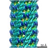
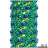
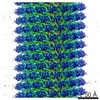
| Title | Negative-staining electron microscopy structure of the Agrobacterium tumefaciens T-complex, composed of the protein VirE2 coating single-stranded DNA. | |||||||||
 Map data Map data | Negative-staining EM reconstruction of the Agrobacterium T-complex | |||||||||
 Sample Sample |
| |||||||||
 Keywords Keywords | T-complex / agrobacterium / helical reconstruction / negative staining | |||||||||
| Biological species |  Agrobacterium fabrum str. C58 (bacteria) Agrobacterium fabrum str. C58 (bacteria) | |||||||||
| Method | helical reconstruction / negative staining / Resolution: 25.0 Å | |||||||||
 Authors Authors | Abu-Arish A / Frenkiel-Krispin D / Fricke T / Tzfira T / Citovsky V / Wolf SG / Elbaum M | |||||||||
 Citation Citation | Journal: J Biol Chem / Year: 2004 Title: Three-dimensional reconstruction of Agrobacterium VirE2 protein with single-stranded DNA. Authors: Asmahan Abu-Arish / Daphna Frenkiel-Krispin / Tobin Fricke / Tzvi Tzfira / Vitaly Citovsky / Sharon Grayer Wolf / Michael Elbaum /  Abstract: Agrobacterium tumefaciens infects plant cells by a unique mechanism involving an interkingdom genetic transfer. A single-stranded DNA substrate is transported across the two cell walls along with the ...Agrobacterium tumefaciens infects plant cells by a unique mechanism involving an interkingdom genetic transfer. A single-stranded DNA substrate is transported across the two cell walls along with the bacterial virulence proteins VirD2 and VirE2. A single VirD2 molecule covalently binds to the 5'-end of the single-stranded DNA, while the VirE2 protein binds stoichiometrically along the length of the DNA, without sequence specificity. An earlier transmission/scanning transmission electron microscopy study indicated a solenoidal ("telephone coil") organization of the VirE2-DNA complex. Here we report a three-dimensional reconstruction of this complex using electron microscopy and single-particle image-processing methods. We find a hollow helical structure of 15.7-nm outer diameter, with a helical rise of 51.5 nm and 4.25 VirE2 proteins/turn. The inner face of the protein units contains a continuous wall and an inward protruding shelf. These structures appear to accommodate the DNA binding. Such a quaternary arrangement naturally sequesters the DNA from cytoplasmic nucleases and suggests a mechanism for its nuclear import by decoration with host cell factors. Coexisting with the helices, we also found VirE2 tetrameric ring structures. A two-dimensional average of the latter confirms the major features of the three-dimensional reconstruction. | |||||||||
| History |
|
- Structure visualization
| Movie |
 Movie viewer Movie viewer |
|---|---|
| Structure viewer | EM map: SurfViewMolmilJmol/JSmol |
| Supplemental images |

- Downloads & links
-EMDB archive
| Map data | emd_2368.map.gz | 1.5 MB | EMDB map data format | |
|---|---|---|---|---|
| Header (meta data) | emd-2368-v30.xmlemd-2368.xml | 9.3 KB 9.3 KB | Display Display | EMDB header |
| Images | 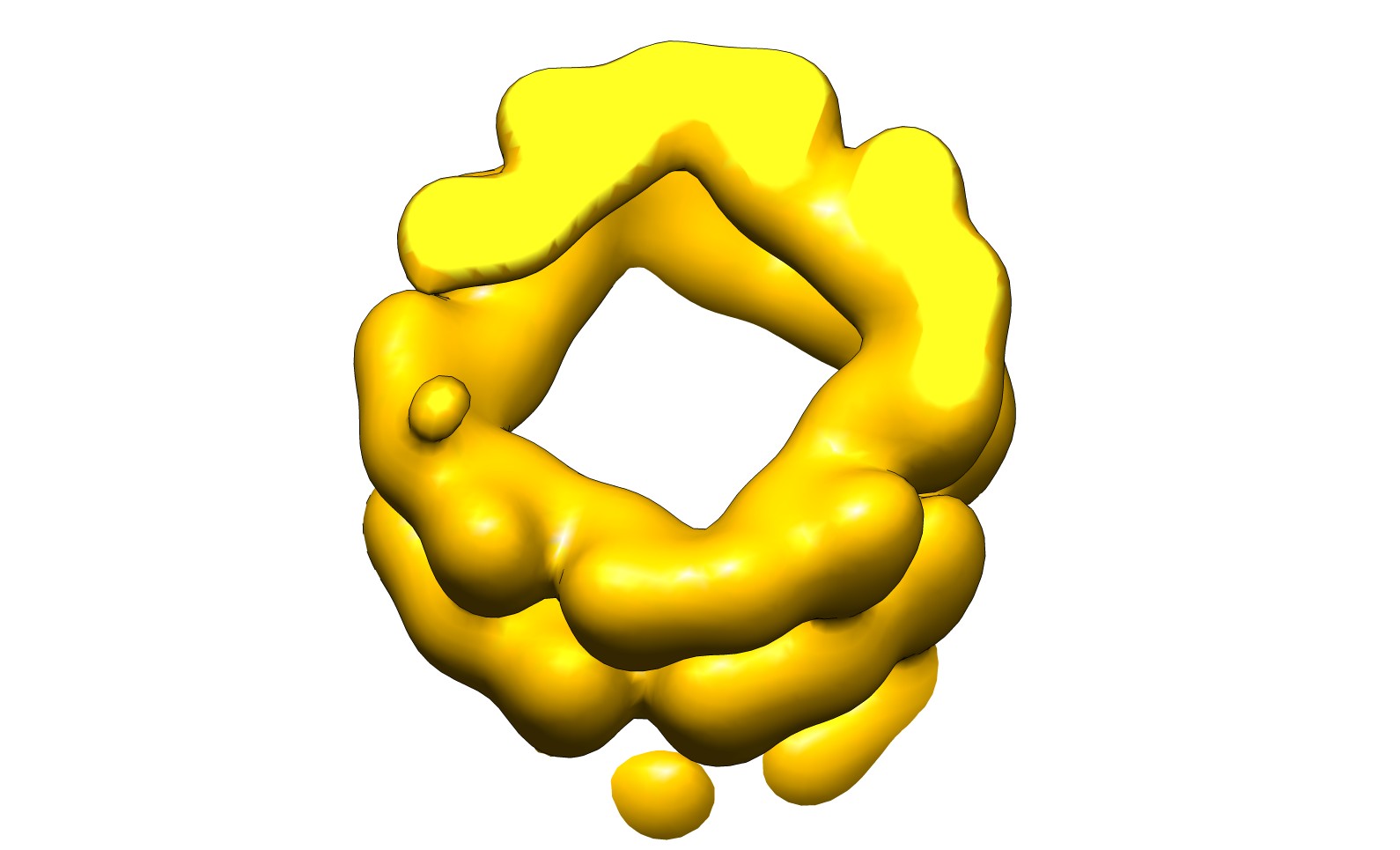 emd_2368.jpg emd_2368.jpg | 97.2 KB | ||
| Archive directory |  http://ftp.pdbj.org/pub/emdb/structures/EMD-2368ftp://ftp.pdbj.org/pub/emdb/structures/EMD-2368 http://ftp.pdbj.org/pub/emdb/structures/EMD-2368ftp://ftp.pdbj.org/pub/emdb/structures/EMD-2368 | HTTPS FTP |
-Related structure data
| Similar structure data |
|---|

-Links
| EMDB pages | EMDB (EBI/PDBe) / EMDataResource |
|---|
-Map
| File | Download / File: emd_2368.map.gz / Format: CCP4 / Size: 1.9 MB / Type: IMAGE STORED AS FLOATING POINT NUMBER (4 BYTES) | ||||||||||||||||||||||||||||||||||||||||||||||||||||||||||||||||||||
|---|---|---|---|---|---|---|---|---|---|---|---|---|---|---|---|---|---|---|---|---|---|---|---|---|---|---|---|---|---|---|---|---|---|---|---|---|---|---|---|---|---|---|---|---|---|---|---|---|---|---|---|---|---|---|---|---|---|---|---|---|---|---|---|---|---|---|---|---|---|
| Annotation | Negative-staining EM reconstruction of the Agrobacterium T-complex | ||||||||||||||||||||||||||||||||||||||||||||||||||||||||||||||||||||
| Projections & slices | Image control
Images are generated by Spider. | ||||||||||||||||||||||||||||||||||||||||||||||||||||||||||||||||||||
| Voxel size | X=Y=Z: 3.2 Å | ||||||||||||||||||||||||||||||||||||||||||||||||||||||||||||||||||||
| Density |
| ||||||||||||||||||||||||||||||||||||||||||||||||||||||||||||||||||||
| Symmetry | Space group: 1 | ||||||||||||||||||||||||||||||||||||||||||||||||||||||||||||||||||||
| Details | EMDB XML:
CCP4 map header:
| ||||||||||||||||||||||||||||||||||||||||||||||||||||||||||||||||||||
 Z (Sec.)
 Y (Row.)
 X (Col.)


-Supplemental data
- Sample components
-Entire : Negative-staining recosntruction of the Agrobacterium T-complex
| Entire | Name: Negative-staining recosntruction of the Agrobacterium T-complex |
|---|---|
| Components |
|
-Supramolecule #1000: Negative-staining recosntruction of the Agrobacterium T-complex
| Supramolecule | Name: Negative-staining recosntruction of the Agrobacterium T-complex type: sample / ID: 1000 / Details: Helical assembly / Oligomeric state: Helical / Number unique components: 2 |
|---|
-Macromolecule #1: Agrobacterium tumefaciens VirE2 protein
| Macromolecule | Name: Agrobacterium tumefaciens VirE2 protein / type: protein_or_peptide / ID: 1 Details: Negative-staining EM reconstruction of the Agrobacterium T-complex Oligomeric state: Helical / Recombinant expression: Yes |
|---|---|
| Source (natural) | Organism: Agrobacterium fabrum str. C58 (bacteria) |
| Molecular weight | Theoretical: 58 KDa |
| Recombinant expression | Organism: |
-Macromolecule #2: short oligomeric 26mer DNA
| Macromolecule | Name: short oligomeric 26mer DNA / type: dna / ID: 2 / Classification: DNA / Structure: DOUBLE HELIX / Synthetic?: Yes |
|---|---|
| Source (natural) | Organism: Agrobacterium fabrum str. C58 (bacteria) |
-Experimental details
-Structure determination
| Method | negative staining |
|---|---|
 Processing Processing | helical reconstruction |
| Aggregation state | helical array |
-Sample preparation
| Staining | Type: NEGATIVE / Details: 1.5% Uranyl acetate staining |
|---|---|
| Grid | Details: Carbon coated grids |
| Vitrification | Cryogen name: NONE / Instrument: OTHER |
| Details | Protein was mixed with M13 circular single-stranded DNA. |
- Electron microscopy
| Microscope | FEI TECNAI F20 |
|---|---|
| Date | Jan 1, 2004 |
| Image recording | Category: CCD / Film or detector model: GENERIC TVIPS |
| Electron beam | Acceleration voltage: 200 kV / Electron source:  FIELD EMISSION GUN FIELD EMISSION GUN |
| Electron optics | Illumination mode: FLOOD BEAM / Imaging mode: BRIGHT FIELD / Cs: 2 mm / Nominal defocus max: 2.7 µm / Nominal defocus min: 0.6 µm |
| Sample stage | Specimen holder model: OTHER |
| Experimental equipment |  Model: Tecnai F20 / Image courtesy: FEI Company |
-Image processing
| Details | Helical reconstruction using IHRSR |
|---|---|
| Final reconstruction | Applied symmetry - Helical parameters - Δz: 12.11 Å Applied symmetry - Helical parameters - Δ&Phi: 84.7 ° Algorithm: OTHER / Resolution.type: BY AUTHOR / Resolution: 25.0 Å / Software - Name: Bsoft, Spider, IHRSR |
| CTF correction | Details: Phase-flipping |
