 Movie
Movie Controller

[English] 日本語
 Yorodumi
- EMDB-23451: Human p97 in complex with Npl4/Ufd1 and polyubiquitinated Ub-Eos ... -
+ Open data

- Basic information
| Entry | Database: EMDB / ID: EMD-23451 | |||||||||
|---|---|---|---|---|---|---|---|---|---|---|
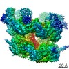
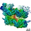
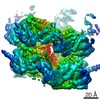
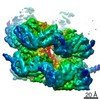
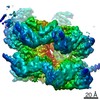
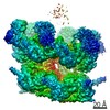
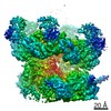
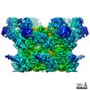
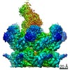
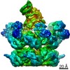
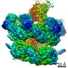
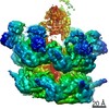
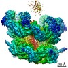
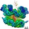
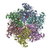
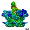
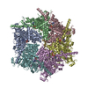
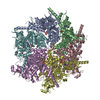
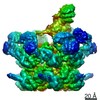
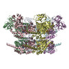
| Title | Human p97 in complex with Npl4/Ufd1 and polyubiquitinated Ub-Eos (Class 3) | |||||||||
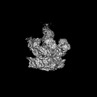
 Map data Map data | ||||||||||
 Sample Sample |
| |||||||||
| Function / homology |  Function and homology information Function and homology informationflavin adenine dinucleotide catabolic process / VCP-NSFL1C complex / endoplasmic reticulum stress-induced pre-emptive quality control / endosome to lysosome transport via multivesicular body sorting pathway / BAT3 complex binding / cytoplasmic ubiquitin ligase complex / cellular response to arsenite ion / protein-DNA covalent cross-linking repair / Derlin-1 retrotranslocation complex / positive regulation of protein K63-linked deubiquitination ...flavin adenine dinucleotide catabolic process / VCP-NSFL1C complex / endoplasmic reticulum stress-induced pre-emptive quality control / endosome to lysosome transport via multivesicular body sorting pathway / BAT3 complex binding / cytoplasmic ubiquitin ligase complex / cellular response to arsenite ion / protein-DNA covalent cross-linking repair / Derlin-1 retrotranslocation complex / positive regulation of protein K63-linked deubiquitination / deubiquitinase activator activity / positive regulation of oxidative phosphorylation / cytoplasm protein quality control / aggresome assembly / ubiquitin-modified protein reader activity / regulation of protein localization to chromatin / mitotic spindle disassembly / VCP-NPL4-UFD1 AAA ATPase complex / cellular response to misfolded protein / positive regulation of mitochondrial membrane potential / vesicle-fusing ATPase / K48-linked polyubiquitin modification-dependent protein binding / regulation of aerobic respiration / NAD+ metabolic process / retrograde protein transport, ER to cytosol / stress granule disassembly / ATPase complex / ubiquitin-specific protease binding / regulation of synapse organization / ciliary transition zone / positive regulation of ATP biosynthetic process / intracellular membrane-bounded organelle / ubiquitin-like protein ligase binding / RHOH GTPase cycle / MHC class I protein binding / autophagosome maturation / negative regulation of hippo signaling / HSF1 activation / endoplasmic reticulum to Golgi vesicle-mediated transport / polyubiquitin modification-dependent protein binding / interstrand cross-link repair / ATP metabolic process / Attachment and Entry / translesion synthesis / negative regulation of protein localization to chromatin / Protein methylation / endoplasmic reticulum unfolded protein response / ERAD pathway / proteasomal protein catabolic process / lipid droplet / ciliary tip / viral genome replication / proteasome complex / Josephin domain DUBs / macroautophagy / negative regulation of smoothened signaling pathway / establishment of protein localization / N-glycan trimming in the ER and Calnexin/Calreticulin cycle / positive regulation of protein-containing complex assembly / ADP binding / Hh mutants are degraded by ERAD / Translesion Synthesis by POLH / positive regulation of non-canonical NF-kappaB signal transduction / Hedgehog ligand biogenesis / Defective CFTR causes cystic fibrosis / autophagy / ABC-family protein mediated transport / cytoplasmic stress granule / Aggrephagy / positive regulation of protein catabolic process / azurophil granule lumen / Ovarian tumor domain proteases / KEAP1-NFE2L2 pathway / positive regulation of canonical Wnt signaling pathway / double-strand break repair / positive regulation of proteasomal ubiquitin-dependent protein catabolic process / cellular response to heat / E3 ubiquitin ligases ubiquitinate target proteins / site of double-strand break / Neddylation / secretory granule lumen / protein phosphatase binding / regulation of apoptotic process / ficolin-1-rich granule lumen / ubiquitin-dependent protein catabolic process / Attachment and Entry / proteasome-mediated ubiquitin-dependent protein catabolic process / ciliary basal body / protein ubiquitination / protein domain specific binding / DNA repair / DNA damage response / Neutrophil degranulation / ubiquitin protein ligase binding / lipid binding / endoplasmic reticulum membrane / perinuclear region of cytoplasm / glutamatergic synapse / endoplasmic reticulum / ATP hydrolysis activity Similarity search - Function | |||||||||
| Biological species |  Homo sapiens (human) Homo sapiens (human) | |||||||||
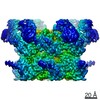
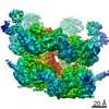
| Method | single particle reconstruction / cryo EM / Resolution: 3.77 Å | |||||||||
 Authors Authors | Pan M / Yu Y / Liu L / Zhao M | |||||||||
 Citation Citation | Journal: Nat Struct Mol Biol / Year: 2021 Title: Mechanistic insight into substrate processing and allosteric inhibition of human p97. Authors: Man Pan / Yuanyuan Yu / Huasong Ai / Qingyun Zheng / Yuan Xie / Lei Liu / Minglei Zhao /   Abstract: p97 processes ubiquitinated substrates and plays a central role in cellular protein homeostasis. Here, we report a series of cryo-EM structures of the substrate-engaged human p97 complex with ...p97 processes ubiquitinated substrates and plays a central role in cellular protein homeostasis. Here, we report a series of cryo-EM structures of the substrate-engaged human p97 complex with resolutions ranging from 2.9 to 3.8 Å that captured 'power-stroke'-like motions of both the D1 and D2 ATPase rings of p97. A key feature of these structures is the critical conformational changes of the intersubunit signaling (ISS) motifs, which tighten the binding of nucleotides and neighboring subunits and contribute to the spiral staircase conformation of the D1 and D2 rings. In addition, we determined the cryo-EM structure of human p97 in complex with NMS-873, a potent p97 inhibitor, at a resolution of 2.4 Å. The structures showed that NMS-873 binds at a cryptic groove in the D2 domain and interacts with the ISS motif, preventing its conformational change and thus blocking substrate translocation allosterically. | |||||||||
| History |
|
- Structure visualization
| Movie |
Movie viewer |
|---|---|
| Structure viewer | EM map: SurfViewMolmilJmol/JSmol |
| Supplemental images |

- Downloads & links
-EMDB archive
| Map data | emd_23451.map.gz | 98 MB | EMDB map data format | |
|---|---|---|---|---|
| Header (meta data) | emd-23451-v30.xmlemd-23451.xml | 20.2 KB 20.2 KB | Display Display | EMDB header |
| FSC (resolution estimation) | emd_23451_fsc.xml | 11.4 KB | Display | FSC data file |
| Images | 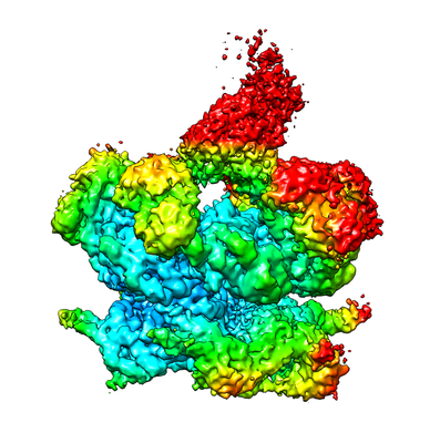 emd_23451.png emd_23451.png | 145.3 KB | ||
| Masks | emd_23451_msk_1.map | 125 MB | Mask map | |
| Others | emd_23451_additional_1.map.gzemd_23451_half_map_1.map.gzemd_23451_half_map_2.map.gz | 116.2 MB 98.4 MB 98.4 MB | ||
| Archive directory |  http://ftp.pdbj.org/pub/emdb/structures/EMD-23451ftp://ftp.pdbj.org/pub/emdb/structures/EMD-23451 http://ftp.pdbj.org/pub/emdb/structures/EMD-23451ftp://ftp.pdbj.org/pub/emdb/structures/EMD-23451 | HTTPS FTP |
-Related structure data
| Related structure data |  7lmyC  7lmzC  7ln0C  7ln1C  7ln2C  7ln3C  7ln4C 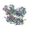 7ln5C 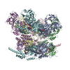 7ln6C C: citing same article ( |
|---|---|
| Similar structure data |

-Links
| EMDB pages | EMDB (EBI/PDBe) / EMDataResource |
|---|---|
| Related items in Molecule of the Month |


-Map
| File | Download / File: emd_23451.map.gz / Format: CCP4 / Size: 125 MB / Type: IMAGE STORED AS FLOATING POINT NUMBER (4 BYTES) | ||||||||||||||||||||||||||||||||||||||||||||||||||||||||||||
|---|---|---|---|---|---|---|---|---|---|---|---|---|---|---|---|---|---|---|---|---|---|---|---|---|---|---|---|---|---|---|---|---|---|---|---|---|---|---|---|---|---|---|---|---|---|---|---|---|---|---|---|---|---|---|---|---|---|---|---|---|---|
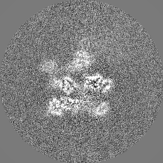
| Projections & slices | Image control
Images are generated by Spider. | ||||||||||||||||||||||||||||||||||||||||||||||||||||||||||||
| Voxel size | X=Y=Z: 1.063 Å | ||||||||||||||||||||||||||||||||||||||||||||||||||||||||||||
| Density |
| ||||||||||||||||||||||||||||||||||||||||||||||||||||||||||||
| Symmetry | Space group: 1 | ||||||||||||||||||||||||||||||||||||||||||||||||||||||||||||
| Details | EMDB XML:
CCP4 map header:
| ||||||||||||||||||||||||||||||||||||||||||||||||||||||||||||
 Z (Sec.)
 Y (Row.)
 X (Col.)


-Supplemental data
-Mask #1
| File | emd_23451_msk_1.map | ||||||||||||
|---|---|---|---|---|---|---|---|---|---|---|---|---|---|
| Projections & Slices |
| ||||||||||||
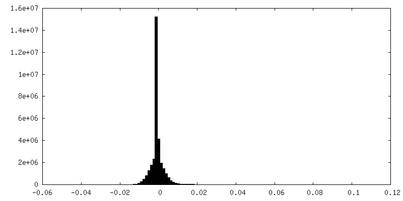
| Density Histograms |


-Additional map: #1
| File | emd_23451_additional_1.map | ||||||||||||
|---|---|---|---|---|---|---|---|---|---|---|---|---|---|
| Projections & Slices |
| ||||||||||||
| Density Histograms |


-Half map: #1
| File | emd_23451_half_map_1.map | ||||||||||||
|---|---|---|---|---|---|---|---|---|---|---|---|---|---|
| Projections & Slices |
| ||||||||||||
| Density Histograms |


-Half map: #2
| File | emd_23451_half_map_2.map | ||||||||||||
|---|---|---|---|---|---|---|---|---|---|---|---|---|---|
| Projections & Slices |
| ||||||||||||
| Density Histograms |


- Sample components
-Entire : Human p97 in complex with Npl4/Ufd1 and K48-linked polyubiquitina...
| Entire | Name: Human p97 in complex with Npl4/Ufd1 and K48-linked polyubiquitinated Ub-Eos in the presence of ATP |
|---|---|
| Components |
|
-Supramolecule #1: Human p97 in complex with Npl4/Ufd1 and K48-linked polyubiquitina...
| Supramolecule | Name: Human p97 in complex with Npl4/Ufd1 and K48-linked polyubiquitinated Ub-Eos in the presence of ATP type: complex / ID: 1 / Parent: 0 / Macromolecule list: all |
|---|---|
| Source (natural) | Organism: Homo sapiens (human) |
| Recombinant expression | Organism:  |
-Macromolecule #1: Human p97 (A232E/E578Q)
| Macromolecule | Name: Human p97 (A232E/E578Q) / type: protein_or_peptide / ID: 1 / Enantiomer: LEVO |
|---|---|
| Source (natural) | Organism: Homo sapiens (human) |
| Recombinant expression | Organism: |
| Sequence | String: MASGADSKGD DLSTAILKQK NRPNRLIVDE AINEDNSVVS LSQPKMDELQ LFRGDTVLLK GKKRREAVC IVLSDDTCSD EKIRMNRVVR NNLRVRLGDV ISIQPCPDVK YGKRIHVLPI D DTVEGITG NLFEVYLKPY FLEAYRPIRK GDIFLVRGGM RAVEFKVVET ...String: MASGADSKGD DLSTAILKQK NRPNRLIVDE AINEDNSVVS LSQPKMDELQ LFRGDTVLLK GKKRREAVC IVLSDDTCSD EKIRMNRVVR NNLRVRLGDV ISIQPCPDVK YGKRIHVLPI D DTVEGITG NLFEVYLKPY FLEAYRPIRK GDIFLVRGGM RAVEFKVVET DPSPYCIVAP DT VIHCEGE PIKREDEEES LNEVGYDDIG GCRKQLAQIK EMVELPLRHP ALFKEIGVKP PRG ILLYGP PGTGKTLIAR AVANETGAFF FLINGPEIMS KLAGESESNL RKAFEEAEKN APAI IFIDE LDAIAPKREK THGEVERRIV SQLLTLMDGL KQRAHVIVMA ATNRPNSIDP ALRRF GRFD REVDIGIPDA TGRLEILQIH TKNMKLADDV DLEQVANETH GHVGADLAAL CSEAAL QAI RKKMDLIDLE DETIDAEVMN SLAVTMDDFR WALSQSNPSA LRETVVEVPQ VTWEDIG GL EDVKRELQEL VQYPVEHPDK FLKFGMTPSK GVLFYGPPGC GKTLLAKAIA NECQANFI S IKGPELLTMW FGESEANVRE IFDKARQAAP CVLFFDQLDS IAKARGGNIG DGGGAADRV INQILTEMDG MSTKKNVFII GATNRPDIID PAILRPGRLD QLIYIPLPDE KSRVAILKAN LRKSPVAKD VDLEFLAKMT NGFSGADLTE ICQRACKLAI RESIESEIRR ERERQTNPSA M EVEEDDPV PEIRRDHFEE AMRFARRSVS DNDIRKYEMF AQTLQQSRGF GSFRFPSGNQ GG AGPSQGS GGGTGGSVYT EDNDDDLYG |
-Experimental details
-Structure determination
| Method | cryo EM |
|---|---|
 Processing Processing | single particle reconstruction |
| Aggregation state | particle |
-Sample preparation
| Concentration | 2.5 mg/mL |
|---|---|
| Buffer | pH: 8 |
| Vitrification | Cryogen name: ETHANE / Chamber humidity: 100 % / Chamber temperature: 281 K |
- Electron microscopy
| Microscope | FEI TITAN KRIOS |
|---|---|
| Image recording | Film or detector model: GATAN K3 BIOQUANTUM (6k x 4k) / Average electron dose: 50.0 e/Å2 |
| Electron beam | Acceleration voltage: 300 kV / Electron source:  FIELD EMISSION GUN FIELD EMISSION GUN |
| Electron optics | C2 aperture diameter: 70.0 µm / Illumination mode: FLOOD BEAM / Imaging mode: BRIGHT FIELD |
| Experimental equipment |  Model: Titan Krios / Image courtesy: FEI Company |
+Image processing

-Atomic model buiding 1
| Refinement | Space: REAL / Protocol: AB INITIO MODEL |
|---|
