ムービー
ムービー コントローラー


+ データを開く

- 基本情報
基本情報
| 登録情報 | データベース: EMDB / ID: EMD-2327 | |||||||||
|---|---|---|---|---|---|---|---|---|---|---|
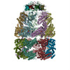
| タイトル | Visualizing GroEL/ES in the Act of Encapsulating a Non-Native Substrate Protein | |||||||||
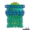
 マップデータ マップデータ | Symmetry-free 3D reconstruction of the bullet-shaped substrate-encapsulated subpopulation sorted from the heterogeneous sample of wild-type GroEL reacted with the substrate protein RuBisCO, GroES and nucleotide ATP | |||||||||
 試料 試料 |
| |||||||||
 キーワード キーワード | protein folding / chaperonin / GroEL / GroEL-GroES / cryo-EM / heterogeneity / substrate / RuBisCO / encapsulation | |||||||||
| 機能・相同性 |  機能・相同性情報 機能・相同性情報GroEL-GroES complex / chaperonin ATPase / virion assembly / : / isomerase activity / protein folding chaperone / ATP-dependent protein folding chaperone / response to radiation / unfolded protein binding / protein folding ...GroEL-GroES complex / chaperonin ATPase / virion assembly / : / isomerase activity / protein folding chaperone / ATP-dependent protein folding chaperone / response to radiation / unfolded protein binding / protein folding / protein-folding chaperone binding / response to heat / protein refolding / magnesium ion binding / ATP hydrolysis activity / ATP binding / metal ion binding / identical protein binding / membrane / cytosol 類似検索 - 分子機能 | |||||||||
| 生物種 |  Rhodospirillum rubrum (バクテリア) / synthetic construct (人工物) Rhodospirillum rubrum (バクテリア) / synthetic construct (人工物) | |||||||||
| 手法 | 単粒子再構成法 / クライオ電子顕微鏡法 / 解像度: 15.9 Å | |||||||||
 データ登録者 データ登録者 | Chen D-H / Madan D / Weaver J / Lin Z / Schroder GF / Chiu W / Rye HS | |||||||||
 引用 引用 | ジャーナル: Cell / 年: 2013 タイトル: Visualizing GroEL/ES in the act of encapsulating a folding protein. 著者: Dong-Hua Chen / Damian Madan / Jeremy Weaver / Zong Lin / Gunnar F Schröder / Wah Chiu / Hays S Rye /  要旨: The GroEL/ES chaperonin system is required for the assisted folding of many proteins. How these substrate proteins are encapsulated within the GroEL-GroES cavity is poorly understood. Using symmetry- ...The GroEL/ES chaperonin system is required for the assisted folding of many proteins. How these substrate proteins are encapsulated within the GroEL-GroES cavity is poorly understood. Using symmetry-free, single-particle cryo-electron microscopy, we have characterized a chemically modified mutant of GroEL (EL43Py) that is trapped at a normally transient stage of substrate protein encapsulation. We show that the symmetric pattern of the GroEL subunits is broken as the GroEL cis-ring apical domains reorient to accommodate the simultaneous binding of GroES and an incompletely folded substrate protein (RuBisCO). The collapsed RuBisCO folding intermediate binds to the lower segment of two apical domains, as well as to the normally unstructured GroEL C-terminal tails. A comparative structural analysis suggests that the allosteric transitions leading to substrate protein release and folding involve concerted shifts of GroES and the GroEL apical domains and C-terminal tails. | |||||||||
| 履歴 |
|
- 構造の表示
構造の表示
| ムービー |
ムービービューア |
|---|---|
| 構造ビューア | EMマップ: SurfViewMolmilJmol/JSmol |
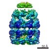
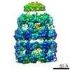
| 添付画像 |

- ダウンロードとリンク
-EMDBアーカイブ
| マップデータ | emd_2327.map.gz | 19.3 MB | EMDBマップデータ形式 | |
|---|---|---|---|---|
| ヘッダ (付随情報) | emd-2327-v30.xmlemd-2327.xml | 16.6 KB 16.6 KB | 表示 表示 | EMDBヘッダ |
| 画像 | 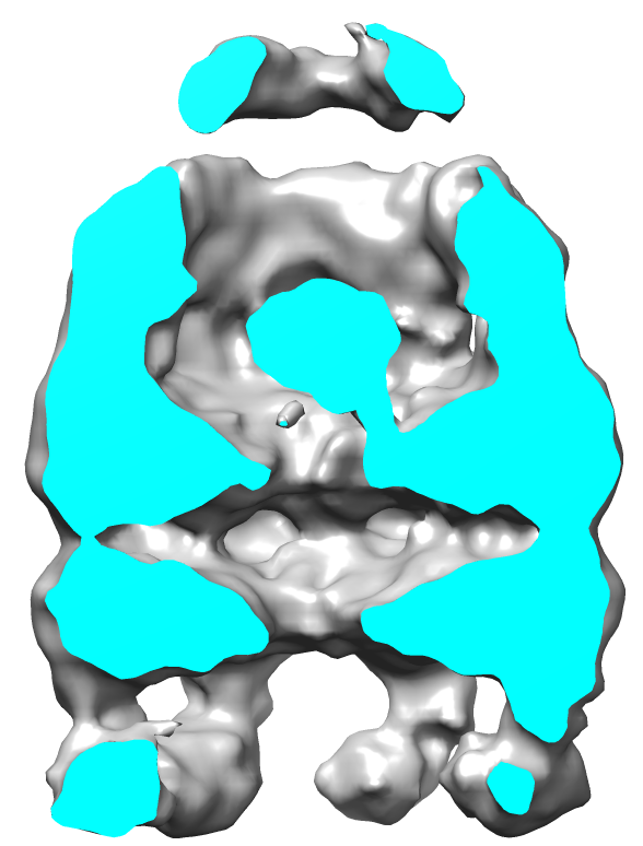 EMD-2327.png EMD-2327.png | 135.8 KB | ||
| アーカイブディレクトリ |  http://ftp.pdbj.org/pub/emdb/structures/EMD-2327ftp://ftp.pdbj.org/pub/emdb/structures/EMD-2327 http://ftp.pdbj.org/pub/emdb/structures/EMD-2327ftp://ftp.pdbj.org/pub/emdb/structures/EMD-2327 | HTTPS FTP |
-検証レポート
| 文書・要旨 | emd_2327_validation.pdf.gz | 198.9 KB | 表示 | EMDB検証レポート |
|---|---|---|---|---|
| 文書・詳細版 | emd_2327_full_validation.pdf.gz | 198 KB | 表示 | |
| XML形式データ | emd_2327_validation.xml.gz | 5.9 KB | 表示 | |
| アーカイブディレクトリ | https://ftp.pdbj.org/pub/emdb/validation_reports/EMD-2327ftp://ftp.pdbj.org/pub/emdb/validation_reports/EMD-2327 | HTTPS FTP |
-関連構造データ


-リンク
| EMDBのページ | EMDB (EBI/PDBe) / EMDataResource |
|---|---|
| 「今月の分子」の関連する項目 |


-マップ
| ファイル | ダウンロード / ファイル: emd_2327.map.gz / 形式: CCP4 / 大きさ: 21.7 MB / タイプ: IMAGE STORED AS FLOATING POINT NUMBER (4 BYTES) | ||||||||||||||||||||||||||||||||||||||||||||||||||||||||||||||||||||
|---|---|---|---|---|---|---|---|---|---|---|---|---|---|---|---|---|---|---|---|---|---|---|---|---|---|---|---|---|---|---|---|---|---|---|---|---|---|---|---|---|---|---|---|---|---|---|---|---|---|---|---|---|---|---|---|---|---|---|---|---|---|---|---|---|---|---|---|---|---|
| 注釈 | Symmetry-free 3D reconstruction of the bullet-shaped substrate-encapsulated subpopulation sorted from the heterogeneous sample of wild-type GroEL reacted with the substrate protein RuBisCO, GroES and nucleotide ATP | ||||||||||||||||||||||||||||||||||||||||||||||||||||||||||||||||||||
| 投影像・断面図 | 画像のコントロール
画像は Spider により作成 | ||||||||||||||||||||||||||||||||||||||||||||||||||||||||||||||||||||
| ボクセルのサイズ | X=Y=Z: 2.12 Å | ||||||||||||||||||||||||||||||||||||||||||||||||||||||||||||||||||||
| 密度 |
| ||||||||||||||||||||||||||||||||||||||||||||||||||||||||||||||||||||
| 対称性 | 空間群: 1 | ||||||||||||||||||||||||||||||||||||||||||||||||||||||||||||||||||||
| 詳細 | EMDB XML:
CCP4マップ ヘッダ情報:
| ||||||||||||||||||||||||||||||||||||||||||||||||||||||||||||||||||||
 Z (Sec.)
 Y (Row.)
 X (Col.)


-添付データ
- 試料の構成要素
-全体 : Non-native RuBisCO substrate protein encapsulated inside the cavi...
| 全体 | 名称: Non-native RuBisCO substrate protein encapsulated inside the cavity of GroEL capped by GroES with the assistance of nucleotide ATP |
|---|---|
| 要素 |
|
-超分子 #1000: Non-native RuBisCO substrate protein encapsulated inside the cavi...
| 超分子 | 名称: Non-native RuBisCO substrate protein encapsulated inside the cavity of GroEL capped by GroES with the assistance of nucleotide ATP タイプ: sample / ID: 1000 / 詳細: The molecule is bullet-shaped 集合状態: Non-native RuBisCO substrate protein was encapsulated inside the cavity formed by one tetradecamer of GroEL, one heptamer of GroES and seven nucleotides of ATP Number unique components: 4 |
|---|---|
| 分子量 | 実験値: 920 KDa / 理論値: 920 KDa 手法: Estimated by the sum of GroEL molecular weight 800kDa, GroES molecular weight 70kDa and one RuBisCO monomer molecular weight 50kDa |
-分子 #1: GroEL-D398A
| 分子 | 名称: GroEL-D398A / タイプ: protein_or_peptide / ID: 1 / Name.synonym: EL398A 詳細: The D398A mutation prevents ATP hydrolysis by GroEL. コピー数: 14 / 集合状態: Tetradecamer / 組換発現: Yes |
|---|---|
| 由来(天然) | 生物種: |
| 分子量 | 実験値: 800 KDa / 理論値: 800 KDa |
| 組換発現 | 生物種: |
| 配列 | UniProtKB: Chaperonin GroEL |
-分子 #2: GroES
| 分子 | 名称: GroES / タイプ: protein_or_peptide / ID: 2 / コピー数: 7 / 集合状態: Heptamer / 組換発現: Yes |
|---|---|
| 由来(天然) | 生物種: |
| 分子量 | 実験値: 70 KDa / 理論値: 70 KDa |
| 組換発現 | 生物種: |
| 配列 | UniProtKB: Co-chaperonin GroES |
-分子 #3: Ribulose-1,5-bisphosphate carboxylase oxygenase
| 分子 | 名称: Ribulose-1,5-bisphosphate carboxylase oxygenase / タイプ: protein_or_peptide / ID: 3 / Name.synonym: RuBisCO 詳細: The native RuBisCO is a dimer, but the encapsulated RuBisCO is a non-native monomer. コピー数: 1 / 集合状態: monomer / 組換発現: Yes |
|---|---|
| 由来(天然) | 生物種: Rhodospirillum rubrum (バクテリア) / 細胞中の位置: cytoplasm |
| 分子量 | 実験値: 50 KDa / 理論値: 50 KDa |
| 組換発現 | 生物種: |
-分子 #4: Adenosine triphosphate
| 分子 | 名称: Adenosine triphosphate / タイプ: ligand / ID: 4 / Name.synonym: ATP / 組換発現: No |
|---|---|
| 由来(天然) | 生物種: synthetic construct (人工物) |
-実験情報
-構造解析
| 手法 | クライオ電子顕微鏡法 |
|---|---|
 解析 解析 | 単粒子再構成法 |
| 試料の集合状態 | particle |
-試料調製
| 濃度 | 2.4 mg/mL |
|---|---|
| 緩衝液 | pH: 7.6 / 詳細: 50 mM Hepes, 50 mM KOAc, 10 mM Mg(OAc)2, 2 mM DTT |
| グリッド | 詳細: 400 mesh R1.2/1.3 Quantifoil grid glow-discharged 10 sec before freezing |
| 凍結 | 凍結剤: ETHANE / チャンバー内湿度: 95 % / チャンバー内温度: 98 K / 装置: FEI VITROBOT MARK III / 手法: Blot for 1 second before plunging |
- 電子顕微鏡法
| 顕微鏡 | JEOL 3200FSC |
|---|---|
| 温度 | 最低: 100.9 K / 最高: 101.1 K / 平均: 101 K |
| アライメント法 | Legacy - 非点収差: Objective lens astigmatism was corrected at 100,000 times magnification |
| 特殊光学系 | エネルギーフィルター - 名称: JEOL エネルギーフィルター - エネルギー下限: 0.0 eV エネルギーフィルター - エネルギー上限: 25.0 eV |
| 日付 | 2008年12月21日 |
| 撮影 | カテゴリ: CCD / フィルム・検出器のモデル: GENERIC GATAN / デジタル化 - サンプリング間隔: 15 µm / 実像数: 1537 / 平均電子線量: 20 e/Å2 / ビット/ピクセル: 8 |
| 電子線 | 加速電圧: 300 kV / 電子線源:  FIELD EMISSION GUN FIELD EMISSION GUN |
| 電子光学系 | 倍率(補正後): 70760 / 照射モード: FLOOD BEAM / 撮影モード: OTHER / 最大 デフォーカス(公称値): 5.0 µm / 最小 デフォーカス(公称値): 1.5 µm / 倍率(公称値): 50000 |
| 試料ステージ | 試料ホルダーモデル: JEOL 3200FSC CRYOHOLDER |
+画像解析
-原子モデル構築 1
| 初期モデル | PDB ID: Chain - #0 - Chain ID: A / Chain - #1 - Chain ID: B / Chain - #2 - Chain ID: C / Chain - #3 - Chain ID: D / Chain - #4 - Chain ID: E / Chain - #5 - Chain ID: F / Chain - #6 - Chain ID: G / Chain - #7 - Chain ID: H / Chain - #8 - Chain ID: I / Chain - #9 - Chain ID: J / Chain - #10 - Chain ID: K / Chain - #11 - Chain ID: L / Chain - #12 - Chain ID: M / Chain - #13 - Chain ID: N / Chain - #14 - Chain ID: O / Chain - #15 - Chain ID: P / Chain - #16 - Chain ID: Q / Chain - #17 - Chain ID: R / Chain - #18 - Chain ID: S / Chain - #19 - Chain ID: T / Chain - #20 - Chain ID: U |
|---|---|
| ソフトウェア | 名称: DireX |
| 精密化 | 空間: REAL / プロトコル: FLEXIBLE FIT 当てはまり具合の基準: cross-correlation coefficient |
| 得られたモデル | 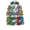 PDB-3zq1: |