 Movie
Movie Controller


+ Open data

- Basic information
| Entry | Database: EMDB / ID: EMD-2313 | |||||||||
|---|---|---|---|---|---|---|---|---|---|---|
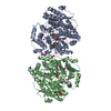
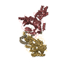
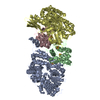
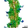
| Title | 3D reconstruction of Mechanosensitive Channel Candidate MCA2 | |||||||||
 Map data Map data | Reconstruction of MCA2 in detergent solubilized state from Zernike Phase Contrast cryoEM images | |||||||||
 Sample Sample |
| |||||||||
 Keywords Keywords | Mechanosensitive channel candidate / membrane protein / Calcium uptake | |||||||||
| Function / homology |  Function and homology information Function and homology informationpost-embryonic root development / calcium channel activity / cell surface receptor signaling pathway / plasma membrane Similarity search - Function | |||||||||
| Biological species |  | |||||||||
| Method | single particle reconstruction / cryo EM / Resolution: 26.0 Å | |||||||||
 Authors Authors | Shigematsu H / Iida K / Nakano M / Chaudhuri P / Iida H / Nagayama K | |||||||||
 Citation Citation | Journal: PLoS One / Year: 2014 Title: Structural characterization of the mechanosensitive channel candidate MCA2 from Arabidopsis thaliana. Authors: Hideki Shigematsu / Kazuko Iida / Masataka Nakano / Pratima Chaudhuri / Hidetoshi Iida / Kuniaki Nagayama /  Abstract: Mechanosensing in plants is thought to be governed by sensory complexes containing a Ca²⁺-permeable, mechanosensitive channel. The plasma membrane protein MCA1 and its paralog MCA2 from ...Mechanosensing in plants is thought to be governed by sensory complexes containing a Ca²⁺-permeable, mechanosensitive channel. The plasma membrane protein MCA1 and its paralog MCA2 from Arabidopsis thaliana are involved in mechanical stress-induced Ca²⁺ influx and are thus considered as candidates for such channels or their regulators. Both MCA1 and MCA2 were functionally expressed in Sf9 cells using a baculovirus system in order to elucidate their molecular natures. Because of the abundance of protein in these cells, MCA2 was chosen for purification. Purified MCA2 in a detergent-solubilized state formed a tetramer, which was confirmed by chemical cross-linking. Single-particle analysis of cryo-electron microscope images was performed to depict the overall shape of the purified protein. The three-dimensional structure of MCA2 was reconstructed at a resolution of 26 Å from 5,500 particles and appears to comprise a small transmembrane region and large cytoplasmic region. | |||||||||
| History |
|
- Structure visualization
| Movie |
Movie viewer |
|---|---|
| Structure viewer | EM map: SurfViewMolmilJmol/JSmol |
| Supplemental images |

- Downloads & links
-EMDB archive
| Map data | emd_2313.map.gz | 1.3 MB | EMDB map data format | |
|---|---|---|---|---|
| Header (meta data) | emd-2313-v30.xmlemd-2313.xml | 10 KB 10 KB | Display Display | EMDB header |
| Images | EMD-2313-For_EMDB.tif | 950 KB | ||
| Archive directory |  http://ftp.pdbj.org/pub/emdb/structures/EMD-2313ftp://ftp.pdbj.org/pub/emdb/structures/EMD-2313 http://ftp.pdbj.org/pub/emdb/structures/EMD-2313ftp://ftp.pdbj.org/pub/emdb/structures/EMD-2313 | HTTPS FTP |
-Related structure data
| Similar structure data |
|---|


-Links
| EMDB pages | EMDB (EBI/PDBe) / EMDataResource |
|---|
-Map
| File | Download / File: emd_2313.map.gz / Format: CCP4 / Size: 1.9 MB / Type: IMAGE STORED AS FLOATING POINT NUMBER (4 BYTES) | ||||||||||||||||||||||||||||||||||||||||||||||||||||||||||||||||||||
|---|---|---|---|---|---|---|---|---|---|---|---|---|---|---|---|---|---|---|---|---|---|---|---|---|---|---|---|---|---|---|---|---|---|---|---|---|---|---|---|---|---|---|---|---|---|---|---|---|---|---|---|---|---|---|---|---|---|---|---|---|---|---|---|---|---|---|---|---|---|
| Annotation | Reconstruction of MCA2 in detergent solubilized state from Zernike Phase Contrast cryoEM images | ||||||||||||||||||||||||||||||||||||||||||||||||||||||||||||||||||||
| Projections & slices | Image control
Images are generated by Spider. | ||||||||||||||||||||||||||||||||||||||||||||||||||||||||||||||||||||
| Voxel size | X=Y=Z: 3 Å | ||||||||||||||||||||||||||||||||||||||||||||||||||||||||||||||||||||
| Density |
| ||||||||||||||||||||||||||||||||||||||||||||||||||||||||||||||||||||
| Symmetry | Space group: 1 | ||||||||||||||||||||||||||||||||||||||||||||||||||||||||||||||||||||
| Details | EMDB XML:
CCP4 map header:
| ||||||||||||||||||||||||||||||||||||||||||||||||||||||||||||||||||||
 Z (Sec.)
 Y (Row.)
 X (Col.)


-Supplemental data
- Sample components
-Entire : MCA2
| Entire | Name: MCA2 |
|---|---|
| Components |
|
-Supramolecule #1000: MCA2
| Supramolecule | Name: MCA2 / type: sample / ID: 1000 Details: The sample was purified with size-exclusion chromatography finally in the center of peak fraction Oligomeric state: homotetramer / Number unique components: 1 |
|---|---|
| Molecular weight | Theoretical: 200 KDa |
-Macromolecule #1: Mid1-complementing activity 2
| Macromolecule | Name: Mid1-complementing activity 2 / type: protein_or_peptide / ID: 1 / Name.synonym: MCA2 Details: recombinantly expressed in Sf9 and purified under ammonium perfluorooctanoate. Number of copies: 1 / Oligomeric state: tetramer / Recombinant expression: Yes |
|---|---|
| Source (natural) | Organism: |
| Molecular weight | Theoretical: 200 KDa |
| Recombinant expression | Organism:  unidentified baculovirus / Recombinant cell: Sf9 / Recombinant plasmid: pFastBac1 unidentified baculovirus / Recombinant cell: Sf9 / Recombinant plasmid: pFastBac1 |
| Sequence | UniProtKB: Protein MID1-COMPLEMENTING ACTIVITY 2 / InterPro: PLAC8 motif-containing protein |
-Experimental details
-Structure determination
| Method | cryo EM |
|---|---|
 Processing Processing | single particle reconstruction |
| Aggregation state | particle |
-Sample preparation
| Concentration | 0.05 mg/mL |
|---|---|
| Buffer | pH: 8 / Details: PBS, 4% ammonium perfluorooctanoate |
| Grid | Details: thin carbon over the Quantifoil R1.2/1.3, glow discharged in air |
| Vitrification | Cryogen name: ETHANE / Chamber humidity: 100 % / Chamber temperature: 293 K / Instrument: FEI VITROBOT MARK IV |
- Electron microscopy
| Microscope | OTHER |
|---|---|
| Temperature | Min: 45 K / Max: 60 K / Average: 55 K |
| Alignment procedure | Legacy - Astigmatism: Objective lens astigmatism was corrected at 100,000 times magnification |
| Specialist optics | Energy filter - Name: JEOL / Energy filter - Lower energy threshold: 0.0 eV / Energy filter - Upper energy threshold: 20.0 eV |
| Date | May 10, 2008 |
| Image recording | Category: CCD / Film or detector model: GENERIC GATAN (2k x 2k) / Digitization - Sampling interval: 30 µm / Number real images: 50 / Average electron dose: 20 e/Å2 / Bits/pixel: 8 |
| Electron beam | Acceleration voltage: 300 kV / Electron source:  FIELD EMISSION GUN FIELD EMISSION GUN |
| Electron optics | Calibrated magnification: 100000 / Illumination mode: FLOOD BEAM / Imaging mode: BRIGHT FIELD / Cs: 3.7 mm / Nominal magnification: 60000 |
| Sample stage | Specimen holder: Liquid Helium cooled stage maintained around 55 K Specimen holder model: JEOL |
-Image processing
| Details | Particles were selected using boxer. |
|---|---|
| Final reconstruction | Applied symmetry - Point group: C4 (4 fold cyclic) / Algorithm: OTHER / Resolution.type: BY AUTHOR / Resolution: 26.0 Å / Resolution method: FSC 0.5 CUT-OFF / Software - Name: EMAN / Number images used: 5700 |
| Final two d classification | Number classes: 133 |
