National Institutes of Health/National Institute Of Allergy and Infectious Diseases (NIH/NIAID)
HHSN272201700060C
United States
National Institutes of Health/National Institute Of Allergy and Infectious Diseases (NIH/NIAID)
75N93019C00062
United States
Citation
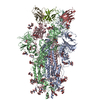
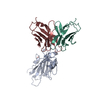
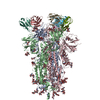
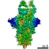
Journal: Cell Rep / Year: 2021 Title: Structural mechanism of SARS-CoV-2 neutralization by two murine antibodies targeting the RBD. Authors: John M Errico / Haiyan Zhao / Rita E Chen / Zhuoming Liu / James Brett Case / Meisheng Ma / Aaron J Schmitz / Michael J Rau / James A J Fitzpatrick / Pei-Yong Shi / Michael S Diamond / Sean ...Authors: John M Errico / Haiyan Zhao / Rita E Chen / Zhuoming Liu / James Brett Case / Meisheng Ma / Aaron J Schmitz / Michael J Rau / James A J Fitzpatrick / Pei-Yong Shi / Michael S Diamond / Sean P J Whelan / Ali H Ellebedy / Daved H Fremont / Abstract: The severe acute respiratory syndrome coronavirus 2 (SARS-CoV-2) pandemic has necessitated the rapid development of antibody-based therapies and vaccines as countermeasures. Here, we use cryoelectron ...The severe acute respiratory syndrome coronavirus 2 (SARS-CoV-2) pandemic has necessitated the rapid development of antibody-based therapies and vaccines as countermeasures. Here, we use cryoelectron microscopy (cryo-EM) to characterize two protective anti-SARS-CoV-2 murine monoclonal antibodies (mAbs) in complex with the spike protein, revealing similarities between epitopes targeted by human and murine B cells. The more neutralizing mAb, 2B04, binds the receptor-binding motif (RBM) of the receptor-binding domain (RBD) and competes with angiotensin-converting enzyme 2 (ACE2). By contrast, 2H04 binds adjacent to the RBM and does not compete for ACE2 binding. Naturally occurring sequence variants of SARS-CoV-2 and corresponding neutralization escape variants selected in vitro map to our structurally defined epitopes, suggesting that SARS-CoV-2 might evade therapeutic antibodies with a limited set of mutations, underscoring the importance of combination mAb therapeutics. Finally, we show that 2B04 neutralizes SARS-CoV-2 infection by preventing ACE2 engagement, whereas 2H04 reduces host cell attachment without directly disrupting ACE2-RBM interactions, providing distinct inhibitory mechanisms used by RBD-specific mAbs.
History
Deposition
Sep 29, 2020
-
Header (metadata) release
Sep 29, 2021
-
Map release
Sep 29, 2021
-
Update
Nov 6, 2024
-
Current status
Nov 6, 2024
Processing site: RCSB / Status: Released
-
Structure visualization
Movie
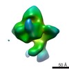
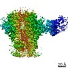
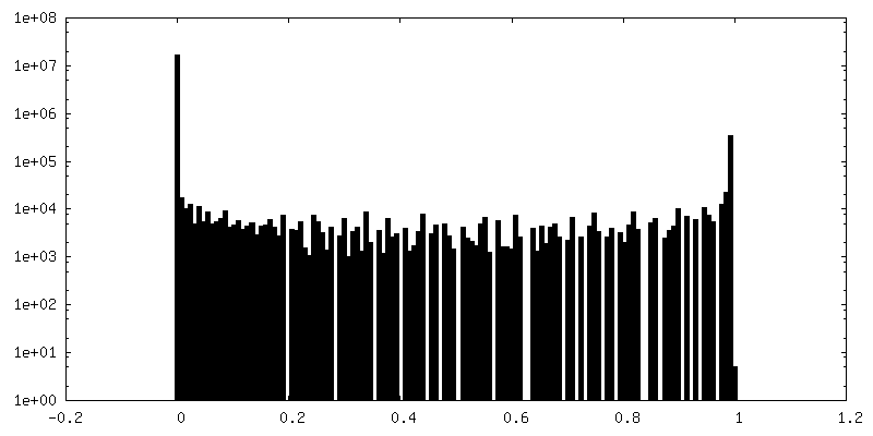
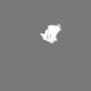
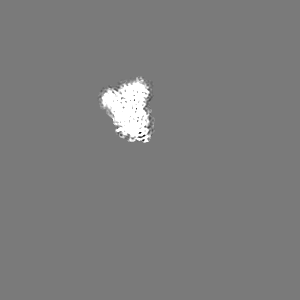
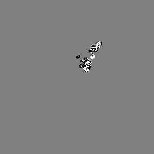
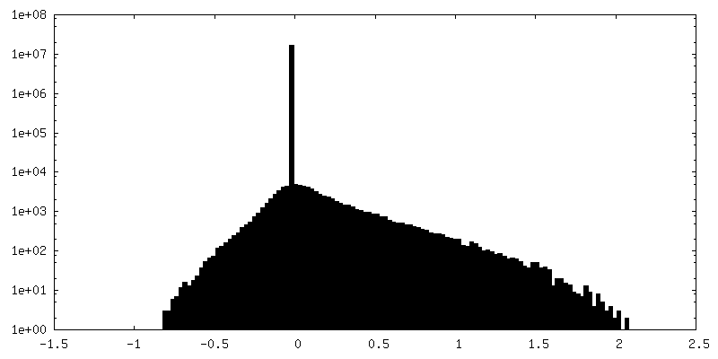
Surface view with section colored by density value
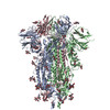
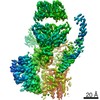
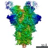
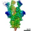
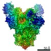
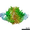
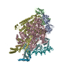
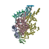
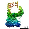
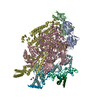
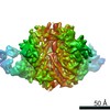
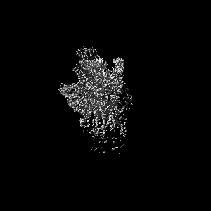
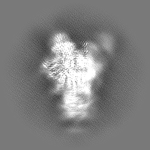
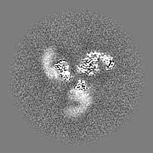
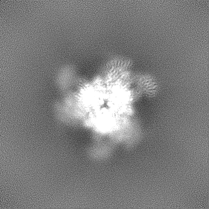
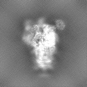
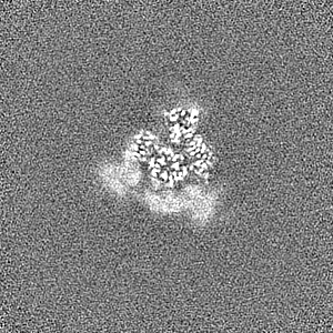
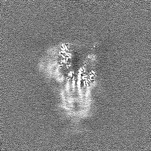
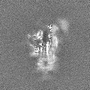
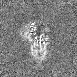
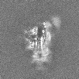
Entire : Complex of SARS-CoV-2 Spike RBD with Fab fragment of monoclonal a...
Entire
Name: Complex of SARS-CoV-2 Spike RBD with Fab fragment of monoclonal antibody 2H04
Components
Complex: Complex of SARS-CoV-2 Spike RBD with Fab fragment of monoclonal antibody 2H04
Complex: SARS-CoV-2 Spike RBD
Protein or peptide: Spike protein S1
Complex: Fab fragment of monoclonal antibody 2H04
Protein or peptide: 2H04 heavy chain
Protein or peptide: 2H04 light chain
-
Supramolecule #1: Complex of SARS-CoV-2 Spike RBD with Fab fragment of monoclonal a...
Supramolecule
Name: Complex of SARS-CoV-2 Spike RBD with Fab fragment of monoclonal antibody 2H04 type: complex / ID: 1 / Parent: 0 / Macromolecule list: all Details: Fab fragments generated by proteolytic cleavage of recombinantly expressed IgG
In the structure databanks used in Yorodumi, some data are registered as the other names, "COVID-19 virus" and "2019-nCoV". Here are the details of the virus and the list of structure data.
Jan 31, 2019. EMDB accession codes are about to change! (news from PDBe EMDB page)
EMDB accession codes are about to change! (news from PDBe EMDB page)
The allocation of 4 digits for EMDB accession codes will soon come to an end. Whilst these codes will remain in use, new EMDB accession codes will include an additional digit and will expand incrementally as the available range of codes is exhausted. The current 4-digit format prefixed with “EMD-” (i.e. EMD-XXXX) will advance to a 5-digit format (i.e. EMD-XXXXX), and so on. It is currently estimated that the 4-digit codes will be depleted around Spring 2019, at which point the 5-digit format will come into force.
The EM Navigator/Yorodumi systems omit the EMD- prefix.
Related info.:Q: What is EMD? / ID/Accession-code notation in Yorodumi/EM Navigator
Yorodumi is a browser for structure data from EMDB, PDB, SASBDB, etc.
This page is also the successor to EM Navigator detail page, and also detail information page/front-end page for Omokage search.
The word "yorodu" (or yorozu) is an old Japanese word meaning "ten thousand". "mi" (miru) is to see.
Related info.:EMDB / PDB / SASBDB / Comparison of 3 databanks / Yorodumi Search / Aug 31, 2016. New EM Navigator & Yorodumi / Yorodumi Papers / Jmol/JSmol / Function and homology information / Changes in new EM Navigator and Yorodumi
 Movie
Movie Controller

 Yorodumi
 Open data
 Basic information
 Map data
 Sample
 Keywords
 Function and homology information
 Severe acute respiratory syndrome coronavirus 2 /
 Authors
 United States, 2 items
  Citation
 Structure visualization
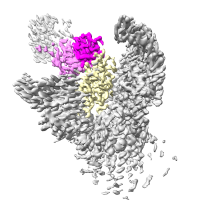
 Downloads & links
 emd_22751.png
 http://ftp.pdbj.org/pub/emdb/structures/EMD-22751
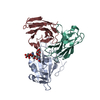


 Z (Sec.)
 Y (Row.)
 X (Col.)


 Sample components
 Homo sapiens (human)
 Processing
 Electron microscopy
 FIELD EMISSION GUN