National Institutes of Health/National Institute Of Allergy and Infectious Diseases (NIH/NIAID)
United States
Citation
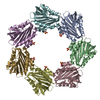
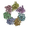
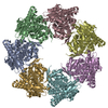
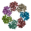
Journal: Sci Adv / Year: 2021 Title: Identification and architecture of a putative secretion tube across mycobacterial outer envelope. Authors: Xiaoying Cai / Lei Liu / Chunhong Qiu / Chongzheng Wen / Yao He / Yanxiang Cui / Siyu Li / Xuan Zhang / Longhua Zhang / Changlin Tian / Lijun Bi / Z Hong Zhou / Weimin Gong / Abstract: Tuberculosis-causing mycobacteria have thick cell-wall and capsule layers that are formed from complex structures. Protein secretion across these barriers depends on a specialized protein secretion ...Tuberculosis-causing mycobacteria have thick cell-wall and capsule layers that are formed from complex structures. Protein secretion across these barriers depends on a specialized protein secretion system, but none has been reported. We show that Rv3705c and its homologous MSMEG_6251 in are tube-forming proteins in the mycobacterial envelope (TiME). Crystallographic and cryo-EM structures of these two proteins show that both proteins form rotationally symmetric rings. Two layers of TiME rings pack together in a tail-to-tail manner into a ring-shaped complex, which, in turn, stacks together to form tubes. TiME was detected mainly in the cell wall and capsule. Knocking out the TiME gene markedly decreased the amount of secreted protein in the culture medium, and expression of this gene in knocked-out strain partially restored the level of secreted protein. Our structure and functional data thus suggest that TiME forms a protein transport tube across the mycobacterial outer envelope.
History
Deposition
Sep 24, 2020
-
Header (metadata) release
Sep 1, 2021
-
Map release
Sep 1, 2021
-
Update
Sep 1, 2021
-
Current status
Sep 1, 2021
Processing site: RCSB / Status: Released
-
Structure visualization
Movie
Surface view with section colored by density value
In the structure databanks used in Yorodumi, some data are registered as the other names, "COVID-19 virus" and "2019-nCoV". Here are the details of the virus and the list of structure data.
Jan 31, 2019. EMDB accession codes are about to change! (news from PDBe EMDB page)
EMDB accession codes are about to change! (news from PDBe EMDB page)
The allocation of 4 digits for EMDB accession codes will soon come to an end. Whilst these codes will remain in use, new EMDB accession codes will include an additional digit and will expand incrementally as the available range of codes is exhausted. The current 4-digit format prefixed with “EMD-” (i.e. EMD-XXXX) will advance to a 5-digit format (i.e. EMD-XXXXX), and so on. It is currently estimated that the 4-digit codes will be depleted around Spring 2019, at which point the 5-digit format will come into force.
The EM Navigator/Yorodumi systems omit the EMD- prefix.
Related info.:Q: What is EMD? / ID/Accession-code notation in Yorodumi/EM Navigator
Yorodumi is a browser for structure data from EMDB, PDB, SASBDB, etc.
This page is also the successor to EM Navigator detail page, and also detail information page/front-end page for Omokage search.
The word "yorodu" (or yorozu) is an old Japanese word meaning "ten thousand". "mi" (miru) is to see.
Related info.:EMDB / PDB / SASBDB / Comparison of 3 databanks / Yorodumi Search / Aug 31, 2016. New EM Navigator & Yorodumi / Yorodumi Papers / Jmol/JSmol / Function and homology information / Changes in new EM Navigator and Yorodumi
 Movie
Movie Controller

 Yorodumi
 Open data
 Basic information
 Map data
 Sample
 Function and homology information
 Mycobacterium tuberculosis (bacteria)
 Authors
 United States, 1 items
  Citation
 Structure visualization
 Downloads & links
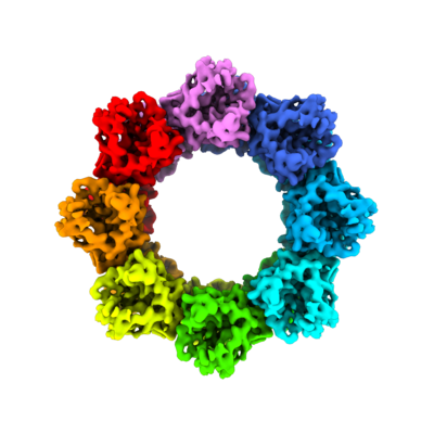 emd_22705.png
 http://ftp.pdbj.org/pub/emdb/structures/EMD-22705
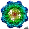


 Z (Sec.)
 Y (Row.)
 X (Col.)


 Sample components
 Processing
 Electron microscopy
 FIELD EMISSION GUN
