 Movie
Movie Controller

[English] 日本語
 Yorodumi
- EMDB-22458: Structure of the Visual Signaling Complex between Transducin and ... -
+ Open data

- Basic information
| Entry | Database: EMDB / ID: EMD-22458 | ||||||||||||
|---|---|---|---|---|---|---|---|---|---|---|---|---|---|
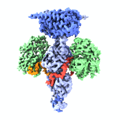
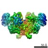
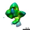
| Title | Structure of the Visual Signaling Complex between Transducin and Phosphodiesterase 6 | ||||||||||||
 Map data Map data | Visual Signaling Complex between Transducin and Phosphodiesterase 6 | ||||||||||||
 Sample Sample |
| ||||||||||||
 Keywords Keywords | G protein / G protein-effector complex / Transducin / phosphodiesterase 6 / phototransduction / GPCR signaling / SIGNALING PROTEIN | ||||||||||||
| Function / homology |  Function and homology information Function and homology informationnegative regulation of cyclic-nucleotide phosphodiesterase activity / transducin-mediated opsin signaling pathway / 3',5'-cyclic-GMP phosphodiesterase / detection of light stimulus involved in visual perception / G protein-coupled receptor complex / Inactivation, recovery and regulation of the phototransduction cascade / Activation of the phototransduction cascade / positive regulation of G protein-coupled receptor signaling pathway / Ca2+ pathway / photoreceptor outer segment membrane ...negative regulation of cyclic-nucleotide phosphodiesterase activity / transducin-mediated opsin signaling pathway / 3',5'-cyclic-GMP phosphodiesterase / detection of light stimulus involved in visual perception / G protein-coupled receptor complex / Inactivation, recovery and regulation of the phototransduction cascade / Activation of the phototransduction cascade / positive regulation of G protein-coupled receptor signaling pathway / Ca2+ pathway / photoreceptor outer segment membrane / G alpha (i) signalling events / entrainment of circadian clock by photoperiod / cGMP binding / 3',5'-cyclic-GMP phosphodiesterase activity / acyl binding / 3',5'-cyclic-AMP phosphodiesterase activity / phototransduction, visible light / phototransduction / response to light stimulus / positive regulation of epidermal growth factor receptor signaling pathway / negative regulation of cAMP/PKA signal transduction / photoreceptor inner segment / visual perception / G protein-coupled receptor binding / G-protein beta/gamma-subunit complex binding / adenylate cyclase-modulating G protein-coupled receptor signaling pathway / photoreceptor disc membrane / GDP binding / heterotrimeric G-protein complex / retina development in camera-type eye / G protein activity / molecular adaptor activity / Hydrolases; Acting on acid anhydrides; Acting on GTP to facilitate cellular and subcellular movement / positive regulation of MAPK cascade / GTPase activity / protein kinase binding / GTP binding / signal transduction / zinc ion binding / metal ion binding / cytoplasm Similarity search - Function | ||||||||||||
| Biological species |  | ||||||||||||
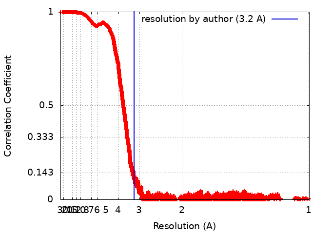
| Method | single particle reconstruction / cryo EM / Resolution: 3.2 Å | ||||||||||||
 Authors Authors | Gao Y / Eskici G | ||||||||||||
| Funding support |  United States, 3 items United States, 3 items
| ||||||||||||
 Citation Citation | Journal: Mol Cell / Year: 2020 Title: Structure of the Visual Signaling Complex between Transducin and Phosphodiesterase 6. Authors: Yang Gao / Gözde Eskici / Sekar Ramachandran / Frédéric Poitevin / Alpay Burak Seven / Ouliana Panova / Georgios Skiniotis / Richard A Cerione / Abstract: Heterotrimeric G proteins communicate signals from activated G protein-coupled receptors to downstream effector proteins. In the phototransduction pathway responsible for vertebrate vision, the G ...Heterotrimeric G proteins communicate signals from activated G protein-coupled receptors to downstream effector proteins. In the phototransduction pathway responsible for vertebrate vision, the G protein-effector complex is composed of the GTP-bound transducin α subunit (Gα·GTP) and the cyclic GMP (cGMP) phosphodiesterase 6 (PDE6), which stimulates cGMP hydrolysis, leading to hyperpolarization of the photoreceptor cell. Here we report a cryo-electron microscopy (cryoEM) structure of PDE6 complexed to GTP-bound Gα. The structure reveals two Gα·GTP subunits engaging the PDE6 hetero-tetramer at both the PDE6 catalytic core and the PDEγ subunits, driving extensive rearrangements to relieve all inhibitory constraints on enzyme catalysis. Analysis of the conformational ensemble in the cryoEM data highlights the dynamic nature of the contacts between the two Gα·GTP subunits and PDE6 that supports an alternating-site catalytic mechanism. | ||||||||||||
| History |
|
- Structure visualization
| Movie |
Movie viewer |
|---|---|
| Structure viewer | EM map: SurfViewMolmilJmol/JSmol |
| Supplemental images |
- Downloads & links
-EMDB archive
| Map data | emd_22458.map.gz | 97.5 MB | EMDB map data format | |
|---|---|---|---|---|
| Header (meta data) | emd-22458-v30.xmlemd-22458.xml | 27.7 KB 27.7 KB | Display Display | EMDB header |
| FSC (resolution estimation) | emd_22458_fsc.xml | 2.5 MB | Display | FSC data file |
| Images |  emd_22458.png emd_22458.png | 140.3 KB | ||
| Filedesc metadata | emd-22458.cif.gz | 8.9 KB | ||
| Archive directory |  http://ftp.pdbj.org/pub/emdb/structures/EMD-22458ftp://ftp.pdbj.org/pub/emdb/structures/EMD-22458 http://ftp.pdbj.org/pub/emdb/structures/EMD-22458ftp://ftp.pdbj.org/pub/emdb/structures/EMD-22458 | HTTPS FTP |
-Related structure data
| Related structure data |  7jsnMC M: atomic model generated by this map C: citing same article ( |
|---|---|
| Similar structure data |


-Links
| EMDB pages | EMDB (EBI/PDBe) / EMDataResource |
|---|---|
| Related items in Molecule of the Month |


-Map
| File | Download / File: emd_22458.map.gz / Format: CCP4 / Size: 125 MB / Type: IMAGE STORED AS FLOATING POINT NUMBER (4 BYTES) | ||||||||||||||||||||||||||||||||||||||||||||||||||||||||||||||||||||
|---|---|---|---|---|---|---|---|---|---|---|---|---|---|---|---|---|---|---|---|---|---|---|---|---|---|---|---|---|---|---|---|---|---|---|---|---|---|---|---|---|---|---|---|---|---|---|---|---|---|---|---|---|---|---|---|---|---|---|---|---|---|---|---|---|---|---|---|---|---|
| Annotation | Visual Signaling Complex between Transducin and Phosphodiesterase 6 | ||||||||||||||||||||||||||||||||||||||||||||||||||||||||||||||||||||
| Projections & slices | Image control
Images are generated by Spider. | ||||||||||||||||||||||||||||||||||||||||||||||||||||||||||||||||||||
| Voxel size | X=Y=Z: 0.85 Å | ||||||||||||||||||||||||||||||||||||||||||||||||||||||||||||||||||||
| Density |
| ||||||||||||||||||||||||||||||||||||||||||||||||||||||||||||||||||||
| Symmetry | Space group: 1 | ||||||||||||||||||||||||||||||||||||||||||||||||||||||||||||||||||||
| Details | EMDB XML:
CCP4 map header:
| ||||||||||||||||||||||||||||||||||||||||||||||||||||||||||||||||||||
 Z (Sec.)
 Y (Row.)
 X (Col.)


-Supplemental data
- Sample components
+Entire : Complex of Transducin and Phosphodiesterase 6
+Supramolecule #1: Complex of Transducin and Phosphodiesterase 6
+Supramolecule #2: Rod cGMP-specific 3',5'-cyclic phosphodiesterase subunit alpha (E...
+Supramolecule #3: Guanine nucleotide-binding protein G(t) subunit alpha-1, Guanine ...
+Macromolecule #1: Rod cGMP-specific 3',5'-cyclic phosphodiesterase subunit alpha
+Macromolecule #2: Rod cGMP-specific 3',5'-cyclic phosphodiesterase subunit beta
+Macromolecule #3: Guanine nucleotide-binding protein G(t) subunit alpha-1, Guanine ...

+Macromolecule #4: Retinal rod rhodopsin-sensitive cGMP 3',5'-cyclic phosphodiestera...
+Macromolecule #5: ZINC ION
+Macromolecule #6: MAGNESIUM ION
+Macromolecule #7: 2-{2-ETHOXY-5-[(4-ETHYLPIPERAZIN-1-YL)SULFONYL]PHENYL}-5-METHYL-7...

+Macromolecule #8: GUANOSINE-3',5'-MONOPHOSPHATE
+Macromolecule #9: GUANOSINE-5'-TRIPHOSPHATE

-Experimental details
-Structure determination
| Method | cryo EM |
|---|---|
 Processing Processing | single particle reconstruction |
| Aggregation state | particle |
-Sample preparation
| Concentration | 2 mg/mL |
|---|---|
| Buffer | pH: 8 / Details: 20 mM Tris pH 8.0, 5 mM MgCl2 and 1 uM vardenafil |
| Grid | Model: Quantifoil R1.2/1.3 / Material: GOLD / Pretreatment - Type: GLOW DISCHARGE |
| Vitrification | Cryogen name: ETHANE / Chamber humidity: 100 % / Chamber temperature: 295 K / Instrument: FEI VITROBOT MARK IV |
| Details | 0.05% octyl glucoside was used as an additive. |
- Electron microscopy
| Microscope | FEI TITAN KRIOS |
|---|---|
| Image recording | Film or detector model: GATAN K3 (6k x 4k) / Average exposure time: 4.0 sec. / Average electron dose: 48.0 e/Å2 |
| Electron beam | Acceleration voltage: 300 kV / Electron source:  FIELD EMISSION GUN FIELD EMISSION GUN |
| Electron optics | Illumination mode: FLOOD BEAM / Imaging mode: BRIGHT FIELD / Cs: 2.7 mm |
| Experimental equipment |  Model: Titan Krios / Image courtesy: FEI Company |
+Image processing
-Atomic model buiding 1
| Refinement | Protocol: BACKBONE TRACE |
|---|---|
| Output model | PDB-7jsn: |
