 Movie
Movie Controller

[English] 日本語
 Yorodumi
- PDB-7jsn: Structure of the Visual Signaling Complex between Transducin and ... -
+ Open data

- Basic information
| Entry | Database: PDB / ID: 7jsn | ||||||||||||||||||||||||||||||||||||||||||||||||
|---|---|---|---|---|---|---|---|---|---|---|---|---|---|---|---|---|---|---|---|---|---|---|---|---|---|---|---|---|---|---|---|---|---|---|---|---|---|---|---|---|---|---|---|---|---|---|---|---|---|
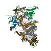
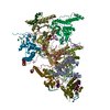
| Title | Structure of the Visual Signaling Complex between Transducin and Phosphodiesterase 6 | ||||||||||||||||||||||||||||||||||||||||||||||||
 Components Components |
| ||||||||||||||||||||||||||||||||||||||||||||||||
 Keywords Keywords | SIGNALING PROTEIN / G protein / G protein-effector complex / Transducin / phosphodiesterase 6 / phototransduction / GPCR signaling | ||||||||||||||||||||||||||||||||||||||||||||||||
| Function / homology |  Function and homology information Function and homology informationnegative regulation of cyclic-nucleotide phosphodiesterase activity / 3',5'-cyclic-GMP phosphodiesterase / detection of light stimulus involved in visual perception / G protein-coupled receptor complex / Inactivation, recovery and regulation of the phototransduction cascade / Activation of the phototransduction cascade / positive regulation of G protein-coupled receptor signaling pathway / Ca2+ pathway / photoreceptor outer segment membrane / G alpha (i) signalling events ...negative regulation of cyclic-nucleotide phosphodiesterase activity / 3',5'-cyclic-GMP phosphodiesterase / detection of light stimulus involved in visual perception / G protein-coupled receptor complex / Inactivation, recovery and regulation of the phototransduction cascade / Activation of the phototransduction cascade / positive regulation of G protein-coupled receptor signaling pathway / Ca2+ pathway / photoreceptor outer segment membrane / G alpha (i) signalling events / entrainment of circadian clock by photoperiod / cGMP binding / 3',5'-cyclic-GMP phosphodiesterase activity / acyl binding / 3',5'-cyclic-AMP phosphodiesterase activity / phototransduction, visible light / phototransduction / response to light stimulus / positive regulation of epidermal growth factor receptor signaling pathway / negative regulation of cAMP/PKA signal transduction / photoreceptor inner segment / visual perception / G protein-coupled receptor binding / G-protein beta/gamma-subunit complex binding / adenylate cyclase-modulating G protein-coupled receptor signaling pathway / photoreceptor disc membrane / GDP binding / heterotrimeric G-protein complex / retina development in camera-type eye / molecular adaptor activity / positive regulation of MAPK cascade / GTPase activity / protein kinase binding / GTP binding / signal transduction / zinc ion binding / metal ion binding / cytoplasm Similarity search - Function | ||||||||||||||||||||||||||||||||||||||||||||||||
| Biological species |  | ||||||||||||||||||||||||||||||||||||||||||||||||
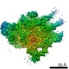
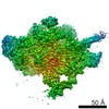
| Method | ELECTRON MICROSCOPY / single particle reconstruction / cryo EM / Resolution: 3.2 Å | ||||||||||||||||||||||||||||||||||||||||||||||||
 Authors Authors | Gao, Y. / Eskici, G. / Ramachandran, S. / Skiniotis, G. / Cerione, R.A. | ||||||||||||||||||||||||||||||||||||||||||||||||
| Funding support |  United States, 3items United States, 3items
| ||||||||||||||||||||||||||||||||||||||||||||||||
 Citation Citation | Journal: Mol Cell / Year: 2020 Title: Structure of the Visual Signaling Complex between Transducin and Phosphodiesterase 6. Authors: Yang Gao / Gözde Eskici / Sekar Ramachandran / Frédéric Poitevin / Alpay Burak Seven / Ouliana Panova / Georgios Skiniotis / Richard A Cerione / Abstract: Heterotrimeric G proteins communicate signals from activated G protein-coupled receptors to downstream effector proteins. In the phototransduction pathway responsible for vertebrate vision, the G ...Heterotrimeric G proteins communicate signals from activated G protein-coupled receptors to downstream effector proteins. In the phototransduction pathway responsible for vertebrate vision, the G protein-effector complex is composed of the GTP-bound transducin α subunit (Gα·GTP) and the cyclic GMP (cGMP) phosphodiesterase 6 (PDE6), which stimulates cGMP hydrolysis, leading to hyperpolarization of the photoreceptor cell. Here we report a cryo-electron microscopy (cryoEM) structure of PDE6 complexed to GTP-bound Gα. The structure reveals two Gα·GTP subunits engaging the PDE6 hetero-tetramer at both the PDE6 catalytic core and the PDEγ subunits, driving extensive rearrangements to relieve all inhibitory constraints on enzyme catalysis. Analysis of the conformational ensemble in the cryoEM data highlights the dynamic nature of the contacts between the two Gα·GTP subunits and PDE6 that supports an alternating-site catalytic mechanism. | ||||||||||||||||||||||||||||||||||||||||||||||||
| History |
|
- Structure visualization
| Movie |
Movie viewer |
|---|---|
| Structure viewer | Molecule: MolmilJmol/JSmol |
- Downloads & links
-Download
| PDBx/mmCIF format | 7jsn.cif.gz | 441.3 KB | Display | PDBx/mmCIF format |
|---|---|---|---|---|
| PDB format | pdb7jsn.ent.gz | 353.4 KB | Display | PDB format |
| PDBx/mmJSON format | 7jsn.json.gz | Tree view | PDBx/mmJSON format | |
| Others |  Other downloads Other downloads |
-Validation report
| Arichive directory | https://data.pdbj.org/pub/pdb/validation_reports/js/7jsnftp://data.pdbj.org/pub/pdb/validation_reports/js/7jsn | HTTPS FTP |
|---|
-Related structure data
| Related structure data |  22458MC M: map data used to model this data C: citing same article ( |
|---|---|
| Similar structure data |

-Links
 PDBj
PDBj


- Assembly
| Deposited unit | 
|
|---|---|
| 1 |
|
-Components
-Rod cGMP-specific 3',5'-cyclic phosphodiesterase subunit ... , 2 types, 2 molecules AB
| #1: Protein | Mass: 99461.789 Da / Num. of mol.: 1 / Source method: isolated from a natural source / Source: (natural) References: UniProt: P11541, 3',5'-cyclic-GMP phosphodiesterase |
|---|---|
| #2: Protein | Mass: 98449.648 Da / Num. of mol.: 1 / Source method: isolated from a natural source / Source: (natural) References: UniProt: P23439, 3',5'-cyclic-GMP phosphodiesterase |
-Protein , 2 types, 4 molecules EFCD
| #3: Protein | Mass: 42218.035 Da / Num. of mol.: 2 / Mutation: R174C,Q200L Source method: isolated from a genetically manipulated source Source: (gene. exp.)  #4: Protein | Mass: 9684.229 Da / Num. of mol.: 2 / Source method: isolated from a natural source / Source: (natural) References: UniProt: P04972, UniProt: P04695*PLUS, 3',5'-cyclic-GMP phosphodiesterase |
|---|
-Non-polymers , 5 types, 10 molecules 


| #5: Chemical |  Mass: 65.409 Da / Num. of mol.: 2 / Source method: obtained synthetically / Formula: Zn Mass: 65.409 Da / Num. of mol.: 2 / Source method: obtained synthetically / Formula: Zn#6: Chemical |  Mass: 24.305 Da / Num. of mol.: 2 / Source method: obtained synthetically / Formula: Mg Mass: 24.305 Da / Num. of mol.: 2 / Source method: obtained synthetically / Formula: Mg#7: Chemical |  Mass: 488.603 Da / Num. of mol.: 2 / Source method: obtained synthetically / Formula: C23H32N6O4S / Feature type: SUBJECT OF INVESTIGATION Mass: 488.603 Da / Num. of mol.: 2 / Source method: obtained synthetically / Formula: C23H32N6O4S / Feature type: SUBJECT OF INVESTIGATION#8: Chemical |  Mass: 345.205 Da / Num. of mol.: 2 / Source method: obtained synthetically / Formula: C10H12N5O7P Mass: 345.205 Da / Num. of mol.: 2 / Source method: obtained synthetically / Formula: C10H12N5O7P#9: Chemical |  Mass: 523.180 Da / Num. of mol.: 2 / Source method: obtained synthetically / Formula: C10H16N5O14P3 / Comment: GTP, energy-carrying molecule*YM Mass: 523.180 Da / Num. of mol.: 2 / Source method: obtained synthetically / Formula: C10H16N5O14P3 / Comment: GTP, energy-carrying molecule*YM |
|---|
-Details
| Has ligand of interest | Y |
|---|---|
| Has protein modification | N |
-Experimental details
-Experiment
| Experiment | Method: ELECTRON MICROSCOPY |
|---|---|
| EM experiment | Aggregation state: PARTICLE / 3D reconstruction method: single particle reconstruction |
- Sample preparation
| Component |
| ||||||||||||||||||||||||
|---|---|---|---|---|---|---|---|---|---|---|---|---|---|---|---|---|---|---|---|---|---|---|---|---|---|
| Molecular weight | Value: 0.3 MDa / Experimental value: NO | ||||||||||||||||||||||||
| Source (natural) |
| ||||||||||||||||||||||||
| Source (recombinant) | Organism: | ||||||||||||||||||||||||
| Buffer solution | pH: 8 / Details: 20 mM Tris pH 8.0, 5 mM MgCl2 and 1 uM vardenafil | ||||||||||||||||||||||||
| Specimen | Conc.: 2 mg/ml / Embedding applied: NO / Shadowing applied: NO / Staining applied: NO / Vitrification applied: YES / Details: 0.05% octyl glucoside was used as an additive. | ||||||||||||||||||||||||
| Specimen support | Grid material: GOLD / Grid type: Quantifoil R1.2/1.3 | ||||||||||||||||||||||||
| Vitrification | Instrument: FEI VITROBOT MARK IV / Cryogen name: ETHANE / Humidity: 100 % / Chamber temperature: 295 K |
- Electron microscopy imaging
| Experimental equipment |  Model: Titan Krios / Image courtesy: FEI Company |
|---|---|
| Microscopy | Model: FEI TITAN KRIOS |
| Electron gun | Electron source:  FIELD EMISSION GUN / Accelerating voltage: 300 kV / Illumination mode: FLOOD BEAM FIELD EMISSION GUN / Accelerating voltage: 300 kV / Illumination mode: FLOOD BEAM |
| Electron lens | Mode: BRIGHT FIELD / Cs: 2.7 mm |
| Image recording | Average exposure time: 4 sec. / Electron dose: 48 e/Å2 / Film or detector model: GATAN K3 (6k x 4k) |
- Processing
| Software | Name: PHENIX / Version: 1.18.2_3874: / Classification: refinement | ||||||||||||||||||||||||
|---|---|---|---|---|---|---|---|---|---|---|---|---|---|---|---|---|---|---|---|---|---|---|---|---|---|
| EM software | Name: PHENIX / Category: model refinement | ||||||||||||||||||||||||
| CTF correction | Type: PHASE FLIPPING AND AMPLITUDE CORRECTION | ||||||||||||||||||||||||
| Symmetry | Point symmetry: C1 (asymmetric) | ||||||||||||||||||||||||
| 3D reconstruction | Resolution: 3.2 Å / Resolution method: FSC 0.143 CUT-OFF / Num. of particles: 143125 / Symmetry type: POINT | ||||||||||||||||||||||||
| Atomic model building | Protocol: BACKBONE TRACE | ||||||||||||||||||||||||
| Refine LS restraints |
|
