- EMDB-2240: 3D structure of myosin filaments isolated from human heart muscle... -
+
データを開く
IDまたはキーワード:
読み込み中...
-
基本情報
登録情報
データベース: EMDB / ID: EMD-2240
タイトル
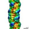
3D structure of myosin filaments isolated from human heart muscles by negative stain electron microscopy and single particle image analysis.
マップデータ
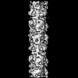
3D map of human cardiac myosin filament
試料
試料: 3D map of human cardiac myosin filament
タンパク質・ペプチド: myosin filament
キーワード
myosin filaments / human cardiac muscles / single particle analysis / electron microscopy / muscle contraction
機能・相同性
機能・相同性情報
myosin II heavy chain binding / muscle cell fate specification / regulation of slow-twitch skeletal muscle fiber contraction / regulation of the force of skeletal muscle contraction / A band / regulation of striated muscle contraction / cardiac myofibril / muscle myosin complex / cardiac myofibril assembly / regulation of the force of heart contraction ...myosin II heavy chain binding / muscle cell fate specification / regulation of slow-twitch skeletal muscle fiber contraction / regulation of the force of skeletal muscle contraction / A band / regulation of striated muscle contraction / cardiac myofibril / muscle myosin complex / cardiac myofibril assembly / regulation of the force of heart contraction / transition between fast and slow fiber / myosin filament / positive regulation of ATP-dependent activity / adult heart development / Striated Muscle Contraction / cardiac muscle hypertrophy in response to stress / muscle filament sliding / myosin complex / myosin II complex / I band / structural constituent of muscle / myosin heavy chain binding / ventricular cardiac muscle tissue morphogenesis / microfilament motor activity / heart contraction / myofibril / positive regulation of the force of heart contraction / cytoskeletal motor activity / actin monomer binding / skeletal muscle contraction / striated muscle contraction / ATP metabolic process / skeletal muscle tissue development / cardiac muscle contraction / stress fiber / muscle contraction / regulation of heart rate / sarcomere / post-embryonic development / negative regulation of cell growth / Z disc / actin filament binding / heart development / cytoskeleton / calmodulin binding / calcium ion binding / ATP binding / cytoplasm / cytosol 類似検索 - 分子機能
: / DNA repair protein XRCC4-like, C-terminal / Myosin tail / Myosin tail / Myosin N-terminal SH3-like domain / Myosin S1 fragment, N-terminal / Myosin, N-terminal, SH3-like / Myosin N-terminal SH3-like domain profile. / Short calmodulin-binding motif containing conserved Ile and Gln residues. / IQ motif, EF-hand binding site ...: / DNA repair protein XRCC4-like, C-terminal / Myosin tail / Myosin tail / Myosin N-terminal SH3-like domain / Myosin S1 fragment, N-terminal / Myosin, N-terminal, SH3-like / Myosin N-terminal SH3-like domain profile. / Short calmodulin-binding motif containing conserved Ile and Gln residues. / IQ motif, EF-hand binding site / Myosin head, motor domain / Myosin head (motor domain) / Myosin motor domain profile. / Myosin. Large ATPases. / IQ motif profile. / Kinesin motor domain superfamily / : / EF-hand domain pair / EF-hand, calcium binding motif / EF-Hand 1, calcium-binding site / EF-hand calcium-binding domain. / EF-hand calcium-binding domain profile. / EF-hand domain / EF-hand domain pair / P-loop containing nucleoside triphosphate hydrolase 類似検索 - ドメイン・相同性
ジャーナル: Proc Natl Acad Sci U S A / 年: 2013 タイトル: Atomic model of the human cardiac muscle myosin filament. 著者: Hind A Al-Khayat / Robert W Kensler / John M Squire / Steven B Marston / Edward P Morris / 要旨: Of all the myosin filaments in muscle, the most important in terms of human health, and so far the least studied, are those in the human heart. Here we report a 3D single-particle analysis of ...Of all the myosin filaments in muscle, the most important in terms of human health, and so far the least studied, are those in the human heart. Here we report a 3D single-particle analysis of electron micrograph images of negatively stained myosin filaments isolated from human cardiac muscle in the normal (undiseased) relaxed state. The resulting 28-Å resolution 3D reconstruction shows axial and azimuthal (no radial) myosin head perturbations within the 429-Å axial repeat, with rotations between successive 132 Å-, 148 Å-, and 149 Å-spaced crowns of heads close to 60°, 35°, and 25° (all would be 40° in an unperturbed three-stranded helix). We have defined the myosin head atomic arrangements within the three crown levels and have modeled the organization of myosin subfragment 2 and the possible locations of the 39 Å-spaced domains of titin and the cardiac isoform of myosin-binding protein-C on the surface of the myosin filament backbone. Best fits were obtained with head conformations on all crowns close to the structure of the two-headed myosin molecule of vertebrate chicken smooth muscle in the dephosphorylated relaxed state. Individual crowns show differences in head-pair tilts and subfragment 2 orientations, which, together with the observed perturbations, result in different intercrown head interactions, including one not reported before. Analysis of the interactions between the myosin heads, the cardiac isoform of myosin-binding protein-C, and titin will aid in understanding of the structural effects of mutations in these proteins known to be associated with human cardiomyopathies.
PDB-5tby: HUMAN BETA CARDIAC HEAVY MEROMYOSIN INTERACTING-HEADS MOTIF OBTAINED BY HOMOLOGY MODELING (USING SWISS-MODEL) OF HUMAN SEQUENCE FROM APHONOPELMA HOMOLOGY MODEL (PDB-3JBH), RIGIDLY FITTED TO HUMAN BETA-CARDIAC NEGATIVELY STAINED THICK FILAMENT 3D-RECONSTRUCTION (EMD-2240)
 ムービー
ムービー コントローラー


 データを開く
 基本情報
基本情報 マップデータ
 試料
試料 キーワード
キーワード 機能・相同性情報
 Homo sapiens (ヒト)
 データ登録者
 引用
引用
 構造の表示
構造の表示
 ダウンロードとリンク
 EMD-2240.jpg
 http://ftp.pdbj.org/pub/emdb/structures/EMD-2240
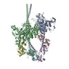
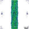


 Z (Sec.)
 Y (Row.)
 X (Col.)


 試料の構成要素
 解析
解析 電子顕微鏡法
 FIELD EMISSION GUN
