National Institutes of Health/National Institute of General Medical Sciences (NIH/NIGMS)
R01 GM30598
United States
Citation
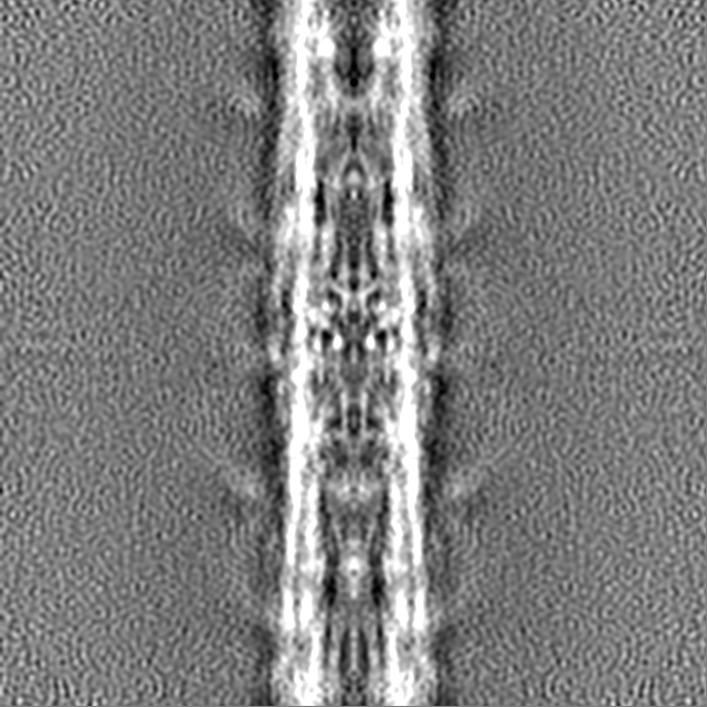
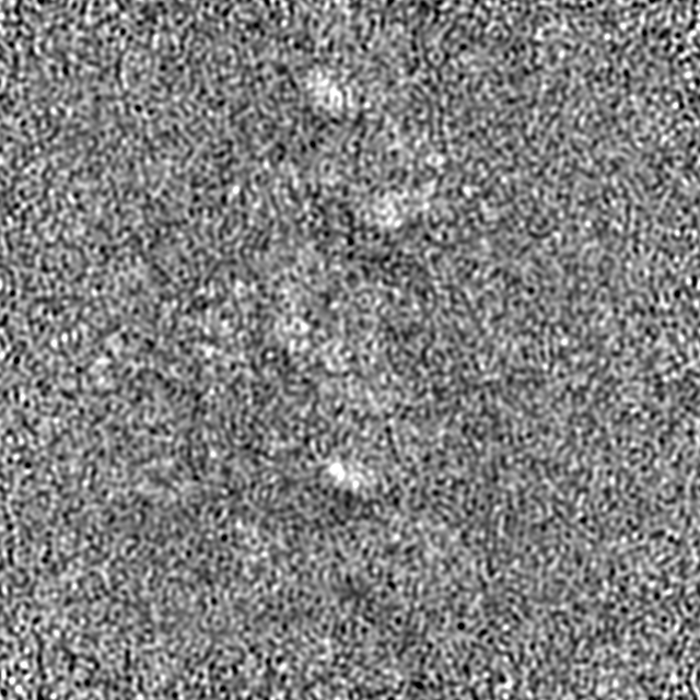
Journal: Life Sci Alliance / Year: 2020 Title: CryoEM structure of flight muscle thick filaments at 7 Å resolution. Authors: Nadia Daneshparvar / Dianne W Taylor / Thomas S O'Leary / Hamidreza Rahmani / Fatemeh Abbasiyeganeh / Michael J Previs / Kenneth A Taylor / Abstract: Striated muscle thick filaments are composed of myosin II and several non-myosin proteins. Myosin II's long α-helical coiled-coil tail forms the dense protein backbone of filaments, whereas its N- ...Striated muscle thick filaments are composed of myosin II and several non-myosin proteins. Myosin II's long α-helical coiled-coil tail forms the dense protein backbone of filaments, whereas its N-terminal globular head containing the catalytic and actin-binding activities extends outward from the backbone. Here, we report the structure of thick filaments of the flight muscle of the fruit fly at 7 Å resolution. Its myosin tails are arranged in curved molecular crystalline layers identical to flight muscles of the giant water bug Four non-myosin densities are observed, three of which correspond to ones found in ; one new density, possibly stretchin-mlck, is found on the backbone outer surface. Surprisingly, the myosin heads are disordered rather than ordered along the filament backbone. Our results show striking myosin tail similarity within flight muscle filaments of two insect orders separated by several hundred million years of evolution.
History
Deposition
Jun 26, 2020
-
Header (metadata) release
Jul 8, 2020
-
Map release
Jul 8, 2020
-
Update
Jul 8, 2020
-
Current status
Jul 8, 2020
Processing site: RCSB / Status: Released
-
Structure visualization
Movie
Surface view with section colored by density value
EMPIAR-10436 (Title: The CryoEM Structure of Drosophila Flight Muscle Thick Filaments at 7Å Resolution Data size: 12.0 TB / Data #1: Wild Type Drosophila [micrographs - multiframe])
In the structure databanks used in Yorodumi, some data are registered as the other names, "COVID-19 virus" and "2019-nCoV". Here are the details of the virus and the list of structure data.
Jan 31, 2019. EMDB accession codes are about to change! (news from PDBe EMDB page)
EMDB accession codes are about to change! (news from PDBe EMDB page)
The allocation of 4 digits for EMDB accession codes will soon come to an end. Whilst these codes will remain in use, new EMDB accession codes will include an additional digit and will expand incrementally as the available range of codes is exhausted. The current 4-digit format prefixed with “EMD-” (i.e. EMD-XXXX) will advance to a 5-digit format (i.e. EMD-XXXXX), and so on. It is currently estimated that the 4-digit codes will be depleted around Spring 2019, at which point the 5-digit format will come into force.
The EM Navigator/Yorodumi systems omit the EMD- prefix.
Related info.:Q: What is EMD? / ID/Accession-code notation in Yorodumi/EM Navigator
Yorodumi is a browser for structure data from EMDB, PDB, SASBDB, etc.
This page is also the successor to EM Navigator detail page, and also detail information page/front-end page for Omokage search.
The word "yorodu" (or yorozu) is an old Japanese word meaning "ten thousand". "mi" (miru) is to see.
Related info.:EMDB / PDB / SASBDB / Comparison of 3 databanks / Yorodumi Search / Aug 31, 2016. New EM Navigator & Yorodumi / Yorodumi Papers / Jmol/JSmol / Function and homology information / Changes in new EM Navigator and Yorodumi
 Movie
Movie Controller


 Open data
 Basic information
 Map data
 Sample
 Authors
 United States, 1 items
  Citation
 Structure visualization
 Movie viewer
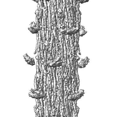
 Downloads & links
 emd_22218.png
 http://ftp.pdbj.org/pub/emdb/structures/EMD-22218
 Z (Sec.)
 Y (Row.)
 X (Col.)


 Sample components
 Processing
 Electron microscopy
 FIELD EMISSION GUN