- EMDB-2240: 3D structure of myosin filaments isolated from human heart muscle... -
+
Open data
ID or keywords:
Loading...
-
Basic information
Entry
Database: EMDB / ID: EMD-2240
Title
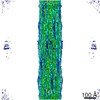
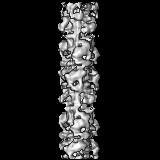
3D structure of myosin filaments isolated from human heart muscles by negative stain electron microscopy and single particle image analysis.
Map data
3D map of human cardiac myosin filament
Sample
Sample: 3D map of human cardiac myosin filament
Protein or peptide: myosin filament
Keywords
myosin filaments / human cardiac muscles / single particle analysis / electron microscopy / muscle contraction
Function / homology
Function and homology information
myosin II heavy chain binding / muscle cell fate specification / regulation of slow-twitch skeletal muscle fiber contraction / regulation of the force of skeletal muscle contraction / regulation of striated muscle contraction / cardiac myofibril / muscle myosin complex / A band / cardiac myofibril assembly / regulation of the force of heart contraction ...myosin II heavy chain binding / muscle cell fate specification / regulation of slow-twitch skeletal muscle fiber contraction / regulation of the force of skeletal muscle contraction / regulation of striated muscle contraction / cardiac myofibril / muscle myosin complex / A band / cardiac myofibril assembly / regulation of the force of heart contraction / transition between fast and slow fiber / myosin filament / Striated Muscle Contraction / muscle filament sliding / cardiac muscle hypertrophy in response to stress / adult heart development / myosin complex / myosin II complex / I band / structural constituent of muscle / ventricular cardiac muscle tissue morphogenesis / microfilament motor activity / myosin heavy chain binding / heart contraction / positive regulation of the force of heart contraction / myofibril / cytoskeletal motor activity / actin monomer binding / skeletal muscle tissue development / ATP metabolic process / striated muscle contraction / skeletal muscle contraction / cardiac muscle contraction / stress fiber / regulation of heart rate / muscle contraction / post-embryonic development / sarcomere / negative regulation of cell growth / Z disc / actin filament binding / heart development / cytoskeleton / calmodulin binding / calcium ion binding / ATP binding / cytoplasm / cytosol Similarity search - Function
: / DNA repair protein XRCC4-like, C-terminal / Myosin tail / Myosin tail / Myosin N-terminal SH3-like domain / Myosin S1 fragment, N-terminal / Myosin, N-terminal, SH3-like / Myosin N-terminal SH3-like domain profile. / Short calmodulin-binding motif containing conserved Ile and Gln residues. / IQ motif, EF-hand binding site ...: / DNA repair protein XRCC4-like, C-terminal / Myosin tail / Myosin tail / Myosin N-terminal SH3-like domain / Myosin S1 fragment, N-terminal / Myosin, N-terminal, SH3-like / Myosin N-terminal SH3-like domain profile. / Short calmodulin-binding motif containing conserved Ile and Gln residues. / IQ motif, EF-hand binding site / Myosin motor domain profile. / Myosin head, motor domain / Myosin head (motor domain) / Myosin. Large ATPases. / IQ motif profile. / Kinesin motor domain superfamily / : / EF-hand domain pair / EF-hand, calcium binding motif / EF-Hand 1, calcium-binding site / EF-hand calcium-binding domain. / EF-hand calcium-binding domain profile. / EF-hand domain / EF-hand domain pair / P-loop containing nucleoside triphosphate hydrolase Similarity search - Domain/homology
Journal: Proc Natl Acad Sci U S A / Year: 2013 Title: Atomic model of the human cardiac muscle myosin filament. Authors: Hind A Al-Khayat / Robert W Kensler / John M Squire / Steven B Marston / Edward P Morris / Abstract: Of all the myosin filaments in muscle, the most important in terms of human health, and so far the least studied, are those in the human heart. Here we report a 3D single-particle analysis of ...Of all the myosin filaments in muscle, the most important in terms of human health, and so far the least studied, are those in the human heart. Here we report a 3D single-particle analysis of electron micrograph images of negatively stained myosin filaments isolated from human cardiac muscle in the normal (undiseased) relaxed state. The resulting 28-Å resolution 3D reconstruction shows axial and azimuthal (no radial) myosin head perturbations within the 429-Å axial repeat, with rotations between successive 132 Å-, 148 Å-, and 149 Å-spaced crowns of heads close to 60°, 35°, and 25° (all would be 40° in an unperturbed three-stranded helix). We have defined the myosin head atomic arrangements within the three crown levels and have modeled the organization of myosin subfragment 2 and the possible locations of the 39 Å-spaced domains of titin and the cardiac isoform of myosin-binding protein-C on the surface of the myosin filament backbone. Best fits were obtained with head conformations on all crowns close to the structure of the two-headed myosin molecule of vertebrate chicken smooth muscle in the dephosphorylated relaxed state. Individual crowns show differences in head-pair tilts and subfragment 2 orientations, which, together with the observed perturbations, result in different intercrown head interactions, including one not reported before. Analysis of the interactions between the myosin heads, the cardiac isoform of myosin-binding protein-C, and titin will aid in understanding of the structural effects of mutations in these proteins known to be associated with human cardiomyopathies.
History
Deposition
Dec 7, 2012
-
Header (metadata) release
Dec 26, 2012
-
Map release
Dec 26, 2012
-
Update
Dec 26, 2012
-
Current status
Dec 26, 2012
Processing site: PDBe / Status: Released
-
Structure visualization
Movie
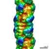
Surface view with section colored by density value
Supramolecule #1000: 3D map of human cardiac myosin filament
Supramolecule
Name: 3D map of human cardiac myosin filament / type: sample / ID: 1000 / Number unique components: 1
-
Macromolecule #1: myosin filament
Macromolecule
Name: myosin filament / type: protein_or_peptide / ID: 1 / Details: 3D map of human cardiac myosin filament / Recombinant expression: No / Database: NCBI
Source (natural)
Organism: Homo sapiens (human) / synonym: human
-
Experimental details
-
Structure determination
Processing
subtomogram averaging
-
Sample preparation
Vitrification
Cryogen name: NONE / Instrument: OTHER
-
Electron microscopy
Microscope
FEI TECNAI 20
Date
May 15, 2012
Image recording
Category: CCD / Film or detector model: TVIPS TEMCAM-F415 (4k x 4k) / Digitization - Sampling interval: 17 µm / Number real images: 237 / Average electron dose: 100 e/Å2 / Bits/pixel: 16
Electron beam
Acceleration voltage: 200 kV / Electron source: FIELD EMISSION GUN
PDB-5tby: HUMAN BETA CARDIAC HEAVY MEROMYOSIN INTERACTING-HEADS MOTIF OBTAINED BY HOMOLOGY MODELING (USING SWISS-MODEL) OF HUMAN SEQUENCE FROM APHONOPELMA HOMOLOGY MODEL (PDB-3JBH), RIGIDLY FITTED TO HUMAN BETA-CARDIAC NEGATIVELY STAINED THICK FILAMENT 3D-RECONSTRUCTION (EMD-2240)
+
About Yorodumi
-
News
-
Feb 9, 2022. New format data for meta-information of EMDB entries
New format data for meta-information of EMDB entries
Version 3 of the EMDB header file is now the official format.
The previous official version 1.9 will be removed from the archive.
In the structure databanks used in Yorodumi, some data are registered as the other names, "COVID-19 virus" and "2019-nCoV". Here are the details of the virus and the list of structure data.
Jan 31, 2019. EMDB accession codes are about to change! (news from PDBe EMDB page)
EMDB accession codes are about to change! (news from PDBe EMDB page)
The allocation of 4 digits for EMDB accession codes will soon come to an end. Whilst these codes will remain in use, new EMDB accession codes will include an additional digit and will expand incrementally as the available range of codes is exhausted. The current 4-digit format prefixed with “EMD-” (i.e. EMD-XXXX) will advance to a 5-digit format (i.e. EMD-XXXXX), and so on. It is currently estimated that the 4-digit codes will be depleted around Spring 2019, at which point the 5-digit format will come into force.
The EM Navigator/Yorodumi systems omit the EMD- prefix.
Related info.:Q: What is EMD? / ID/Accession-code notation in Yorodumi/EM Navigator
Yorodumi is a browser for structure data from EMDB, PDB, SASBDB, etc.
This page is also the successor to EM Navigator detail page, and also detail information page/front-end page for Omokage search.
The word "yorodu" (or yorozu) is an old Japanese word meaning "ten thousand". "mi" (miru) is to see.
Related info.:EMDB / PDB / SASBDB / Comparison of 3 databanks / Yorodumi Search / Aug 31, 2016. New EM Navigator & Yorodumi / Yorodumi Papers / Jmol/JSmol / Function and homology information / Changes in new EM Navigator and Yorodumi
 Movie
Movie Controller

 Yorodumi
 Open data
 Basic information
 Map data
 Sample
 Keywords
 Function and homology information
 Homo sapiens (human)
 Authors
 Citation
 Structure visualization
 Downloads & links
 EMD-2240.jpg
 http://ftp.pdbj.org/pub/emdb/structures/EMD-2240
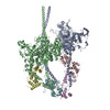


 Z (Sec.)
 Y (Row.)
 X (Col.)


 Sample components
 Processing
 Electron microscopy
 FIELD EMISSION GUN
