 ムービー
ムービー コントローラー


+ データを開く

- 基本情報
基本情報
| 登録情報 | データベース: EMDB / ID: EMD-2239 | |||||||||
|---|---|---|---|---|---|---|---|---|---|---|
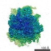
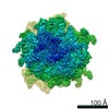
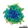
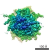
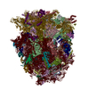
| タイトル | Cryo-electron microscopy structure of the Trypanosoma brucei 80S ribosome | |||||||||
 マップデータ マップデータ | Trypanosoma Brucei 80S Ribosome cryo-em reconstruction filtered according to the local FSC at 0.5 | |||||||||
 試料 試料 |
| |||||||||
 キーワード キーワード | Trypanosoma / brucei / 80S / ribosome / eukaryotic / kinetoplastids / expansion segments / high-resolution | |||||||||
| 機能・相同性 |  機能・相同性情報 機能・相同性情報organellar small ribosomal subunit / organellar large ribosomal subunit / ciliary transition zone / mitochondrial large ribosomal subunit / nuclear lumen / phosphate ion binding / negative regulation of translational frameshifting / endonucleolytic cleavage to generate mature 3'-end of SSU-rRNA from (SSU-rRNA, 5.8S rRNA, LSU-rRNA) / endonucleolytic cleavage in ITS1 to separate SSU-rRNA from 5.8S rRNA and LSU-rRNA from tricistronic rRNA transcript (SSU-rRNA, 5.8S rRNA, LSU-rRNA) / protein-RNA complex assembly ...organellar small ribosomal subunit / organellar large ribosomal subunit / ciliary transition zone / mitochondrial large ribosomal subunit / nuclear lumen / phosphate ion binding / negative regulation of translational frameshifting / endonucleolytic cleavage to generate mature 3'-end of SSU-rRNA from (SSU-rRNA, 5.8S rRNA, LSU-rRNA) / endonucleolytic cleavage in ITS1 to separate SSU-rRNA from 5.8S rRNA and LSU-rRNA from tricistronic rRNA transcript (SSU-rRNA, 5.8S rRNA, LSU-rRNA) / protein-RNA complex assembly / maturation of LSU-rRNA / translation regulator activity / rescue of stalled ribosome / protein kinase C binding / maturation of LSU-rRNA from tricistronic rRNA transcript (SSU-rRNA, 5.8S rRNA, LSU-rRNA) / ribosomal large subunit biogenesis / ribosome assembly / maturation of SSU-rRNA from tricistronic rRNA transcript (SSU-rRNA, 5.8S rRNA, LSU-rRNA) / maturation of SSU-rRNA / small-subunit processome / regulation of cell growth / modification-dependent protein catabolic process / protein tag activity / maintenance of translational fidelity / rRNA processing / ribosome biogenesis / regulation of cell population proliferation / ribosome binding / ribosomal small subunit biogenesis / ribosomal small subunit assembly / small ribosomal subunit / 5S rRNA binding / small ribosomal subunit rRNA binding / ribosomal large subunit assembly / large ribosomal subunit rRNA binding / cytosolic small ribosomal subunit / cytosolic large ribosomal subunit / cytoplasmic translation / protein ubiquitination / negative regulation of translation / rRNA binding / structural constituent of ribosome / ribosome / translation / ribonucleoprotein complex / mRNA binding / ubiquitin protein ligase binding / nucleolus / RNA binding / zinc ion binding / nucleoplasm / nucleus / cytosol / cytoplasm 類似検索 - 分子機能 | |||||||||
| 生物種 |  | |||||||||
| 手法 | 単粒子再構成法 / クライオ電子顕微鏡法 / 解像度: 5.57 Å | |||||||||
 データ登録者 データ登録者 | Hashem Y / des Georges A / Fu J / Buss SN / Jossinet F / Jobe A / Zhang Q / Liao HY / Grassucci B / Bajaj C ...Hashem Y / des Georges A / Fu J / Buss SN / Jossinet F / Jobe A / Zhang Q / Liao HY / Grassucci B / Bajaj C / Westhof E / Madison-Antenucci S / Frank J | |||||||||
 引用 引用 | ジャーナル: Nature / 年: 2013 タイトル: High-resolution cryo-electron microscopy structure of the Trypanosoma brucei ribosome. 著者: Yaser Hashem / Amedee des Georges / Jie Fu / Sarah N Buss / Fabrice Jossinet / Amy Jobe / Qin Zhang / Hstau Y Liao / Robert A Grassucci / Chandrajit Bajaj / Eric Westhof / Susan Madison- ...著者: Yaser Hashem / Amedee des Georges / Jie Fu / Sarah N Buss / Fabrice Jossinet / Amy Jobe / Qin Zhang / Hstau Y Liao / Robert A Grassucci / Chandrajit Bajaj / Eric Westhof / Susan Madison-Antenucci / Joachim Frank /  要旨: Ribosomes, the protein factories of living cells, translate genetic information carried by messenger RNAs into proteins, and are thus involved in virtually all aspects of cellular development and ...Ribosomes, the protein factories of living cells, translate genetic information carried by messenger RNAs into proteins, and are thus involved in virtually all aspects of cellular development and maintenance. The few available structures of the eukaryotic ribosome reveal that it is more complex than its prokaryotic counterpart, owing mainly to the presence of eukaryote-specific ribosomal proteins and additional ribosomal RNA insertions, called expansion segments. The structures also differ among species, partly in the size and arrangement of these expansion segments. Such differences are extreme in kinetoplastids, unicellular eukaryotic parasites often infectious to humans. Here we present a high-resolution cryo-electron microscopy structure of the ribosome of Trypanosoma brucei, the parasite that is transmitted by the tsetse fly and that causes African sleeping sickness. The atomic model reveals the unique features of this ribosome, characterized mainly by the presence of unusually large expansion segments and ribosomal-protein extensions leading to the formation of four additional inter-subunit bridges. We also find additional rRNA insertions, including one large rRNA domain that is not found in other eukaryotes. Furthermore, the structure reveals the five cleavage sites of the kinetoplastid large ribosomal subunit (LSU) rRNA chain, which is known to be cleaved uniquely into six pieces, and suggests that the cleavage is important for the maintenance of the T. brucei ribosome in the observed structure. We discuss several possible implications of the large rRNA expansion segments for the translation-regulation process. The structure could serve as a basis for future experiments aimed at understanding the functional importance of these kinetoplastid-specific ribosomal features in protein-translation regulation, an essential step towards finding effective and safe kinetoplastid-specific drugs. | |||||||||
| 履歴 |
|
- 構造の表示
構造の表示
| ムービー |
ムービービューア |
|---|---|
| 構造ビューア | EMマップ: SurfViewMolmilJmol/JSmol |
| 添付画像 |

- ダウンロードとリンク
-EMDBアーカイブ
| マップデータ | emd_2239.map.gz | 7 MB | EMDBマップデータ形式 | |
|---|---|---|---|---|
| ヘッダ (付随情報) | emd-2239-v30.xmlemd-2239.xml | 13.2 KB 13.2 KB | 表示 表示 | EMDBヘッダ |
| FSC (解像度算出) | emd_2239_fsc.xml | 22.9 KB | 表示 | FSCデータファイル |
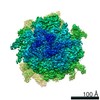
| 画像 | 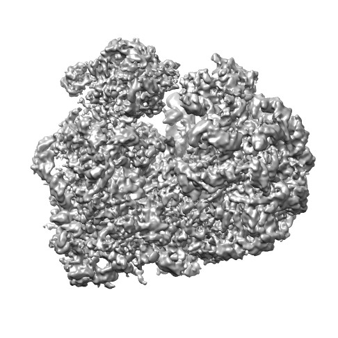 emd_2239.jpg emd_2239.jpg | 59.9 KB | ||
| アーカイブディレクトリ |  http://ftp.pdbj.org/pub/emdb/structures/EMD-2239ftp://ftp.pdbj.org/pub/emdb/structures/EMD-2239 http://ftp.pdbj.org/pub/emdb/structures/EMD-2239ftp://ftp.pdbj.org/pub/emdb/structures/EMD-2239 | HTTPS FTP |
-検証レポート
| 文書・要旨 | emd_2239_validation.pdf.gz | 285.1 KB | 表示 | EMDB検証レポート |
|---|---|---|---|---|
| 文書・詳細版 | emd_2239_full_validation.pdf.gz | 284.2 KB | 表示 | |
| XML形式データ | emd_2239_validation.xml.gz | 17.7 KB | 表示 | |
| アーカイブディレクトリ | https://ftp.pdbj.org/pub/emdb/validation_reports/EMD-2239ftp://ftp.pdbj.org/pub/emdb/validation_reports/EMD-2239 | HTTPS FTP |
-関連構造データ


-リンク
| EMDBのページ | EMDB (EBI/PDBe) / EMDataResource |
|---|---|
| 「今月の分子」の関連する項目 |


-マップ
| ファイル | ダウンロード / ファイル: emd_2239.map.gz / 形式: CCP4 / 大きさ: 172.4 MB / タイプ: IMAGE STORED AS FLOATING POINT NUMBER (4 BYTES) | ||||||||||||||||||||||||||||||||||||||||||||||||||||||||||||||||||||
|---|---|---|---|---|---|---|---|---|---|---|---|---|---|---|---|---|---|---|---|---|---|---|---|---|---|---|---|---|---|---|---|---|---|---|---|---|---|---|---|---|---|---|---|---|---|---|---|---|---|---|---|---|---|---|---|---|---|---|---|---|---|---|---|---|---|---|---|---|---|
| 注釈 | Trypanosoma Brucei 80S Ribosome cryo-em reconstruction filtered according to the local FSC at 0.5 | ||||||||||||||||||||||||||||||||||||||||||||||||||||||||||||||||||||
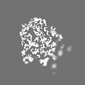
| 投影像・断面図 | 画像のコントロール
画像は Spider により作成 | ||||||||||||||||||||||||||||||||||||||||||||||||||||||||||||||||||||
| ボクセルのサイズ | X=Y=Z: 1.09 Å | ||||||||||||||||||||||||||||||||||||||||||||||||||||||||||||||||||||
| 密度 |
| ||||||||||||||||||||||||||||||||||||||||||||||||||||||||||||||||||||
| 対称性 | 空間群: 1 | ||||||||||||||||||||||||||||||||||||||||||||||||||||||||||||||||||||
| 詳細 | EMDB XML:
CCP4マップ ヘッダ情報:
| ||||||||||||||||||||||||||||||||||||||||||||||||||||||||||||||||||||
 Z (Sec.)
 Y (Row.)
 X (Col.)


-添付データ
- 試料の構成要素
-全体 : Trypanosoma Brucei 80S Ribosome
| 全体 | 名称: Trypanosoma Brucei 80S Ribosome |
|---|---|
| 要素 |
|
-超分子 #1000: Trypanosoma Brucei 80S Ribosome
| 超分子 | 名称: Trypanosoma Brucei 80S Ribosome / タイプ: sample / ID: 1000 / 集合状態: monomer / Number unique components: 1 |
|---|---|
| 分子量 | 理論値: 3.305 MDa 手法: Molecular weight estimated form the protein and RNA sequence. |
-超分子 #1: 80S Ribosome
| 超分子 | 名称: 80S Ribosome / タイプ: complex / ID: 1 / 組換発現: No / Ribosome-details: ribosome-eukaryote: ALL |
|---|---|
| 由来(天然) | 生物種: |
| 分子量 | 理論値: 3.3 MDa |
-実験情報
-構造解析
| 手法 | クライオ電子顕微鏡法 |
|---|---|
 解析 解析 | 単粒子再構成法 |
| 試料の集合状態 | particle |
-試料調製
| 濃度 | 0.105 mg/mL |
|---|---|
| 緩衝液 | pH: 7.2 詳細: 20 mM Tris pH 7.2, 100mM MgCl2, 500 mM KCl, 5 mM beta-mercaptoethanol |
| グリッド | 詳細: 300 mesh Copper/Molbydenum holey carbon-coated Quantifoil 2/4 grid (Quantifoil Micro Tools GmbH) containing an additional continuous thin layer of carbon |
| 凍結 | 凍結剤: ETHANE / チャンバー内湿度: 100 % / チャンバー内温度: 100 K / 装置: FEI VITROBOT MARK IV / 手法: Wait 30 sec, Blot 6 seconds, plunge |
- 電子顕微鏡法
| 顕微鏡 | FEI POLARA 300 |
|---|---|
| 日付 | 2011年1月1日 |
| 撮影 | カテゴリ: FILM / フィルム・検出器のモデル: KODAK SO-163 FILM デジタル化 - スキャナー: NIKON SUPER COOLSCAN 9000 実像数: 1000 / 平均電子線量: 25 e/Å2 / ビット/ピクセル: 32 |
| 電子線 | 加速電圧: 300 kV / 電子線源:  FIELD EMISSION GUN FIELD EMISSION GUN |
| 電子光学系 | 照射モード: FLOOD BEAM / 撮影モード: BRIGHT FIELD / Cs: 2.26 mm / 最大 デフォーカス(公称値): 4.0 µm / 最小 デフォーカス(公称値): 1.5 µm / 倍率(公称値): 59000 |
| 試料ステージ | 試料ホルダーモデル: SIDE ENTRY, EUCENTRIC |
| 実験機器 |  モデル: Tecnai Polara / 画像提供: FEI Company |
-画像解析
| 詳細 | Data were processed using SPIDER. The particles windows were automatically extracted from 1000 film-recorded micrographs and inspected manually. Standard SPIDER protocols for reference-based reconstruction, except that contrast transfer function (CTF) of the reconstructions was corrected by phase-flipping the particles using the defocus value estimated for each micrograph and a single reconstruction was obtained from the entire dataset using conjugate gradients with regularization (BP CG in SPIDER). |
|---|---|
| CTF補正 | 詳細: Phase-flip on each particle |
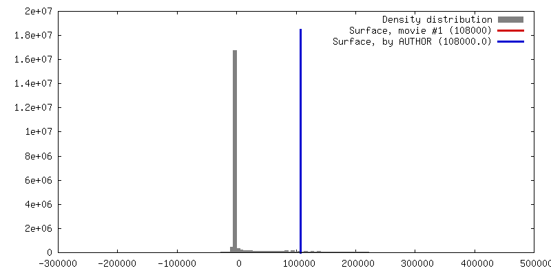
| 最終 再構成 | 想定した対称性 - 点群: C1 (非対称) / アルゴリズム: OTHER / 解像度のタイプ: BY AUTHOR / 解像度: 5.57 Å / 解像度の算出法: FSC 0.5 CUT-OFF / ソフトウェア - 名称: Spider / 使用した粒子像数: 164000 |
| FSC曲線 (解像度の算出) |  |
-原子モデル構築 1
| 初期モデル | PDB ID:  3u5b |
|---|---|
| ソフトウェア | 名称: Chimera |
| 詳細 | Protocol: Rigid body. The structure of the 80S from Yeast (3U5B and others) as well as the structure of the 60S from Tetrahymena thermophila (4A17 and others) were used as starting model for the 60S subunit model. The 40S from Tetrahymena thermophila (2XZM and 2XZN) as well as the 80S from Yeast were used as starting model for the 40S subunit model. The 80S model of Triticum aestivum (3IZR and others) was used to fit missing proteins form the two X-ray structures. |
| 精密化 | 空間: REAL / プロトコル: RIGID BODY FIT / 当てはまり具合の基準: cross correlation |
| 得られたモデル |  PDB-4v8m: |
-原子モデル構築 2
| 初期モデル | PDB ID: 2xzm |
|---|---|
| ソフトウェア | 名称: Chimera |
| 詳細 | Protocol: Rigid body. The structure of the 80S from Yeast (3U5B and others) as well as the structure of the 60S from Tetrahymena thermophila (4A17 and others) were used as starting model for the 60S subunit model. The 40S from Tetrahymena thermophila (2XZM and 2XZN) as well as the 80S from Yeast were used as starting model for the 40S subunit model. The 80S model of Triticum aestivum (3IZR and others) was used to fit missing proteins form the two X-ray structures. |
| 精密化 | 空間: REAL / プロトコル: RIGID BODY FIT / 当てはまり具合の基準: cross correlation |
| 得られたモデル | PDB-4v8m: |
-原子モデル構築 3
| 初期モデル | PDB ID: 3izr |
|---|---|
| ソフトウェア | 名称: Chimera |
| 詳細 | Protocol: Rigid body. The structure of the 80S from Yeast (3U5B and others) as well as the structure of the 60S from Tetrahymena thermophila (4A17 and others) were used as starting model for the 60S subunit model. The 40S from Tetrahymena thermophila (2XZM and 2XZN) as well as the 80S from Yeast were used as starting model for the 40S subunit model. The 80S model of Triticum aestivum (3IZR and others) was used to fit missing proteins form the two X-ray structures. |
| 精密化 | 空間: REAL / プロトコル: RIGID BODY FIT / 当てはまり具合の基準: cross correlation |
| 得られたモデル | PDB-4v8m: |
