 ムービー
ムービー コントローラー


+ データを開く

- 基本情報
基本情報
| 登録情報 | データベース: EMDB / ID: EMD-22050 | |||||||||
|---|---|---|---|---|---|---|---|---|---|---|
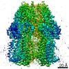
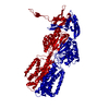
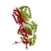
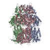
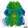
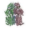
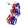
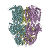
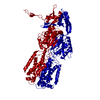
| タイトル | 3.9 Angstrom reconstruction of E.coli AcrB embedded in the liposome | |||||||||
 マップデータ マップデータ | 3D reconstruction of AcrB after Density Modification | |||||||||
 試料 試料 |
| |||||||||
| 機能・相同性 |  機能・相同性情報 機能・相同性情報alkane transmembrane transporter activity / alkane transport / enterobactin transport / enterobactin transmembrane transporter activity / xenobiotic detoxification by transmembrane export across the cell outer membrane / periplasmic side of plasma membrane / efflux pump complex / bile acid transmembrane transporter activity / xenobiotic transport / bile acid and bile salt transport ...alkane transmembrane transporter activity / alkane transport / enterobactin transport / enterobactin transmembrane transporter activity / xenobiotic detoxification by transmembrane export across the cell outer membrane / periplasmic side of plasma membrane / efflux pump complex / bile acid transmembrane transporter activity / xenobiotic transport / bile acid and bile salt transport / efflux transmembrane transporter activity / xenobiotic transmembrane transporter activity / fatty acid transport / response to toxic substance / response to xenobiotic stimulus / response to antibiotic / identical protein binding / membrane / plasma membrane 類似検索 - 分子機能 | |||||||||
| 生物種 |  | |||||||||
| 手法 | 単粒子再構成法 / クライオ電子顕微鏡法 / 解像度: 3.9 Å | |||||||||
 データ登録者 データ登録者 | Xia Y / Fan X / Yan N | |||||||||
 引用 引用 | ジャーナル: Proc Natl Acad Sci U S A / 年: 2020 タイトル: Cryo-EM analysis of a membrane protein embedded in the liposome. 著者: Xia Yao / Xiao Fan / Nieng Yan /  要旨: Membrane proteins (MPs) used to be the most difficult targets for structural biology when X-ray crystallography was the mainstream approach. With the resolution revolution of single-particle electron ...Membrane proteins (MPs) used to be the most difficult targets for structural biology when X-ray crystallography was the mainstream approach. With the resolution revolution of single-particle electron cryo-microscopy (cryo-EM), rapid progress has been made for structural elucidation of isolated MPs. The next challenge is to preserve the electrochemical gradients and membrane curvature for a comprehensive structural elucidation of MPs that rely on these chemical and physical properties for their biological functions. Toward this goal, here we present a convenient workflow for cryo-EM structural analysis of MPs embedded in liposomes, using the well-characterized AcrB as a prototype. Combining optimized proteoliposome isolation, cryo-sample preparation on graphene grids, and an efficient particle selection strategy, the three-dimensional (3D) reconstruction of AcrB embedded in liposomes was obtained at 3.9 Å resolution. The conformation of the homotrimeric AcrB remains the same when the surrounding membranes display different curvatures. Our approach, which can be widely applied to cryo-EM analysis of MPs with distinctive soluble domains, lays out the foundation for cryo-EM analysis of integral or peripheral MPs whose functions are affected by transmembrane electrochemical gradients or/and membrane curvatures. | |||||||||
| 履歴 |
|
- 構造の表示
構造の表示
| ムービー |
ムービービューア |
|---|---|
| 構造ビューア | EMマップ: SurfViewMolmilJmol/JSmol |
| 添付画像 |

- ダウンロードとリンク
-EMDBアーカイブ
| マップデータ | emd_22050.map.gz | 6.7 MB | EMDBマップデータ形式 | |
|---|---|---|---|---|
| ヘッダ (付随情報) | emd-22050-v30.xmlemd-22050.xml | 15 KB 15 KB | 表示 表示 | EMDBヘッダ |
| FSC (解像度算出) | emd_22050_fsc.xml | 7.1 KB | 表示 | FSCデータファイル |
| 画像 |  emd_22050.png emd_22050.png | 184.3 KB | ||
| その他 | emd_22050_additional_1.map.gzemd_22050_additional_2.map.gz | 28.1 MB 23 MB | ||
| アーカイブディレクトリ |  http://ftp.pdbj.org/pub/emdb/structures/EMD-22050ftp://ftp.pdbj.org/pub/emdb/structures/EMD-22050 http://ftp.pdbj.org/pub/emdb/structures/EMD-22050ftp://ftp.pdbj.org/pub/emdb/structures/EMD-22050 | HTTPS FTP |
-検証レポート
| 文書・要旨 | emd_22050_validation.pdf.gz | 78.4 KB | 表示 | EMDB検証レポート |
|---|---|---|---|---|
| 文書・詳細版 | emd_22050_full_validation.pdf.gz | 77.5 KB | 表示 | |
| XML形式データ | emd_22050_validation.xml.gz | 492 B | 表示 | |
| アーカイブディレクトリ | https://ftp.pdbj.org/pub/emdb/validation_reports/EMD-22050ftp://ftp.pdbj.org/pub/emdb/validation_reports/EMD-22050 | HTTPS FTP |
-関連構造データ
| 類似構造データ | |
|---|---|
| 電子顕微鏡画像生データ | EMPIAR-10426 (タイトル: 3.9 Angstrom reconstruction of E.coli AcrB embedded in the liposome Data size: 9.6 TB Data #1: Unaligned multi-frame micrographs of AcrB embedded in the liposome. [micrographs - multiframe]) |

-リンク
| EMDBのページ | EMDB (EBI/PDBe) / EMDataResource |
|---|---|
| 「今月の分子」の関連する項目 |

-マップ
| ファイル | ダウンロード / ファイル: emd_22050.map.gz / 形式: CCP4 / 大きさ: 7.2 MB / タイプ: IMAGE STORED AS FLOATING POINT NUMBER (4 BYTES) | ||||||||||||||||||||||||||||||||||||||||||||||||||||||||||||||||||||
|---|---|---|---|---|---|---|---|---|---|---|---|---|---|---|---|---|---|---|---|---|---|---|---|---|---|---|---|---|---|---|---|---|---|---|---|---|---|---|---|---|---|---|---|---|---|---|---|---|---|---|---|---|---|---|---|---|---|---|---|---|---|---|---|---|---|---|---|---|---|
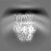
| 注釈 | 3D reconstruction of AcrB after Density Modification | ||||||||||||||||||||||||||||||||||||||||||||||||||||||||||||||||||||
| 投影像・断面図 | 画像のコントロール
画像は Spider により作成 これらの図は立方格子座標系で作成されたものです | ||||||||||||||||||||||||||||||||||||||||||||||||||||||||||||||||||||
| ボクセルのサイズ | X=Y=Z: 1.114 Å | ||||||||||||||||||||||||||||||||||||||||||||||||||||||||||||||||||||
| 密度 |
| ||||||||||||||||||||||||||||||||||||||||||||||||||||||||||||||||||||
| 対称性 | 空間群: 1 | ||||||||||||||||||||||||||||||||||||||||||||||||||||||||||||||||||||
| 詳細 | EMDB XML:
CCP4マップ ヘッダ情報:
| ||||||||||||||||||||||||||||||||||||||||||||||||||||||||||||||||||||
 X (Sec.)
 Y (Row.)
 Z (Col.)


-添付データ
-追加マップ: 3D reconstruction of AcrB after postprocess
| ファイル | emd_22050_additional_1.map | ||||||||||||
|---|---|---|---|---|---|---|---|---|---|---|---|---|---|
| 注釈 | 3D reconstruction of AcrB after postprocess | ||||||||||||
| 投影像・断面図 |
| ||||||||||||
| 密度ヒストグラム |


-追加マップ: Unfiltered 3D reconstruction of AcrB
| ファイル | emd_22050_additional_2.map | ||||||||||||
|---|---|---|---|---|---|---|---|---|---|---|---|---|---|
| 注釈 | Unfiltered 3D reconstruction of AcrB | ||||||||||||
| 投影像・断面図 |
| ||||||||||||
| 密度ヒストグラム |


- 試料の構成要素
-全体 : E.coli AcrB
| 全体 | 名称: E.coli AcrB |
|---|---|
| 要素 |
|
-超分子 #1: E.coli AcrB
| 超分子 | 名称: E.coli AcrB / タイプ: complex / ID: 1 / 親要素: 0 |
|---|---|
| 由来(天然) | 生物種: |
| 組換発現 | 生物種: |
| 分子量 | 実験値: 350 kDa/nm |
-実験情報
-構造解析
| 手法 | クライオ電子顕微鏡法 |
|---|---|
 解析 解析 | 単粒子再構成法 |
| 試料の集合状態 | particle |
-試料調製
| 緩衝液 | pH: 7.5 |
|---|---|
| グリッド | モデル: Quantifoil R1.2/1.3 / 材質: GOLD / メッシュ: 300 / 支持フィルム - 材質: GRAPHENE / 支持フィルム - トポロジー: CONTINUOUS / 詳細: Ozonated |
| 凍結 | 凍結剤: ETHANE / チャンバー内湿度: 100 % / チャンバー内温度: 283 K |
- 電子顕微鏡法
| 顕微鏡 | FEI TITAN KRIOS |
|---|---|
| 特殊光学系 | 球面収差補正装置: Cs corrector applied / エネルギーフィルター - 名称: GIF Bioquantum / エネルギーフィルター - スリット幅: 20 eV |
| 撮影 | フィルム・検出器のモデル: GATAN K2 SUMMIT (4k x 4k) 検出モード: SUPER-RESOLUTION / デジタル化 - サイズ - 横: 3838 pixel / デジタル化 - サイズ - 縦: 3710 pixel / デジタル化 - サンプリング間隔: 4.0 µm / デジタル化 - 画像ごとのフレーム数: 1-32 / 撮影したグリッド数: 1 / 実像数: 5757 / 平均露光時間: 5.6 sec. / 平均電子線量: 50.0 e/Å2 |
| 電子線 | 加速電圧: 300 kV / 電子線源:  FIELD EMISSION GUN FIELD EMISSION GUN |
| 電子光学系 | C2レンズ絞り径: 70.0 µm / 照射モード: FLOOD BEAM / 撮影モード: BRIGHT FIELD / Cs: 0.01 mm / 倍率(公称値): 105000 |
| 試料ステージ | 試料ホルダーモデル: FEI TITAN KRIOS AUTOGRID HOLDER ホルダー冷却材: NITROGEN |
| 実験機器 |  モデル: Titan Krios / 画像提供: FEI Company |

