Movie
Movie Controller

[English] 日本語
 Yorodumi
- EMDB-21919: 14-protofilament, GMPCPP stabilized microtubule with nucleotide f... -
+ Open data

- Basic information
| Entry | Database: EMDB / ID: EMD-21919 | |||||||||
|---|---|---|---|---|---|---|---|---|---|---|
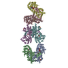
| Title | 14-protofilament, GMPCPP stabilized microtubule with nucleotide free kinesin | |||||||||
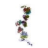
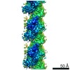
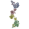
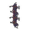
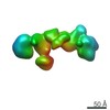
 Map data Map data | Reconstruction from a 14-protofilament, GMPCPP-stabilized microtubule fully decorated with kinesin. | |||||||||
 Sample Sample |
| |||||||||
| Biological species |  | |||||||||
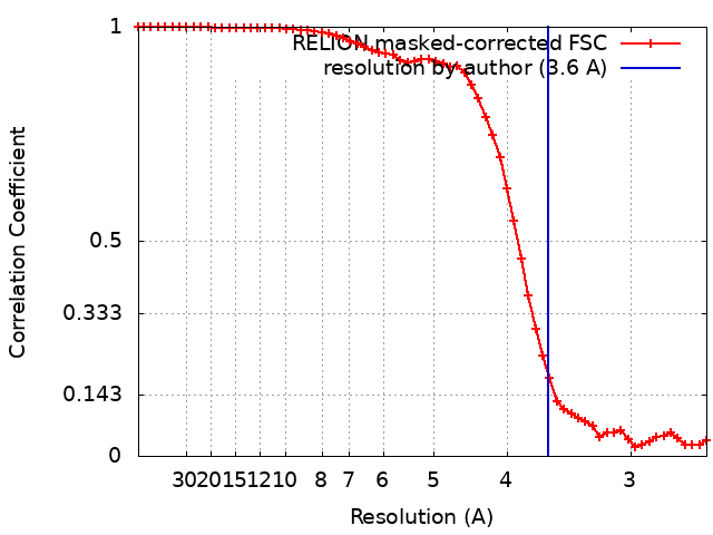
| Method | single particle reconstruction / cryo EM / Resolution: 3.6 Å | |||||||||
 Authors Authors | Debs GE / Cha M / Sindelar CV | |||||||||
| Funding support |  United States, 1 items United States, 1 items
| |||||||||
 Citation Citation | Journal: Proc Natl Acad Sci U S A / Year: 2020 Title: Dynamic and asymmetric fluctuations in the microtubule wall captured by high-resolution cryoelectron microscopy. Authors: Garrett E Debs / Michael Cha / Xueqi Liu / Andrew R Huehn / Charles V Sindelar / Abstract: Microtubules are tubular polymers with essential roles in numerous cellular activities. Structures of microtubules have been captured at increasing resolution by cryo-EM. However, dynamic properties ...Microtubules are tubular polymers with essential roles in numerous cellular activities. Structures of microtubules have been captured at increasing resolution by cryo-EM. However, dynamic properties of the microtubule are key to its function, and this behavior has proved difficult to characterize at a structural level due to limitations in existing structure determination methods. We developed a high-resolution cryo-EM refinement method that divides an imaged microtubule into its constituent protofilaments, enabling deviations from helicity and other sources of heterogeneity to be quantified and corrected for at the single-subunit level. We demonstrate that this method improves the resolution of microtubule 3D reconstructions and substantially reduces anisotropic blurring artifacts, compared with methods that utilize helical symmetry averaging. Moreover, we identified an unexpected, discrete behavior of the m-loop, which mediates lateral interactions between neighboring protofilaments and acts as a flexible hinge between them. The hinge angle adopts preferred values corresponding to distinct conformations of the m-loop that are incompatible with helical symmetry. These hinge angles fluctuate in a stochastic manner, and perfectly cylindrical microtubule conformations are thus energetically and entropically penalized. The hinge angle can diverge further from helical symmetry at the microtubule seam, generating a subpopulation of highly distorted microtubules. However, the seam-distorted subpopulation disappears in the presence of Taxol, a microtubule stabilizing agent. These observations provide clues into the structural origins of microtubule flexibility and dynamics and highlight the role of structural polymorphism in defining microtubule behavior. | |||||||||
| History |
|
- Structure visualization
| Movie |
 Movie viewer Movie viewer |
|---|---|
| Structure viewer | EM map: SurfViewMolmilJmol/JSmol |
| Supplemental images |

- Downloads & links
-EMDB archive
| Map data | emd_21919.map.gz | 14.5 MB | EMDB map data format | |
|---|---|---|---|---|
| Header (meta data) | emd-21919-v30.xmlemd-21919.xml | 14.7 KB 14.7 KB | Display Display | EMDB header |
| FSC (resolution estimation) | emd_21919_fsc.xml | 5.8 KB | Display | FSC data file |
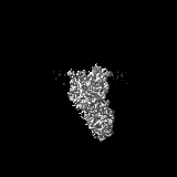
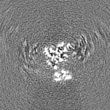
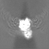
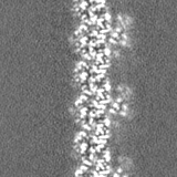
| Images | 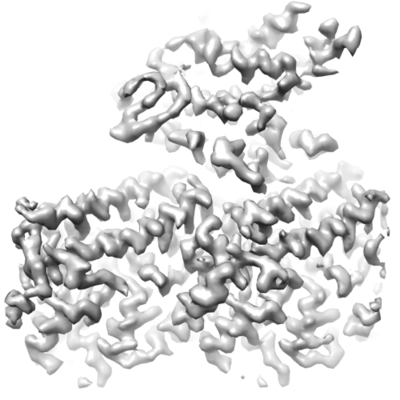 emd_21919.png emd_21919.png | 76.2 KB | ||
| Others | emd_21919_half_map_1.map.gzemd_21919_half_map_2.map.gz | 14.5 MB 14.5 MB | ||
| Archive directory |  http://ftp.pdbj.org/pub/emdb/structures/EMD-21919ftp://ftp.pdbj.org/pub/emdb/structures/EMD-21919 http://ftp.pdbj.org/pub/emdb/structures/EMD-21919ftp://ftp.pdbj.org/pub/emdb/structures/EMD-21919 | HTTPS FTP |
-Related structure data


-Links
| EMDB pages | EMDB (EBI/PDBe) / EMDataResource |
|---|
-Map
| File | Download / File: emd_21919.map.gz / Format: CCP4 / Size: 15.6 MB / Type: IMAGE STORED AS FLOATING POINT NUMBER (4 BYTES) | ||||||||||||||||||||||||||||||||||||||||||||||||||||||||||||||||||||
|---|---|---|---|---|---|---|---|---|---|---|---|---|---|---|---|---|---|---|---|---|---|---|---|---|---|---|---|---|---|---|---|---|---|---|---|---|---|---|---|---|---|---|---|---|---|---|---|---|---|---|---|---|---|---|---|---|---|---|---|---|---|---|---|---|---|---|---|---|---|
| Annotation | Reconstruction from a 14-protofilament, GMPCPP-stabilized microtubule fully decorated with kinesin. | ||||||||||||||||||||||||||||||||||||||||||||||||||||||||||||||||||||
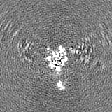
| Projections & slices | Image control
Images are generated by Spider. | ||||||||||||||||||||||||||||||||||||||||||||||||||||||||||||||||||||
| Voxel size | X=Y=Z: 1.3 Å | ||||||||||||||||||||||||||||||||||||||||||||||||||||||||||||||||||||
| Density |
| ||||||||||||||||||||||||||||||||||||||||||||||||||||||||||||||||||||
| Symmetry | Space group: 1 | ||||||||||||||||||||||||||||||||||||||||||||||||||||||||||||||||||||
| Details | EMDB XML:
CCP4 map header:
| ||||||||||||||||||||||||||||||||||||||||||||||||||||||||||||||||||||
 Z (Sec.)
 Y (Row.)
 X (Col.)


-Supplemental data
-Half map: #1
| File | emd_21919_half_map_1.map | ||||||||||||
|---|---|---|---|---|---|---|---|---|---|---|---|---|---|
| Projections & Slices |
| ||||||||||||
| Density Histograms |


-Half map: #2
| File | emd_21919_half_map_2.map | ||||||||||||
|---|---|---|---|---|---|---|---|---|---|---|---|---|---|
| Projections & Slices |
| ||||||||||||
| Density Histograms |


- Sample components
-Entire : Microtubules stabilized with GMPCPP, fully decorated with kinesin...
| Entire | Name: Microtubules stabilized with GMPCPP, fully decorated with kinesin in the nucleotide free state |
|---|---|
| Components |
|
-Supramolecule #1: Microtubules stabilized with GMPCPP, fully decorated with kinesin...
| Supramolecule | Name: Microtubules stabilized with GMPCPP, fully decorated with kinesin in the nucleotide free state type: organelle_or_cellular_component / ID: 1 / Parent: 0 / Macromolecule list: all |
|---|---|
| Source (natural) | Organism: |
-Macromolecule #1: alpha-Tubulin
| Macromolecule | Name: alpha-Tubulin / type: protein_or_peptide / ID: 1 / Enantiomer: LEVO |
|---|---|
| Source (natural) | Organism: |
| Sequence | String: MRECISIHVG QAGVQIGNAC WELYCLEHGI QPDGQMPSDK TIGGGDDSFN TFFSETGAG KHVPRAVFVD LEPTVIDEVR TGTYRQLFHP EQLITGKEDA A NNYARGHY TIGKEIIDLV LDRIRKLADQ CTGLQGFLVF HSFGGGTGSG FT SLLMERL SVDYGKKSKL ...String: MRECISIHVG QAGVQIGNAC WELYCLEHGI QPDGQMPSDK TIGGGDDSFN TFFSETGAG KHVPRAVFVD LEPTVIDEVR TGTYRQLFHP EQLITGKEDA A NNYARGHY TIGKEIIDLV LDRIRKLADQ CTGLQGFLVF HSFGGGTGSG FT SLLMERL SVDYGKKSKL EFSIYPAPQV STAVVEPYNS ILTTHTTLEH SDC AFMVDN EAIYDICRRN LDIERPTYTN LNRLISQIVS SITASLRFDG ALNV DLTEF QTNLVPYPRI HFPLATYAPV ISAEKAYHEQ LSVAEITNAC FEPAN QMVK CDPRHGKYMA CCLLYRGDVV PKDVNAAIAT IKTKRSIQFV DWCPTG FKV GINYQPPTVV PGGDLAKVQR AVCMLSNTTA IAEAWARLDH KFDLMYA KR AFVHWYVGEG MEEGEFSEAR EDMAALEKDY EEVGVDSVEG EGEEEGEE Y |
-Macromolecule #2: beta-Tubulin
| Macromolecule | Name: beta-Tubulin / type: protein_or_peptide / ID: 2 / Enantiomer: LEVO |
|---|---|
| Source (natural) | Organism: |
| Sequence | String: MREIVHIQAG QCGNQIGAKF WEVISDEHGI DPTGSYHGDS DLQLERINVY YNEATGNKY VPRAILVDLE PGTMDSVRSG PFGQIFRPDN FVFGQSGAGN N WAKGHYTE GAELVDSVLD VVRKESESCD CLQGFQLTHS LGGGTGSGMG TL LISKIRE EYPDRIMNTF ...String: MREIVHIQAG QCGNQIGAKF WEVISDEHGI DPTGSYHGDS DLQLERINVY YNEATGNKY VPRAILVDLE PGTMDSVRSG PFGQIFRPDN FVFGQSGAGN N WAKGHYTE GAELVDSVLD VVRKESESCD CLQGFQLTHS LGGGTGSGMG TL LISKIRE EYPDRIMNTF SVMPSPKVSD TVVEPYNATL SVHQLVENTD ETY SIDNEA LYDICFRTLK LTTPTYGDLN HLVSATMSGV TTCLRFPGQL NADL RKLAV NMVPFPRLHF FMPGFAPLTS RGSQQYRALT VPELTQQMFD SKNMM AACD PRHGRYLTVA AIFRGRMSMK EVDEQMLNVQ NKNSSYFVEW IPNNVK TAV CDIPPRGLKM SATFIGNSTA IQELFKRISE QFTAMFRRKA FLHWYTG EG MDEMEFTEAE SNMNDLVSEY QQYQDATADE QGEFEEEEGE DEA |
-Experimental details
-Structure determination
| Method | cryo EM |
|---|---|
 Processing Processing | single particle reconstruction |
| Aggregation state | filament |
-Sample preparation
| Buffer | pH: 6.8 |
|---|---|
| Vitrification | Cryogen name: ETHANE |
- Electron microscopy
| Microscope | FEI TITAN KRIOS |
|---|---|
| Image recording | Film or detector model: GATAN K2 SUMMIT (4k x 4k) / Detector mode: SUPER-RESOLUTION / Digitization - Frames/image: 4-20 / Average electron dose: 65.0 e/Å2 |
| Electron beam | Acceleration voltage: 300 kV / Electron source:  FIELD EMISSION GUN FIELD EMISSION GUN |
| Electron optics | Illumination mode: FLOOD BEAM / Imaging mode: BRIGHT FIELD |
| Experimental equipment |  Model: Titan Krios / Image courtesy: FEI Company |