 Movie
Movie Controller


+ Open data

- Basic information
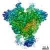
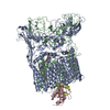
| Entry | Database: EMDB / ID: EMD-21850 | |||||||||
|---|---|---|---|---|---|---|---|---|---|---|
| Title | F. tularensis RNAPs70-iglA DNA complex | |||||||||
 Map data Map data | F. tularensis RNAPs70-iglA sharpened map | |||||||||
 Sample Sample |
| |||||||||
 Keywords Keywords | RNA polymerase complex / TRANSCRIPTION | |||||||||
| Function / homology |  Function and homology information Function and homology informationDNA-directed RNA polymerase complex / ribonucleoside binding / DNA-directed RNA polymerase / DNA-directed RNA polymerase activity / protein dimerization activity / magnesium ion binding / DNA-templated transcription / DNA binding / zinc ion binding / cytoplasm Similarity search - Function | |||||||||
| Biological species |  Francisella tularensis subsp. holarctica LVS (bacteria) / Francisella tularensis subsp. holarctica (strain LVS) (bacteria) Francisella tularensis subsp. holarctica LVS (bacteria) / Francisella tularensis subsp. holarctica (strain LVS) (bacteria) | |||||||||
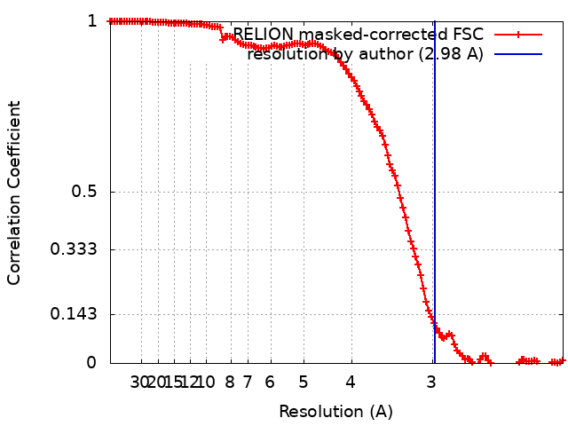
| Method | single particle reconstruction / cryo EM / Resolution: 2.98 Å | |||||||||
 Authors Authors | Travis BA / Brennan RG | |||||||||
| Funding support |  United States, 1 items United States, 1 items
| |||||||||
 Citation Citation | Journal: Mol Cell / Year: 2021 Title: Structural Basis for Virulence Activation of Francisella tularensis. Authors: Brady A Travis / Kathryn M Ramsey / Samantha M Prezioso / Thomas Tallo / Jamie M Wandzilak / Allen Hsu / Mario Borgnia / Alberto Bartesaghi / Simon L Dove / Richard G Brennan / Maria A Schumacher / Abstract: The bacterium Francisella tularensis (Ft) is one of the most infectious agents known. Ft virulence is controlled by a unique combination of transcription regulators: the MglA-SspA heterodimer, PigR, ...The bacterium Francisella tularensis (Ft) is one of the most infectious agents known. Ft virulence is controlled by a unique combination of transcription regulators: the MglA-SspA heterodimer, PigR, and the stress signal, ppGpp. MglA-SspA assembles with the σ-associated RNAP holoenzyme (RNAPσ), forming a virulence-specialized polymerase. These factors activate Francisella pathogenicity island (FPI) gene expression, which is required for virulence, but the mechanism is unknown. Here we report FtRNAPσ-promoter-DNA, FtRNAPσ-(MglA-SspA)-promoter DNA, and FtRNAPσ-(MglA-SspA)-ppGpp-PigR-promoter DNA cryo-EM structures. Structural and genetic analyses show MglA-SspA facilitates σ binding to DNA to regulate virulence and virulence-enhancing genes. Our Escherichia coli RNAPσhomodimeric EcSspA structure suggests this is a general SspA-transcription regulation mechanism. Strikingly, our FtRNAPσ-(MglA-SspA)-ppGpp-PigR-DNA structure reveals ppGpp binding to MglA-SspA tethers PigR to promoters. PigR in turn recruits FtRNAP αCTDs to DNA UP elements. Thus, these studies unveil a unique mechanism for Ft pathogenesis involving a virulence-specialized RNAP that employs two (MglA-SspA)-based strategies to activate virulence genes. | |||||||||
| History |
|
- Structure visualization
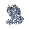
| Movie |
Movie viewer |
|---|---|
| Structure viewer | EM map: SurfViewMolmilJmol/JSmol |
| Supplemental images |

- Downloads & links
-EMDB archive
| Map data | emd_21850.map.gz | 155.9 MB | EMDB map data format | |
|---|---|---|---|---|
| Header (meta data) | emd-21850-v30.xmlemd-21850.xml | 22.6 KB 22.6 KB | Display Display | EMDB header |
| FSC (resolution estimation) | emd_21850_fsc.xml | 12.5 KB | Display | FSC data file |
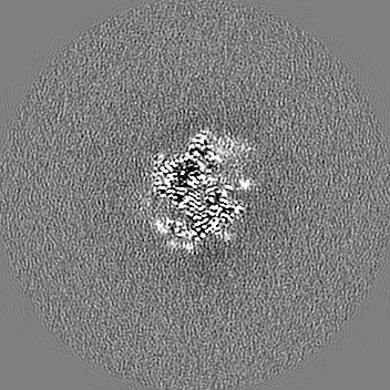
| Images | 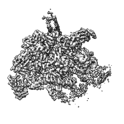 emd_21850.png emd_21850.png | 58.6 KB | ||
| Filedesc metadata | emd-21850.cif.gz | 7.9 KB | ||
| Others | emd_21850_additional_1.map.gz | 131.6 MB | ||
| Archive directory |  http://ftp.pdbj.org/pub/emdb/structures/EMD-21850ftp://ftp.pdbj.org/pub/emdb/structures/EMD-21850 http://ftp.pdbj.org/pub/emdb/structures/EMD-21850ftp://ftp.pdbj.org/pub/emdb/structures/EMD-21850 | HTTPS FTP |
-Related structure data
| Related structure data | 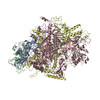 6wmpMC  6wegC  6wmrC  6wmtC  6wmuC M: atomic model generated by this map C: citing same article ( |
|---|---|
| Similar structure data |


-Links
| EMDB pages | EMDB (EBI/PDBe) / EMDataResource |
|---|---|
| Related items in Molecule of the Month |


-Map
| File | Download / File: emd_21850.map.gz / Format: CCP4 / Size: 166.4 MB / Type: IMAGE STORED AS FLOATING POINT NUMBER (4 BYTES) | ||||||||||||||||||||||||||||||||||||||||||||||||||||||||||||||||||||
|---|---|---|---|---|---|---|---|---|---|---|---|---|---|---|---|---|---|---|---|---|---|---|---|---|---|---|---|---|---|---|---|---|---|---|---|---|---|---|---|---|---|---|---|---|---|---|---|---|---|---|---|---|---|---|---|---|---|---|---|---|---|---|---|---|---|---|---|---|---|
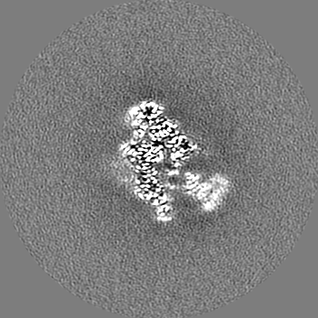
| Annotation | F. tularensis RNAPs70-iglA sharpened map | ||||||||||||||||||||||||||||||||||||||||||||||||||||||||||||||||||||
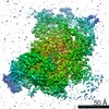
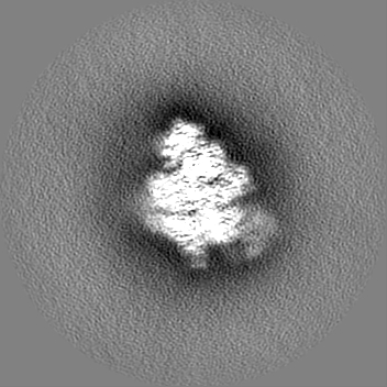
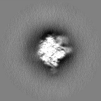
| Projections & slices | Image control
Images are generated by Spider. | ||||||||||||||||||||||||||||||||||||||||||||||||||||||||||||||||||||
| Voxel size | X=Y=Z: 1.07 Å | ||||||||||||||||||||||||||||||||||||||||||||||||||||||||||||||||||||
| Density |
| ||||||||||||||||||||||||||||||||||||||||||||||||||||||||||||||||||||
| Symmetry | Space group: 1 | ||||||||||||||||||||||||||||||||||||||||||||||||||||||||||||||||||||
| Details | EMDB XML:
CCP4 map header:
| ||||||||||||||||||||||||||||||||||||||||||||||||||||||||||||||||||||
 Z (Sec.)
 Y (Row.)
 X (Col.)


-Supplemental data
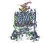
-Additional map: F. tularensis RNAPs70-iglA unsharpened map
| File | emd_21850_additional_1.map | ||||||||||||
|---|---|---|---|---|---|---|---|---|---|---|---|---|---|
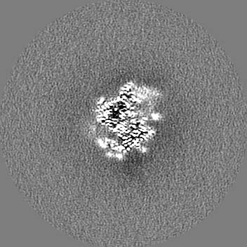
| Annotation | F. tularensis RNAPs70-iglA unsharpened map | ||||||||||||
| Projections & Slices |
| ||||||||||||
| Density Histograms |

- Sample components
+Entire : Francisella RNAPs70-iglA complex
+Supramolecule #1: Francisella RNAPs70-iglA complex
+Macromolecule #1: DNA-directed RNA polymerase subunit alpha 1
+Macromolecule #2: DNA-directed RNA polymerase subunit alpha 2
+Macromolecule #3: DNA-directed RNA polymerase subunit beta
+Macromolecule #4: DNA-directed RNA polymerase subunit beta'
+Macromolecule #5: DNA-directed RNA polymerase subunit omega
+Macromolecule #6: DNA NT-strand
+Macromolecule #7: DNA T-strand
+Macromolecule #8: RNA
+Macromolecule #9: MAGNESIUM ION
+Macromolecule #10: ZINC ION
-Experimental details
-Structure determination
| Method | cryo EM |
|---|---|
 Processing Processing | single particle reconstruction |
| Aggregation state | particle |
-Sample preparation
| Concentration | 0.4 mg/mL |
|---|---|
| Buffer | pH: 8 |
| Vitrification | Cryogen name: ETHANE |
- Electron microscopy
| Microscope | FEI TITAN KRIOS |
|---|---|
| Image recording | Film or detector model: GATAN K3 (6k x 4k) / Detector mode: COUNTING / Average electron dose: 60.0 e/Å2 |
| Electron beam | Acceleration voltage: 300 kV / Electron source:  FIELD EMISSION GUN FIELD EMISSION GUN |
| Electron optics | Illumination mode: OTHER / Imaging mode: BRIGHT FIELD / Cs: 2.7 mm |
| Experimental equipment |  Model: Titan Krios / Image courtesy: FEI Company |
+Image processing
-Atomic model buiding 1
| Refinement | Space: REAL |
|---|---|
| Output model | PDB-6wmp: |
