 Movie
Movie Controller

[English] 日本語
 Yorodumi
- EMDB-2160: Electron cryo-microscopy of the empty (DNA-lacking) head of the y... -
+ Open data

- Basic information
| Entry | Database: EMDB / ID: EMD-2160 | |||||||||
|---|---|---|---|---|---|---|---|---|---|---|
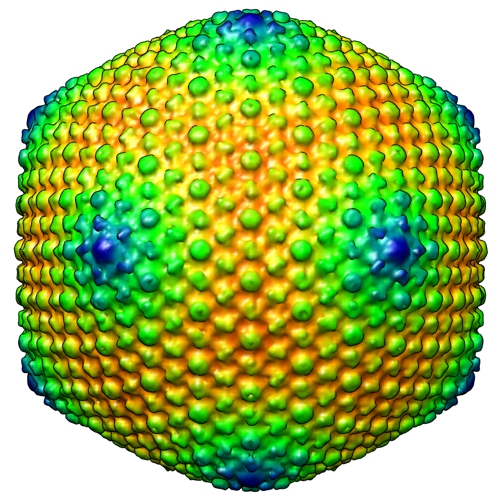
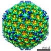
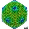
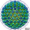
| Title | Electron cryo-microscopy of the empty (DNA-lacking) head of the yersiniophage phiR1-37 | |||||||||
 Map data Map data | Three-dimensional reconstruction of the empty (DNA-lacking) yersiniophage phiR1-37 | |||||||||
 Sample Sample |
| |||||||||
 Keywords Keywords | virus / Yersinia | |||||||||
| Biological species |  Yersinia phage phiR1-37 (virus) Yersinia phage phiR1-37 (virus) | |||||||||
| Method | single particle reconstruction / cryo EM / Resolution: 23.4 Å | |||||||||
 Authors Authors | Skurnik M / Hyytiainen H / Happonen LJ / Kiljunen S / Datta N / Mattinen L / Williamson K / Kristo P / Szeliga M / Kalin-Manttari L ...Skurnik M / Hyytiainen H / Happonen LJ / Kiljunen S / Datta N / Mattinen L / Williamson K / Kristo P / Szeliga M / Kalin-Manttari L / Ahola-Iivarinen E / Kalkkinen N / Butcher SJ | |||||||||
 Citation Citation | Journal: J Virol / Year: 2012 Title: Characterization of the genome, proteome, and structure of yersiniophage ϕR1-37. Authors: Mikael Skurnik / Heidi J Hyytiäinen / Lotta J Happonen / Saija Kiljunen / Neeta Datta / Laura Mattinen / Kirsty Williamson / Paula Kristo / Magdalena Szeliga / Laura Kalin-Mänttäri / ...Authors: Mikael Skurnik / Heidi J Hyytiäinen / Lotta J Happonen / Saija Kiljunen / Neeta Datta / Laura Mattinen / Kirsty Williamson / Paula Kristo / Magdalena Szeliga / Laura Kalin-Mänttäri / Elina Ahola-Iivarinen / Nisse Kalkkinen / Sarah J Butcher /  Abstract: The bacteriophage vB_YecM-ϕR1-37 (ϕR1-37) is a lytic yersiniophage that can propagate naturally in different Yersinia species carrying the correct lipopolysaccharide receptor. This large-tailed ...The bacteriophage vB_YecM-ϕR1-37 (ϕR1-37) is a lytic yersiniophage that can propagate naturally in different Yersinia species carrying the correct lipopolysaccharide receptor. This large-tailed phage has deoxyuridine (dU) instead of thymidine in its DNA. In this study, we determined the genomic sequence of phage ϕR1-37, mapped parts of the phage transcriptome, characterized the phage particle proteome, and characterized the virion structure by cryo-electron microscopy and image reconstruction. The 262,391-bp genome of ϕR1-37 is one of the largest sequenced phage genomes, and it contains 367 putative open reading frames (ORFs) and 5 tRNA genes. Mass-spectrometric analysis identified 69 phage particle structural proteins with the genes scattered throughout the genome. A total of 269 of the ORFs (73%) lack homologues in sequence databases. Based on terminator and promoter sequences identified from the intergenic regions, the phage genome was predicted to consist of 40 to 60 transcriptional units. Image reconstruction revealed that the ϕR1-37 capsid consists of hexameric capsomers arranged on a T=27 lattice similar to the bacteriophage ϕKZ. The tail of ϕR1-37 has a contractile sheath. We conclude that phage ϕR1-37 is a representative of a novel phage type that carries the dU-containing genome in a ϕKZ-like head. | |||||||||
| History |
|
- Structure visualization
| Movie |
 Movie viewer Movie viewer |
|---|---|
| Structure viewer | EM map: SurfViewMolmilJmol/JSmol |
| Supplemental images |
- Downloads & links
-EMDB archive
| Map data | emd_2160.map.gz | 57.4 MB | EMDB map data format | |
|---|---|---|---|---|
| Header (meta data) | emd-2160-v30.xmlemd-2160.xml | 8.7 KB 8.7 KB | Display Display | EMDB header |
| Images |  EMDB-2160.jpg EMDB-2160.jpg | 252.9 KB | ||
| Archive directory |  http://ftp.pdbj.org/pub/emdb/structures/EMD-2160ftp://ftp.pdbj.org/pub/emdb/structures/EMD-2160 http://ftp.pdbj.org/pub/emdb/structures/EMD-2160ftp://ftp.pdbj.org/pub/emdb/structures/EMD-2160 | HTTPS FTP |
-Related structure data

-Links
| EMDB pages | EMDB (EBI/PDBe) / EMDataResource |
|---|
-Map
| File | Download / File: emd_2160.map.gz / Format: CCP4 / Size: 120.1 MB / Type: IMAGE STORED AS SIGNED INTEGER (2 BYTES) | ||||||||||||||||||||||||||||||||||||||||||||||||||||||||||||||||||||
|---|---|---|---|---|---|---|---|---|---|---|---|---|---|---|---|---|---|---|---|---|---|---|---|---|---|---|---|---|---|---|---|---|---|---|---|---|---|---|---|---|---|---|---|---|---|---|---|---|---|---|---|---|---|---|---|---|---|---|---|---|---|---|---|---|---|---|---|---|---|
| Annotation | Three-dimensional reconstruction of the empty (DNA-lacking) yersiniophage phiR1-37 | ||||||||||||||||||||||||||||||||||||||||||||||||||||||||||||||||||||
| Projections & slices | Image control
Images are generated by Spider. | ||||||||||||||||||||||||||||||||||||||||||||||||||||||||||||||||||||
| Voxel size | X=Y=Z: 4.516 Å | ||||||||||||||||||||||||||||||||||||||||||||||||||||||||||||||||||||
| Density |
| ||||||||||||||||||||||||||||||||||||||||||||||||||||||||||||||||||||
| Symmetry | Space group: 1 | ||||||||||||||||||||||||||||||||||||||||||||||||||||||||||||||||||||
| Details | EMDB XML:
CCP4 map header:
| ||||||||||||||||||||||||||||||||||||||||||||||||||||||||||||||||||||
 Z (Sec.)
 Y (Row.)
 X (Col.)


-Supplemental data
- Sample components
-Entire : Electron cryo-microscopy of the empty (DNA-lacking) head of the y...
| Entire | Name: Electron cryo-microscopy of the empty (DNA-lacking) head of the yersiniophage phiR1-37 |
|---|---|
| Components |
|
-Supramolecule #1000: Electron cryo-microscopy of the empty (DNA-lacking) head of the y...
| Supramolecule | Name: Electron cryo-microscopy of the empty (DNA-lacking) head of the yersiniophage phiR1-37 type: sample / ID: 1000 / Number unique components: 1 |
|---|
-Supramolecule #1: Yersinia phage phiR1-37
| Supramolecule | Name: Yersinia phage phiR1-37 / type: virus / ID: 1 / NCBI-ID: 331278 / Sci species name: Yersinia phage phiR1-37 / Virus type: VIRION / Virus isolate: STRAIN / Virus enveloped: No / Virus empty: Yes |
|---|---|
| Host (natural) | Organism:  Yersinia enterocolitica (bacteria) / synonym: BACTERIA(EUBACTERIA) Yersinia enterocolitica (bacteria) / synonym: BACTERIA(EUBACTERIA) |
| Virus shell | Shell ID: 1 / Diameter: 1380 Å / T number (triangulation number): 27 |
-Experimental details
-Structure determination
| Method | cryo EM |
|---|---|
 Processing Processing | single particle reconstruction |
| Aggregation state | particle |
-Sample preparation
| Buffer | Details: 50 mM Tris pH 7.8, 10 mM MgSO4 |
|---|---|
| Vitrification | Cryogen name: ETHANE / Instrument: HOMEMADE PLUNGER |
- Electron microscopy
| Microscope | FEI TECNAI F20 |
|---|---|
| Details | Low dose conditions |
| Date | Sep 7, 2009 |
| Image recording | Category: FILM / Film or detector model: KODAK SO-163 FILM / Digitization - Scanner: ZEISS SCAI / Number real images: 322 / Average electron dose: 15 e/Å2 |
| Electron beam | Acceleration voltage: 200 kV / Electron source:  FIELD EMISSION GUN FIELD EMISSION GUN |
| Electron optics | Illumination mode: FLOOD BEAM / Imaging mode: BRIGHT FIELD / Cs: 2.0 mm / Nominal defocus max: 5.15 µm / Nominal defocus min: 0.7 µm / Nominal magnification: 62000 |
| Sample stage | Specimen holder: Side entry liquid nitrogen-cooled cryo specimen holder Specimen holder model: GATAN LIQUID NITROGEN |
| Experimental equipment |  Model: Tecnai F20 / Image courtesy: FEI Company |
-Image processing
| CTF correction | Details: Each micrograph |
|---|---|
| Final reconstruction | Applied symmetry - Point group: I (icosahedral) / Resolution.type: BY AUTHOR / Resolution: 23.4 Å / Resolution method: FSC 0.5 CUT-OFF / Software - Name: AUTO3DEM / Number images used: 1022 |
