Herpesvirus large tegument protein deneddylase / Herpesvirus UL36 tegument protein / Herpesvirus UL35 / Herpesvirus UL35 family / Herpesvirus capsid vertex component 1 / Herpesvirus UL17 protein / Large tegument protein deneddylase / Herpesvirus tegument ubiquitin-specific protease (htUSP) domain profile. / Herpesvirus large tegument protein, USP domain / Herpesvirus tegument protein, N-terminal conserved region ...Herpesvirus large tegument protein deneddylase / Herpesvirus UL36 tegument protein / Herpesvirus UL35 / Herpesvirus UL35 family / Herpesvirus capsid vertex component 1 / Herpesvirus UL17 protein / Large tegument protein deneddylase / Herpesvirus tegument ubiquitin-specific protease (htUSP) domain profile. / Herpesvirus large tegument protein, USP domain / Herpesvirus tegument protein, N-terminal conserved region / Herpesvirus UL25 / Herpesvirus UL25 family / Herpesvirus capsid shell protein 1 / Herpesvirus capsid shell protein VP19C / Herpesvirus capsid protein 2 / Herpesvirus VP23 like capsid protein / Herpesvirus major capsid protein / Herpesvirus major capsid protein, upper domain superfamily / Herpes virus major capsid protein / Papain-like cysteine peptidase superfamily Similarity search - Domain/homology
Capsid triplex subunit 1 / UL17 / Capsid triplex subunit 2 / Major capsid protein / DNA packaging tegument protein UL25 / Large tegument protein / Small capsomere-interacting protein Similarity search - Component
Biological species
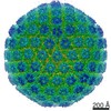
Herpes simplex virus (type 1 / strain F)
Method
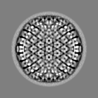
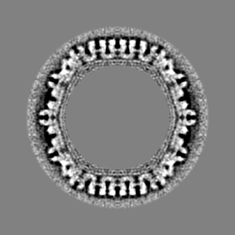
single particle reconstruction / cryo EM / Resolution: 35.0 Å
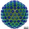
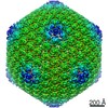
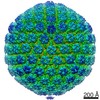
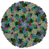
Journal: mBio / Year: 2017 Title: The Primary Enveloped Virion of Herpes Simplex Virus 1: Its Role in Nuclear Egress. Authors: William W Newcomb / Juan Fontana / Dennis C Winkler / Naiqian Cheng / J Bernard Heymann / Alasdair C Steven / Abstract: Many viruses migrate between different cellular compartments for successive stages of assembly. The HSV-1 capsid assembles in the nucleus and then transfers into the cytoplasm. First, the capsid buds ...Many viruses migrate between different cellular compartments for successive stages of assembly. The HSV-1 capsid assembles in the nucleus and then transfers into the cytoplasm. First, the capsid buds through the inner nuclear membrane, becoming coated with nuclear egress complex (NEC) protein. This yields a primary enveloped virion (PEV) whose envelope fuses with the outer nuclear membrane, releasing the capsid into the cytoplasm. We investigated the associated molecular mechanisms by isolating PEVs from US3-null-infected cells and imaging them by cryo-electron microscopy and tomography. (pUS3 is a viral protein kinase in whose absence PEVs accumulate in the perinuclear space.) Unlike mature extracellular virions, PEVs have very few glycoprotein spikes. PEVs are ~20% smaller than mature virions, and the little space available between the capsid and the NEC layer suggests that most tegument proteins are acquired later in the egress pathway. Previous studies have proposed that NEC is organized as hexamers in honeycomb arrays in PEVs, but we find arrays of heptameric rings in extracts from US3-null-infected cells. In a PEV, NEC contacts the capsid predominantly via the pUL17/pUL25 complexes which are located close to the capsid vertices. Finally, the NEC layer dissociates from the capsid as it leaves the nucleus, possibly in response to pUS3-mediated phosphorylation. Overall, nuclear egress emerges as a process driven by a program of multiple weak interactions. On its maturation pathway, the newly formed HSV-1 nucleocapsid must traverse the nuclear envelope, while respecting the integrity of that barrier. Nucleocapsids (125 nm in diameter) are too large to pass through the nuclear pore complexes that conduct most nucleocytoplasmic traffic. It is now widely accepted that the process involves envelopment/de-envelopment of a key intermediate-the primary enveloped virion. In wild-type infections, PEVs are short-lived, which has impeded study. Using a mutant that accumulates PEVs in the perinuclear space, we were able to isolate PEVs in sufficient quantity for structural analysis by cryo-electron microscopy and tomography. The findings not only elucidate the maturation pathway of an important human pathogen but also have implications for cellular processes that involve the trafficking of large macromolecular complexes.
History
Deposition
Feb 19, 2017
-
Header (metadata) release
Mar 1, 2017
-
Map release
Jan 24, 2018
-
Update
Feb 14, 2018
-
Current status
Feb 14, 2018
Processing site: RCSB / Status: Released
-
Structure visualization
Movie
Surface view with section colored by density value
Name: Herpes simplex virus (type 1 / strain F) / type: virus / ID: 1 / Parent: 0 Details: US3 null mutant, a gift from Dr. Richard Roller, Dept. of Microbiology and Immunology, Carver College of Medicine, Univ. of Iowa NCBI-ID: 10304 / Sci species name: Herpes simplex virus (type 1 / strain F) / Virus type: VIRION / Virus isolate: OTHER / Virus enveloped: Yes / Virus empty: No
-
Experimental details
-
Structure determination
Method
cryo EM
Processing
single particle reconstruction
Aggregation state
particle
-
Sample preparation
Buffer
pH: 7.5 / Details: PBS
Vitrification
Cryogen name: ETHANE / Instrument: LEICA KF80
-
Electron microscopy
Microscope
FEI/PHILIPS CM200FEG
Image recording
Film or detector model: GATAN ULTRASCAN 1000 (2k x 2k) / Average electron dose: 15.0 e/Å2
Electron beam
Acceleration voltage: 120 kV / Electron source: FIELD EMISSION GUN
Electron optics
Illumination mode: FLOOD BEAM / Imaging mode: BRIGHT FIELD
+
Image processing
Details
Images (2048 by 2048 pixels) were recorded using a CCD camera (Gatan) at a magnification of ~25,000x (giving a pixel size of 5.7 A) and processed using the Bsoft package (Heymann and Belnap, 2007) as previously described (McHugh et al., 2014).
Particle selection
Number selected: 123 Details: PEV images of were picked manually, yielding a total of 123 particles.
Startup model
Type of model: OTHER Details: Origins and orientations were determined by projection matching, using as a starting model a map of B capsids obtained by in vitro maturation of purified procapsids (Aksyuk et al., 2015).
Final reconstruction
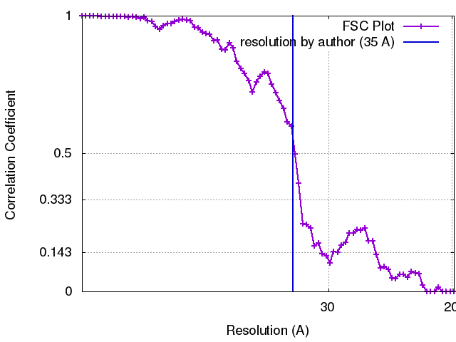
Applied symmetry - Point group: I (icosahedral) / Resolution.type: BY AUTHOR / Resolution: 35.0 Å / Resolution method: FSC 0.33 CUT-OFF Details: Reconstructions were calculated using breconstruct (from the Bsoft package), an algorithm that integrates images as central sections of Fourier space. An inverse Fourier transform was then ...Details: Reconstructions were calculated using breconstruct (from the Bsoft package), an algorithm that integrates images as central sections of Fourier space. An inverse Fourier transform was then calculated. Using an FSC cut-off of 0.3, the resolution of the reconstruction of C capsid-containing PEVs was estimated at ~3.5 nm for the entire particle and calculated (Cardone et al., 2013) at ~2 nm for the region around the capsid shell. Number images used: 123
Initial angle assignment
Type: RANDOM ASSIGNMENT
Final angle assignment
Type: PROJECTION MATCHING
FSC plot (resolution estimation)
+
About Yorodumi
-
News
-
Feb 9, 2022. New format data for meta-information of EMDB entries
New format data for meta-information of EMDB entries
Version 3 of the EMDB header file is now the official format.
The previous official version 1.9 will be removed from the archive.
In the structure databanks used in Yorodumi, some data are registered as the other names, "COVID-19 virus" and "2019-nCoV". Here are the details of the virus and the list of structure data.
Jan 31, 2019. EMDB accession codes are about to change! (news from PDBe EMDB page)
EMDB accession codes are about to change! (news from PDBe EMDB page)
The allocation of 4 digits for EMDB accession codes will soon come to an end. Whilst these codes will remain in use, new EMDB accession codes will include an additional digit and will expand incrementally as the available range of codes is exhausted. The current 4-digit format prefixed with “EMD-” (i.e. EMD-XXXX) will advance to a 5-digit format (i.e. EMD-XXXXX), and so on. It is currently estimated that the 4-digit codes will be depleted around Spring 2019, at which point the 5-digit format will come into force.
The EM Navigator/Yorodumi systems omit the EMD- prefix.
Related info.:Q: What is EMD? / ID/Accession-code notation in Yorodumi/EM Navigator
Yorodumi is a browser for structure data from EMDB, PDB, SASBDB, etc.
This page is also the successor to EM Navigator detail page, and also detail information page/front-end page for Omokage search.
The word "yorodu" (or yorozu) is an old Japanese word meaning "ten thousand". "mi" (miru) is to see.
Related info.:EMDB / PDB / SASBDB / Comparison of 3 databanks / Yorodumi Search / Aug 31, 2016. New EM Navigator & Yorodumi / Yorodumi Papers / Jmol/JSmol / Function and homology information / Changes in new EM Navigator and Yorodumi
 Movie
Movie Controller


 Open data
 Basic information
 Map data
 Sample
 Function and homology information
 Herpes simplex virus (type 1 / strain F)
 Authors
 Citation

 Structure visualization
 Downloads & links
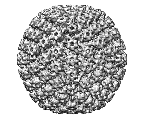 emd_8607.png
 http://ftp.pdbj.org/pub/emdb/structures/EMD-8607

 Z (Sec.)
 Y (Row.)
 X (Col.)


 Sample components
 Processing
 Electron microscopy
 FIELD EMISSION GUN