 Movie
Movie Controller


+ Open data

- Basic information
| Entry | Database: EMDB / ID: EMD-5696 | |||||||||
|---|---|---|---|---|---|---|---|---|---|---|
| Title | Electron cryo-microscopy of human cytomegalovirus virion | |||||||||
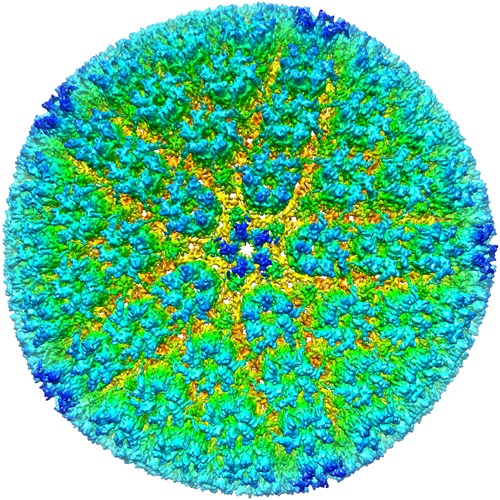
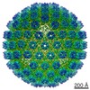
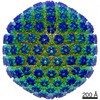
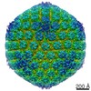
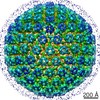
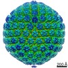
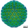
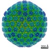
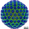
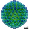
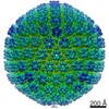
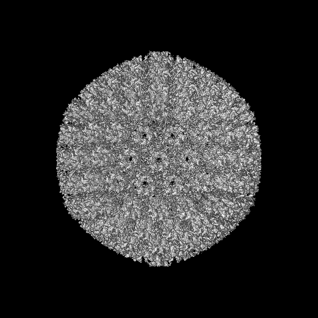
 Map data Map data | Reconstruction of human cytomegalovirus (HCMV) virion. Only the icosahedral capsid and orderly bound tegument proteins are visible in the density map. Other tegument proteins and the envelope are amorphous and thus not visible. | |||||||||
 Sample Sample |
| |||||||||
 Keywords Keywords | herpesvirus / betaherpesvirus / human cytomegalovirus | |||||||||
| Biological species |   Human herpesvirus 5 Human herpesvirus 5 | |||||||||
| Method | single particle reconstruction / cryo EM / Resolution: 8.3 Å | |||||||||
 Authors Authors | Dai XH / Yu XK / Gong H / Jiang XH / Abenes G / Liu HR / Shivakoti S / Britt W / Zhu H / Liu FY / Zhou ZH | |||||||||
 Citation Citation | Journal: PLoS Pathog / Year: 2013 Title: The smallest capsid protein mediates binding of the essential tegument protein pp150 to stabilize DNA-containing capsids in human cytomegalovirus. Authors: Xinghong Dai / Xuekui Yu / Hao Gong / Xiaohong Jiang / Gerrado Abenes / Hongrong Liu / Sakar Shivakoti / William J Britt / Hua Zhu / Fenyong Liu / Z Hong Zhou /  Abstract: Human cytomegalovirus (HCMV) is a ubiquitous herpesvirus that causes birth defects in newborns and life-threatening complications in immunocompromised individuals. Among all human herpesviruses, HCMV ...Human cytomegalovirus (HCMV) is a ubiquitous herpesvirus that causes birth defects in newborns and life-threatening complications in immunocompromised individuals. Among all human herpesviruses, HCMV contains a much larger dsDNA genome within a similarly-sized capsid compared to the others, and it was proposed to require pp150, a tegument protein only found in cytomegaloviruses, to stabilize its genome-containing capsid. However, little is known about how pp150 interacts with the underlying capsid. Moreover, the smallest capsid protein (SCP), while dispensable in herpes simplex virus type 1, was shown to play essential, yet undefined, role in HCMV infection. Here, by cryo electron microscopy (cryoEM), we determine three-dimensional structures of HCMV capsid (no pp150) and virion (with pp150) at sub-nanometer resolution. Comparison of these two structures reveals that each pp150 tegument density is composed of two helix bundles connected by a long central helix. Correlation between the resolved helices and sequence-based secondary structure prediction maps the tegument density to the N-terminal half of pp150. The structures also show that SCP mediates interactions between the capsid and pp150 at the upper helix bundle of pp150. Consistent with this structural observation, ribozyme inhibition of SCP expression in HCMV-infected cells impairs the formation of DNA-containing viral particles and reduces viral yield by 10,000 fold. By cryoEM reconstruction of the resulting "SCP-deficient" viral particles, we further demonstrate that SCP is required for pp150 functionally binding to the capsid. Together, our structural and biochemical results point to a mechanism whereby SCP recruits pp150 to stabilize genome-containing capsid for the production of infectious HCMV virion. | |||||||||
| History |
|
- Structure visualization
| Movie |
 Movie viewer Movie viewer |
|---|---|
| Structure viewer | EM map: SurfViewMolmilJmol/JSmol |
| Supplemental images |
- Downloads & links
-EMDB archive
| Map data | emd_5696.map.gz | 909.6 MB | EMDB map data format | |
|---|---|---|---|---|
| Header (meta data) | emd-5696-v30.xmlemd-5696.xml | 10.3 KB 10.3 KB | Display Display | EMDB header |
| Images | emd_5696.tif | 755.3 KB | ||
| Archive directory |  http://ftp.pdbj.org/pub/emdb/structures/EMD-5696ftp://ftp.pdbj.org/pub/emdb/structures/EMD-5696 http://ftp.pdbj.org/pub/emdb/structures/EMD-5696ftp://ftp.pdbj.org/pub/emdb/structures/EMD-5696 | HTTPS FTP |
-Related structure data

-Links
| EMDB pages | EMDB (EBI/PDBe) / EMDataResource |
|---|
-Map
| File | Download / File: emd_5696.map.gz / Format: CCP4 / Size: 976.6 MB / Type: IMAGE STORED AS FLOATING POINT NUMBER (4 BYTES) | ||||||||||||||||||||||||||||||||||||||||||||||||||||||||||||||||||||
|---|---|---|---|---|---|---|---|---|---|---|---|---|---|---|---|---|---|---|---|---|---|---|---|---|---|---|---|---|---|---|---|---|---|---|---|---|---|---|---|---|---|---|---|---|---|---|---|---|---|---|---|---|---|---|---|---|---|---|---|---|---|---|---|---|---|---|---|---|---|
| Annotation | Reconstruction of human cytomegalovirus (HCMV) virion. Only the icosahedral capsid and orderly bound tegument proteins are visible in the density map. Other tegument proteins and the envelope are amorphous and thus not visible. | ||||||||||||||||||||||||||||||||||||||||||||||||||||||||||||||||||||
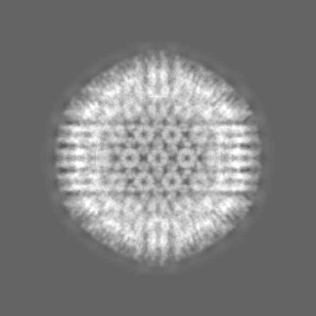
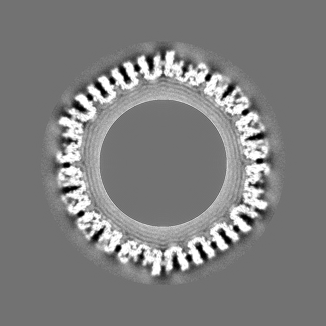
| Projections & slices | Image control
Images are generated by Spider. | ||||||||||||||||||||||||||||||||||||||||||||||||||||||||||||||||||||
| Voxel size | X=Y=Z: 3.076 Å | ||||||||||||||||||||||||||||||||||||||||||||||||||||||||||||||||||||
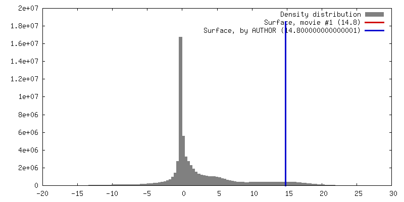
| Density |
| ||||||||||||||||||||||||||||||||||||||||||||||||||||||||||||||||||||
| Symmetry | Space group: 1 | ||||||||||||||||||||||||||||||||||||||||||||||||||||||||||||||||||||
| Details | EMDB XML:
CCP4 map header:
| ||||||||||||||||||||||||||||||||||||||||||||||||||||||||||||||||||||
 Z (Sec.)
 Y (Row.)
 X (Col.)


-Supplemental data
- Sample components
-Entire : Human cytomegalovirus (HCMV) virion
| Entire | Name: Human cytomegalovirus (HCMV) virion |
|---|---|
| Components |
|
-Supramolecule #1000: Human cytomegalovirus (HCMV) virion
| Supramolecule | Name: Human cytomegalovirus (HCMV) virion / type: sample / ID: 1000 / Number unique components: 1 |
|---|
-Supramolecule #1: Human herpesvirus 5
| Supramolecule | Name: Human herpesvirus 5 / type: virus / ID: 1 / Name.synonym: human cytomegalovirus / NCBI-ID: 10359 / Sci species name: Human herpesvirus 5 / Database: NCBI / Virus type: VIRION / Virus isolate: SPECIES / Virus enveloped: Yes / Virus empty: No / Syn species name: human cytomegalovirus |
|---|---|
| Host (natural) | Organism:  Homo sapiens (human) / synonym: VERTEBRATES Homo sapiens (human) / synonym: VERTEBRATES |
| Virus shell | Shell ID: 1 / Name: capsid / Diameter: 1350 Å / T number (triangulation number): 16 |
-Experimental details
-Structure determination
| Method | cryo EM |
|---|---|
 Processing Processing | single particle reconstruction |
| Aggregation state | particle |
-Sample preparation
| Buffer | pH: 7.4 Details: Phosphate buffered saline (pH=7.4): 144 mg/L KH2PO4, 795 mg/L Na2HPO4-7H2O, 0.9% (w/w) NaCl |
|---|---|
| Grid | Details: Quantifoil R2/1 Cu-200mesh grids |
| Vitrification | Cryogen name: ETHANE / Chamber humidity: 100 % / Instrument: FEI VITROBOT MARK IV / Method: Blot force = 0, blot time = 20s |
- Electron microscopy
| Microscope | FEI TITAN KRIOS |
|---|---|
| Temperature | Average: 80 K |
| Alignment procedure | Legacy - Astigmatism: Objective lens astigmatism was corrected at 135,000 times magnification. Astigmatism correction was also carried out for each particle during the data processing. |
| Details | Parallel beam illumination |
| Date | Oct 1, 2010 |
| Image recording | Category: CCD / Film or detector model: GATAN ULTRASCAN 4000 (4k x 4k) / Digitization - Sampling interval: 15 µm / Number real images: 8000 / Average electron dose: 25 e/Å2 |
| Electron beam | Acceleration voltage: 300 kV / Electron source:  FIELD EMISSION GUN FIELD EMISSION GUN |
| Electron optics | Illumination mode: FLOOD BEAM / Imaging mode: BRIGHT FIELD / Cs: 2.7 mm / Nominal defocus max: 3.0 µm / Nominal defocus min: 1.0 µm / Nominal magnification: 59000 |
| Sample stage | Specimen holder model: FEI TITAN KRIOS AUTOGRID HOLDER |
| Experimental equipment |  Model: Titan Krios / Image courtesy: FEI Company |
-Image processing
| CTF correction | Details: Each particle |
|---|---|
| Final reconstruction | Algorithm: OTHER / Resolution.type: BY AUTHOR / Resolution: 8.3 Å / Resolution method: FSC 0.143 CUT-OFF / Software - Name: IMIRS / Number images used: 1 |
