 Movie
Movie Controller

[English] 日本語
 Yorodumi
- EMDB-21538: Structure of TRPA1 (ligand-free and calcium-free), PMAL-C8, class 2 -
+ Open data

- Basic information
| Entry | Database: EMDB / ID: EMD-21538 | |||||||||
|---|---|---|---|---|---|---|---|---|---|---|
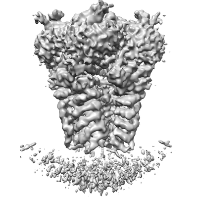
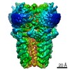
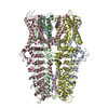
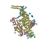
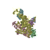
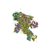
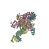
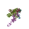
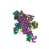
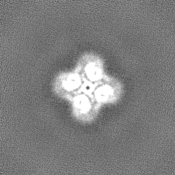
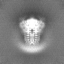
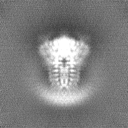
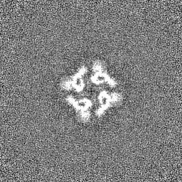
| Title | Structure of TRPA1 (ligand-free and calcium-free), PMAL-C8, class 2 | |||||||||
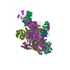
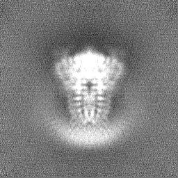
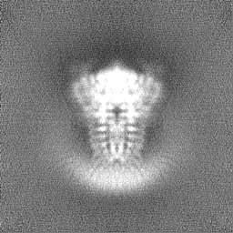
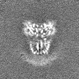
 Map data Map data | TRPA1 (apo) in PMAL-C8, class 2 (open) | |||||||||
 Sample Sample |
| |||||||||
| Biological species |  Homo sapiens (human) Homo sapiens (human) | |||||||||
| Method | single particle reconstruction / cryo EM / Resolution: 4.3 Å | |||||||||
 Authors Authors | Zhao J / Lin King JV / Paulsen CE / Cheng Y / Julius D | |||||||||
| Funding support |  United States, 2 items United States, 2 items
| |||||||||
 Citation Citation | Journal: Nature / Year: 2020 Title: Irritant-evoked activation and calcium modulation of the TRPA1 receptor. Authors: Jianhua Zhao / John V Lin King / Candice E Paulsen / Yifan Cheng / David Julius / Abstract: The transient receptor potential ion channel TRPA1 is expressed by primary afferent nerve fibres, in which it functions as a low-threshold sensor for structurally diverse electrophilic irritants, ...The transient receptor potential ion channel TRPA1 is expressed by primary afferent nerve fibres, in which it functions as a low-threshold sensor for structurally diverse electrophilic irritants, including small volatile environmental toxicants and endogenous algogenic lipids. TRPA1 is also a 'receptor-operated' channel whose activation downstream of metabotropic receptors elicits inflammatory pain or itch, making it an attractive target for novel analgesic therapies. However, the mechanisms by which TRPA1 recognizes and responds to electrophiles or cytoplasmic second messengers remain unknown. Here we use strutural studies and electrophysiology to show that electrophiles act through a two-step process in which modification of a highly reactive cysteine residue (C621) promotes reorientation of a cytoplasmic loop to enhance nucleophilicity and modification of a nearby cysteine (C665), thereby stabilizing the loop in an activating configuration. These actions modulate two restrictions controlling ion permeation, including widening of the selectivity filter to enhance calcium permeability and opening of a canonical gate at the cytoplasmic end of the pore. We propose a model to explain functional coupling between electrophile action and these control points. We also characterize a calcium-binding pocket that is highly conserved across TRP channel subtypes and accounts for all aspects of calcium-dependent TRPA1 regulation, including potentiation, desensitization and activation by metabotropic receptors. These findings provide a structural framework for understanding how a broad-spectrum irritant receptor is controlled by endogenous and exogenous agents that elicit or exacerbate pain and itch. | |||||||||
| History |
|
- Structure visualization
| Movie |
 Movie viewer Movie viewer |
|---|---|
| Structure viewer | EM map: SurfViewMolmilJmol/JSmol |
| Supplemental images |
- Downloads & links
-EMDB archive
| Map data | emd_21538.map.gz | 57.7 MB | EMDB map data format | |
|---|---|---|---|---|
| Header (meta data) | emd-21538-v30.xmlemd-21538.xml | 16.1 KB 16.1 KB | Display Display | EMDB header |
| FSC (resolution estimation) | emd_21538_fsc.xml | 9.1 KB | Display | FSC data file |
| Images |  emd_21538.png emd_21538.png | 73.4 KB | ||
| Masks | emd_21538_msk_1.map | 64 MB | Mask map | |
| Others | emd_21538_half_map_1.map.gzemd_21538_half_map_2.map.gz | 58.9 MB 58.9 MB | ||
| Archive directory |  http://ftp.pdbj.org/pub/emdb/structures/EMD-21538ftp://ftp.pdbj.org/pub/emdb/structures/EMD-21538 http://ftp.pdbj.org/pub/emdb/structures/EMD-21538ftp://ftp.pdbj.org/pub/emdb/structures/EMD-21538 | HTTPS FTP |
-Related structure data


-Links
| EMDB pages | EMDB (EBI/PDBe) / EMDataResource |
|---|
-Map
| File | Download / File: emd_21538.map.gz / Format: CCP4 / Size: 64 MB / Type: IMAGE STORED AS FLOATING POINT NUMBER (4 BYTES) | ||||||||||||||||||||||||||||||||||||||||||||||||||||||||||||||||||||
|---|---|---|---|---|---|---|---|---|---|---|---|---|---|---|---|---|---|---|---|---|---|---|---|---|---|---|---|---|---|---|---|---|---|---|---|---|---|---|---|---|---|---|---|---|---|---|---|---|---|---|---|---|---|---|---|---|---|---|---|---|---|---|---|---|---|---|---|---|---|
| Annotation | TRPA1 (apo) in PMAL-C8, class 2 (open) | ||||||||||||||||||||||||||||||||||||||||||||||||||||||||||||||||||||
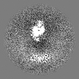
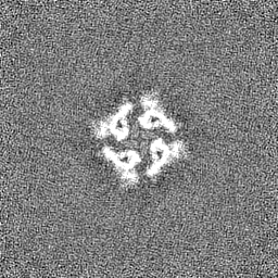
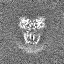
| Projections & slices | Image control
Images are generated by Spider. | ||||||||||||||||||||||||||||||||||||||||||||||||||||||||||||||||||||
| Voxel size | X=Y=Z: 1.2156 Å | ||||||||||||||||||||||||||||||||||||||||||||||||||||||||||||||||||||
| Density |
| ||||||||||||||||||||||||||||||||||||||||||||||||||||||||||||||||||||
| Symmetry | Space group: 1 | ||||||||||||||||||||||||||||||||||||||||||||||||||||||||||||||||||||
| Details | EMDB XML:
CCP4 map header:
| ||||||||||||||||||||||||||||||||||||||||||||||||||||||||||||||||||||
 Z (Sec.)
 Y (Row.)
 X (Col.)


-Supplemental data
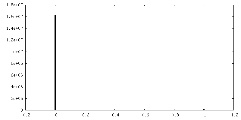
-Mask #1
| File | emd_21538_msk_1.map | ||||||||||||
|---|---|---|---|---|---|---|---|---|---|---|---|---|---|
| Projections & Slices |
| ||||||||||||
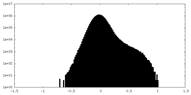
| Density Histograms |


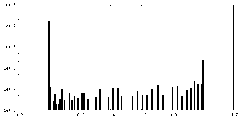
-Half map: #1
| File | emd_21538_half_map_1.map | ||||||||||||
|---|---|---|---|---|---|---|---|---|---|---|---|---|---|
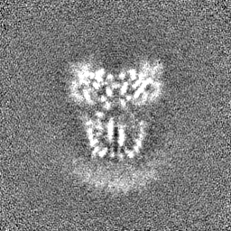
| Projections & Slices |
| ||||||||||||
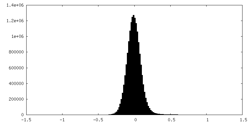
| Density Histograms |
-Half map: #2
| File | emd_21538_half_map_2.map | ||||||||||||
|---|---|---|---|---|---|---|---|---|---|---|---|---|---|
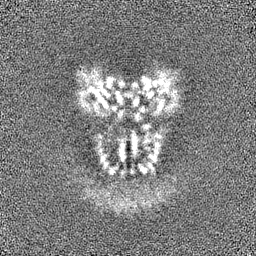
| Projections & Slices |
| ||||||||||||
| Density Histograms |


- Sample components
-Entire : TRPA1 (ligand-free and calcium free), solubilized in PMAL-C8, class 2
| Entire | Name: TRPA1 (ligand-free and calcium free), solubilized in PMAL-C8, class 2 |
|---|---|
| Components |
|
-Supramolecule #1: TRPA1 (ligand-free and calcium free), solubilized in PMAL-C8, class 2
| Supramolecule | Name: TRPA1 (ligand-free and calcium free), solubilized in PMAL-C8, class 2 type: complex / ID: 1 / Parent: 0 / Macromolecule list: all |
|---|---|
| Source (natural) | Organism: Homo sapiens (human) |
| Recombinant expression | Organism: Homo sapiens (human) / Recombinant cell: HEK293F |
-Macromolecule #1: Transient receptor potential A1 (TRPA1)
| Macromolecule | Name: Transient receptor potential A1 (TRPA1) / type: protein_or_peptide / ID: 1 / Enantiomer: LEVO |
|---|---|
| Source (natural) | Organism: Homo sapiens (human) |
| Recombinant expression | Organism: Homo sapiens (human) |
| Sequence | String: MGSSHHHHHH HHGSSMKIEE GKLVIWINGD KGYNGLAEVG KKFEKDTGIK VTVEHPDKLE EKFPQVAATG DGPDIIFWAH DRFGGYAQSG LLAEITPDKA FQDKLYPFTW DAVRYNGKLI AYPIAVEALS LIYNKDLLPN PPKTWEEIPA LDKELKAKGK SALMFNLQEP ...String: MGSSHHHHHH HHGSSMKIEE GKLVIWINGD KGYNGLAEVG KKFEKDTGIK VTVEHPDKLE EKFPQVAATG DGPDIIFWAH DRFGGYAQSG LLAEITPDKA FQDKLYPFTW DAVRYNGKLI AYPIAVEALS LIYNKDLLPN PPKTWEEIPA LDKELKAKGK SALMFNLQEP YFTWPLIAAD GGYAFKYENG KYDIKDVGVD NAGAKAGLTF LVDLIKNKHM NADTDYSIAE AAFNKGETAM TINGPWAWSN IDTSKVNYGV TVLPTFKGQP SKPFVGVLSA GINAASPNKE LAKEFLENYL LTDEGLEAVN KDKPLGAVAL KSYEEELAKD PRIAATMENA QKGEIMPNIP QMSAFWYAVR TAVINAASGR QTVDEALKDA QTNSSSNNNN NNNNNNLGIE ENLYFQGAMG SMKRSLRKMW RPGEKKEPQG VVYEDVPDDT EDFKESLKVV FEGSAYGLQN FNKQKKLKRC DDMDTFFLHY AAAEGQIELM EKITRDSSLE VLHEMDDYGN TPLHCAVEKN QIESVKFLLS RGANPNLRNF NMMAPLHIAV QGMNNEVMKV LLEHRTIDVN LEGENGNTAV IIACTTNNSE ALQILLKKGA KPCKSNKWGC FPIHQAAFSG SKECMEIILR FGEEHGYSRQ LHINFMNNGK ATPLHLAVQN GDLEMIKMCL DNGAQIDPVE KGRCTAIHFA ATQGATEIVK LMISSYSGSV DIVNTTDGCH ETMLHRASLF DHHELADYLI SVGADINKID SEGRSPLILA TASASWNIVN LLLSKGAQVD IKDNFGRNFL HLTVQQPYGL KNLRPEFMQM QQIKELVMDE DNDGCTPLHY ACRQGGPGSV NNLLGFNVSI HSKSKDKKSP LHFAASYGRI NTCQRLLQDI SDTRLLNEGD LHGMTPLHLA AKNGHDKVVQ LLLKKGALFL SDHNGWTALH HASMGGYTQT MKVILDTNLK CTDRLDEDGN TALHFAAREG HAKAVALLLS HNADIVLNKQ QASFLHLALH NKRKEVVLTI IRSKRWDECL KIFSHNSPGN KCPITEMIEY LPECMKVLLD FCMLHSTEDK SCRDYYIEYN FKYLQCPLEF TKKTPTQDVI YEPLTALNAM VQNNRIELLN HPVCKEYLLM KWLAYGFRAH MMNLGSYCLG LIPMTILVVN IKPGMAFNST GIINETSDHS EILDTTNSYL IKTCMILVFL SSIFGYCKEA GQIFQQKRNY FMDISNVLEW IIYTTGIIFV LPLFVEIPAH LQWQCGAIAV YFYWMNFLLY LQRFENCGIF IVMLEVILKT LLRSTVVFIF LLLAFGLSFY ILLNLQDPFS SPLLSIIQTF SMMLGDINYR ESFLEPYLRN ELAHPVLSFA QLVSFTIFVP IVLMNLLIGL AVGDIADVQK HASLKRIAMQ VELHTSLEKK LPLWFLRKVD QKSTIVYPNK PRSGGMLFHI FCFLFCTGEI RQEIPNADKS LEMEILKQKY RLKDLTFLLE KQHELIKLII QKMEIISETE DDDSHCSFQD RFKKEQMEQR NSRWNTVLRA VKAKTHHLEP |
-Experimental details
-Structure determination
| Method | cryo EM |
|---|---|
 Processing Processing | single particle reconstruction |
| Aggregation state | particle |
-Sample preparation
| Buffer | pH: 8 |
|---|---|
| Grid | Details: unspecified |
| Vitrification | Cryogen name: ETHANE |
- Electron microscopy
| Microscope | FEI POLARA 300 |
|---|---|
| Image recording | Film or detector model: GATAN K2 SUMMIT (4k x 4k) / Detector mode: SUPER-RESOLUTION / Average electron dose: 40.0 e/Å2 |
| Electron beam | Acceleration voltage: 300 kV / Electron source:  FIELD EMISSION GUN FIELD EMISSION GUN |
| Electron optics | Illumination mode: FLOOD BEAM / Imaging mode: BRIGHT FIELD |
| Experimental equipment |  Model: Tecnai Polara / Image courtesy: FEI Company |
-Image processing
| Final reconstruction | Applied symmetry - Point group: C4 (4 fold cyclic) / Resolution.type: BY AUTHOR / Resolution: 4.3 Å / Resolution method: FSC 0.143 CUT-OFF / Number images used: 28558 |
|---|---|
| Initial angle assignment | Type: NOT APPLICABLE |
| Final angle assignment | Type: PROJECTION MATCHING |
| FSC plot (resolution estimation) | 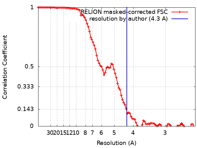 |
