 Movie
Movie Controller

[English] 日本語
 Yorodumi
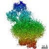
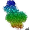
- EMDB-21384: Map of an acid-sensing ion channel in a desensitized state after ... -
+ Open data

- Basic information
| Entry | Database: EMDB / ID: EMD-21384 | |||||||||||||||
|---|---|---|---|---|---|---|---|---|---|---|---|---|---|---|---|---|
| Title | Map of an acid-sensing ion channel in a desensitized state after photo-uncaging protons. | |||||||||||||||
 Map data Map data | Full map of an acid-sensing ion channel in a desensitized state after photo-uncaging protons. | |||||||||||||||
 Sample Sample |
| |||||||||||||||
| Biological species |  | |||||||||||||||
| Method | single particle reconstruction / cryo EM / Resolution: 3.5 Å | |||||||||||||||
 Authors Authors | Yoder N / Jalali-Yazdi F / Gouaux E | |||||||||||||||
| Funding support |  United States, 4 items United States, 4 items
| |||||||||||||||
 Citation Citation | Journal: J Struct Biol / Year: 2020 Title: Light-coupled cryo-plunger for time-resolved cryo-EM. Authors: Nate Yoder / Farzad Jalali-Yazdi / Sigrid Noreng / Alexandra Houser / Isabelle Baconguis / Eric Gouaux / Abstract: Proteins are dynamic molecules that can undergo rapid conformational rearrangements in response to stimuli. These structural changes are often critical to protein function, and thus elucidating time- ...Proteins are dynamic molecules that can undergo rapid conformational rearrangements in response to stimuli. These structural changes are often critical to protein function, and thus elucidating time-dependent conformational landscapes has been a long-standing goal of structural biology. To harness the power of single particle cryo-EM methods to enable 'time-resolved' structure determination, we have developed a light-coupled cryo-plunger that pairs flash-photolysis of caged ligands with rapid sample vitrification. The 'flash-plunger' consists of a high-power ultraviolet LED coupled with focusing optics and a motorized linear actuator, enabling the user to immobilize protein targets in vitreous ice within a programmable time window - as short as tens of milliseconds - after stimulus delivery. The flash-plunger is a simple, inexpensive and flexible tool to explore short-lived conformational states previously unobtainable by conventional sample preparation methods. | |||||||||||||||
| History |
|
- Structure visualization
| Movie |
 Movie viewer Movie viewer |
|---|---|
| Structure viewer | EM map: SurfViewMolmilJmol/JSmol |
| Supplemental images |

- Downloads & links
-EMDB archive
| Map data | emd_21384.map.gz | 230.2 MB | EMDB map data format | |
|---|---|---|---|---|
| Header (meta data) | emd-21384-v30.xmlemd-21384.xml | 16.9 KB 16.9 KB | Display Display | EMDB header |
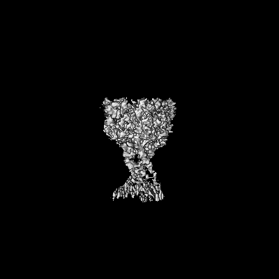
| Images |  emd_21384.png emd_21384.png | 34.3 KB | ||
| Others | emd_21384_additional_1.map.gz | 230.1 MB | ||
| Archive directory |  http://ftp.pdbj.org/pub/emdb/structures/EMD-21384ftp://ftp.pdbj.org/pub/emdb/structures/EMD-21384 http://ftp.pdbj.org/pub/emdb/structures/EMD-21384ftp://ftp.pdbj.org/pub/emdb/structures/EMD-21384 | HTTPS FTP |
-Related structure data
| Related structure data | C: citing same article ( |
|---|---|
| Similar structure data |


-Links
| EMDB pages | EMDB (EBI/PDBe) / EMDataResource |
|---|
-Map
| File | Download / File: emd_21384.map.gz / Format: CCP4 / Size: 244.1 MB / Type: IMAGE STORED AS FLOATING POINT NUMBER (4 BYTES) | ||||||||||||||||||||||||||||||||||||||||||||||||||||||||||||
|---|---|---|---|---|---|---|---|---|---|---|---|---|---|---|---|---|---|---|---|---|---|---|---|---|---|---|---|---|---|---|---|---|---|---|---|---|---|---|---|---|---|---|---|---|---|---|---|---|---|---|---|---|---|---|---|---|---|---|---|---|---|
| Annotation | Full map of an acid-sensing ion channel in a desensitized state after photo-uncaging protons. | ||||||||||||||||||||||||||||||||||||||||||||||||||||||||||||
| Projections & slices | Image control
Images are generated by Spider. | ||||||||||||||||||||||||||||||||||||||||||||||||||||||||||||
| Voxel size | X=Y=Z: 0.83 Å | ||||||||||||||||||||||||||||||||||||||||||||||||||||||||||||
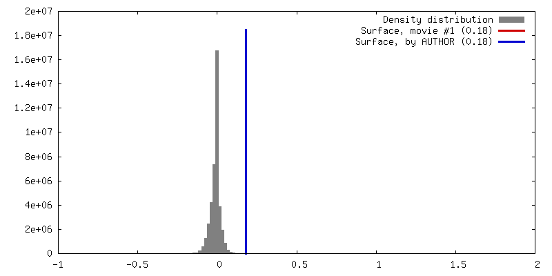
| Density |
| ||||||||||||||||||||||||||||||||||||||||||||||||||||||||||||
| Symmetry | Space group: 1 | ||||||||||||||||||||||||||||||||||||||||||||||||||||||||||||
| Details | EMDB XML:
CCP4 map header:
| ||||||||||||||||||||||||||||||||||||||||||||||||||||||||||||
 Z (Sec.)
 Y (Row.)
 X (Col.)


-Supplemental data
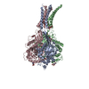
-Additional map: A map of the extracellular domain of an...
| File | emd_21384_additional_1.map | ||||||||||||
|---|---|---|---|---|---|---|---|---|---|---|---|---|---|
| Annotation | A map of the extracellular domain of an acid-sensing ion channel in a proton-bound conformation after photo-uncaging protons. | ||||||||||||
| Projections & Slices |
| ||||||||||||
| Density Histograms |


- Sample components
-Entire : Acid-sensing ion channel
| Entire | Name: Acid-sensing ion channel |
|---|---|
| Components |
|
-Supramolecule #1: Acid-sensing ion channel
| Supramolecule | Name: Acid-sensing ion channel / type: complex / ID: 1 / Parent: 0 / Macromolecule list: all / Details: Map of the entire channel. |
|---|---|
| Source (natural) | Organism: |
| Recombinant expression | Organism:  Homo sapiens (human) Homo sapiens (human) |
| Molecular weight | Theoretical: 180 KDa |
-Macromolecule #1: Acid-sensing ion channel
| Macromolecule | Name: Acid-sensing ion channel / type: protein_or_peptide / ID: 1 / Enantiomer: LEVO |
|---|---|
| Source (natural) | Organism: |
| Recombinant expression | Organism: Homo sapiens (human) |
| Sequence | String: MMDLKVDEEE VDSGQPVSIQ AFASSSTLHG ISHIFSYERL SLKRVVWALC FMGSLALLAL VCTNRIQYY FLYPHVTKLD EVAATRLTFP AVTFCNLNEF RFSRVTKNDL YHAGELLALL N NRYEIPDT QTADEKQLEI LQDKANFRNF KPKPFNMLEF YDRAGHDIRE ...String: MMDLKVDEEE VDSGQPVSIQ AFASSSTLHG ISHIFSYERL SLKRVVWALC FMGSLALLAL VCTNRIQYY FLYPHVTKLD EVAATRLTFP AVTFCNLNEF RFSRVTKNDL YHAGELLALL N NRYEIPDT QTADEKQLEI LQDKANFRNF KPKPFNMLEF YDRAGHDIRE MLLSCFFRGE QC SPEDFKV VFTRYGKCYT FNAGQDGKPR LITMKGGTGN GLEIMLDIQQ DEYLPVWGET DET SFEAGI KVQIHSQDEP PLIDQLGFGV APGFQTFVSC QEQRLIYLPP PWGDCKATTG DSEF YDTYS ITACRIDCET RYLVENCNCR MVHMPGDAPY CTPEQYKECA DPALDFLVEK DNEYC VCEM PCNVTRYGKE LSMVKIPSKA SAKYLAKKYN KSEQYIGENI LVLDIFFEAL NYETIE QKK AYEVAGLLGD IGGQMGLFIG ASILTVLELF DYAYEVIKHR LCRRGKCRKN HKRNNTD KG VALSMDDVKR HNPCESLRGH PAGMTYAANI LPHHPARGTF EDFTC |
-Experimental details
-Structure determination
| Method | cryo EM |
|---|---|
 Processing Processing | single particle reconstruction |
| Aggregation state | particle |
-Sample preparation
| Concentration | 1.00 mg/mL |
|---|---|
| Buffer | pH: 8 |
| Grid | Model: Quantifoil R2/1 / Material: GOLD / Mesh: 200 / Support film - Material: CARBON / Support film - topology: HOLEY / Pretreatment - Type: GLOW DISCHARGE / Pretreatment - Atmosphere: AIR |
| Vitrification | Cryogen name: ETHANE-PROPANE / Chamber humidity: 70 % / Chamber temperature: 277.15 K / Instrument: HOMEMADE PLUNGER Details: Specimen was frozen on a custom-made manual plunge apparatus in a cold room environment maintained at high humidity. Vitrification occurred following 25 ms of exposure to high power UV irradiation.. |
- Electron microscopy
| Microscope | FEI TITAN KRIOS |
|---|---|
| Image recording | Film or detector model: GATAN K3 (6k x 4k) / Detector mode: SUPER-RESOLUTION / Average electron dose: 50.0 e/Å2 |
| Electron beam | Acceleration voltage: 300 kV / Electron source:  FIELD EMISSION GUN FIELD EMISSION GUN |
| Electron optics | Illumination mode: FLOOD BEAM / Imaging mode: BRIGHT FIELD |
| Experimental equipment |  Model: Titan Krios / Image courtesy: FEI Company |
