 ムービー
ムービー コントローラー


+ データを開く

- 基本情報
基本情報
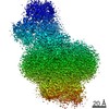
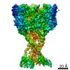
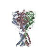
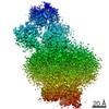
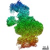
| 登録情報 | データベース: PDB / ID: 6l6i | ||||||
|---|---|---|---|---|---|---|---|
| タイトル | hASIC1a co-crystallized with Mamb-1 | ||||||
 要素 要素 | Acid-sensing ion channel 1 | ||||||
 キーワード キーワード | TRANSPORT PROTEIN / Ion channel / complex | ||||||
| 機能・相同性 |  機能・相同性情報 機能・相同性情報monoatomic ion-gated channel activity / sensory perception of sour taste / pH-gated monoatomic ion channel activity / cellular response to pH / negative regulation of neurotransmitter secretion / neurotransmitter secretion / response to acidic pH / ligand-gated sodium channel activity / sodium ion transport / associative learning ...monoatomic ion-gated channel activity / sensory perception of sour taste / pH-gated monoatomic ion channel activity / cellular response to pH / negative regulation of neurotransmitter secretion / neurotransmitter secretion / response to acidic pH / ligand-gated sodium channel activity / sodium ion transport / associative learning / regulation of postsynapse assembly / behavioral fear response / sodium ion transmembrane transport / response to amphetamine / regulation of membrane potential / postsynaptic density membrane / calcium ion transmembrane transport / Stimuli-sensing channels / memory / presynapse / dendrite / glutamatergic synapse / cell surface / Golgi apparatus / plasma membrane 類似検索 - 分子機能 | ||||||
| 生物種 |  Homo sapiens (ヒト) Homo sapiens (ヒト) | ||||||
| 手法 |  X線回折 / シンクロトロン / 分子置換 / 解像度: 3.24 Å X線回折 / シンクロトロン / 分子置換 / 解像度: 3.24 Å | ||||||
 データ登録者 データ登録者 | Lei, F. / Jian, S. | ||||||
| 資金援助 |  中国, 1件 中国, 1件
| ||||||
 引用 引用 | ジャーナル: To Be Published タイトル: hASIC1a co-crystallized with Mamb-1 著者: Lei, F. / Jian, S. | ||||||
| 履歴 |
|
- 構造の表示
構造の表示
| 構造ビューア | 分子: MolmilJmol/JSmol |
|---|
- ダウンロードとリンク
-ダウンロード
| PDBx/mmCIF形式 | 6l6i.cif.gz | 266.7 KB | 表示 | PDBx/mmCIF形式 |
|---|---|---|---|---|
| PDB形式 | pdb6l6i.ent.gz | 214.8 KB | 表示 | PDB形式 |
| PDBx/mmJSON形式 | 6l6i.json.gz | ツリー表示 | PDBx/mmJSON形式 | |
| その他 |  その他のダウンロード その他のダウンロード |
-検証レポート
| アーカイブディレクトリ | https://data.pdbj.org/pub/pdb/validation_reports/l6/6l6iftp://data.pdbj.org/pub/pdb/validation_reports/l6/6l6i | HTTPS FTP |
|---|
-関連構造データ


-リンク
 PDBj
PDBj- 集合体
集合体
| 登録構造単位 | 
| |||||||||||||||||||||||||||||||||||||||||||||||||||||||||||||||||||||||||||||||||||||||||||||||
|---|---|---|---|---|---|---|---|---|---|---|---|---|---|---|---|---|---|---|---|---|---|---|---|---|---|---|---|---|---|---|---|---|---|---|---|---|---|---|---|---|---|---|---|---|---|---|---|---|---|---|---|---|---|---|---|---|---|---|---|---|---|---|---|---|---|---|---|---|---|---|---|---|---|---|---|---|---|---|---|---|---|---|---|---|---|---|---|---|---|---|---|---|---|---|---|---|
| 1 |
| |||||||||||||||||||||||||||||||||||||||||||||||||||||||||||||||||||||||||||||||||||||||||||||||
| 単位格子 |
| |||||||||||||||||||||||||||||||||||||||||||||||||||||||||||||||||||||||||||||||||||||||||||||||
| 非結晶学的対称性 (NCS) | NCSドメイン:
NCSドメイン領域: Component-ID: _ / Refine code: _
NCSアンサンブル:
|
-要素
| #1: タンパク質 | 分子量: 51701.871 Da / 分子数: 3 / 由来タイプ: 組換発現 / 由来: (組換発現) Homo sapiens (ヒト) / 遺伝子: ASIC1, ACCN2, BNAC2発現宿主:   Spodoptera frugiperda (ツマジロクサヨトウ) Spodoptera frugiperda (ツマジロクサヨトウ)参照: UniProt: P78348 #2: 糖 |   タイプ: D-saccharide, beta linking / 分子量: 221.208 Da / 分子数: 3 / 由来タイプ: 組換発現 / 式: C8H15NO6 タイプ: D-saccharide, beta linking / 分子量: 221.208 Da / 分子数: 3 / 由来タイプ: 組換発現 / 式: C8H15NO6#3: 水 | ChemComp-HOH / |  分子量: 18.015 Da / 分子数: 9 / 由来タイプ: 天然 / 式: H2O 分子量: 18.015 Da / 分子数: 9 / 由来タイプ: 天然 / 式: H2O研究の焦点であるリガンドがあるか | N | Has protein modification | Y | |
|---|
-実験情報
-実験
| 実験 | 手法: X線回折 / 使用した結晶の数: 1 |
|---|
- 試料調製
試料調製
| 結晶 | マシュー密度: 3.97 Å3/Da / 溶媒含有率: 69.05 % |
|---|---|
| 結晶化 | 温度: 293 K / 手法: 蒸気拡散法, ハンギングドロップ法 / 詳細: 0.2M magnesium formate dehydrate, 14% PEG3350 |
-データ収集
| 回折 | 平均測定温度: 100 K / Serial crystal experiment: N | ||||||||||||||||||||||||
|---|---|---|---|---|---|---|---|---|---|---|---|---|---|---|---|---|---|---|---|---|---|---|---|---|---|
| 放射光源 | 由来: シンクロトロン / サイト: SSRF / ビームライン: BL18U1 / 波長: 0.97853 Å | ||||||||||||||||||||||||
| 検出器 | タイプ: DECTRIS PILATUS3 S 6M / 検出器: PIXEL / 日付: 2018年12月2日 | ||||||||||||||||||||||||
| 放射 | プロトコル: SINGLE WAVELENGTH / 単色(M)・ラウエ(L): M / 散乱光タイプ: x-ray | ||||||||||||||||||||||||
| 放射波長 | 波長: 0.97853 Å / 相対比: 1 | ||||||||||||||||||||||||
| 反射 | 解像度: 3.24→49.581 Å / Num. obs: 40093 / % possible obs: 99.8 % / 冗長度: 6.5 % / CC1/2: 0.997 / Rmerge(I) obs: 0.166 / Rpim(I) all: 0.07 / Rrim(I) all: 0.181 / Net I/σ(I): 9.7 | ||||||||||||||||||||||||
| 反射 シェル | Diffraction-ID: 1
|
- 解析
解析
| ソフトウェア |
| ||||||||||||||||||||||||||||||||||||||||||||||||||||||||||||
|---|---|---|---|---|---|---|---|---|---|---|---|---|---|---|---|---|---|---|---|---|---|---|---|---|---|---|---|---|---|---|---|---|---|---|---|---|---|---|---|---|---|---|---|---|---|---|---|---|---|---|---|---|---|---|---|---|---|---|---|---|---|
| 精密化 | 構造決定の手法: 分子置換 開始モデル: 3S3W 解像度: 3.24→49.581 Å / Cor.coef. Fo:Fc: 0.913 / Cor.coef. Fo:Fc free: 0.902 / SU B: 23.362 / SU ML: 0.365 / 交差検証法: THROUGHOUT / σ(F): 0 / ESU R Free: 0.43 詳細: HYDROGENS HAVE BEEN ADDED IN THE RIDING POSITIONS U VALUES : REFINED INDIVIDUALLY
| ||||||||||||||||||||||||||||||||||||||||||||||||||||||||||||
| 溶媒の処理 | イオンプローブ半径: 0.8 Å / 減衰半径: 0.8 Å / VDWプローブ半径: 1.2 Å | ||||||||||||||||||||||||||||||||||||||||||||||||||||||||||||
| 原子変位パラメータ | Biso max: 276.02 Å2 / Biso mean: 94.465 Å2 / Biso min: 27.98 Å2
| ||||||||||||||||||||||||||||||||||||||||||||||||||||||||||||
| 精密化ステップ | サイクル: final / 解像度: 3.24→49.581 Å
| ||||||||||||||||||||||||||||||||||||||||||||||||||||||||||||
| 拘束条件 |
| ||||||||||||||||||||||||||||||||||||||||||||||||||||||||||||
| Refine LS restraints NCS | Refine-ID: X-RAY DIFFRACTION / タイプ: interatomic distance / Rms dev position: 0.13 Å / Weight position: 0.05
| ||||||||||||||||||||||||||||||||||||||||||||||||||||||||||||
| LS精密化 シェル | 解像度: 3.24→3.324 Å / Rfactor Rfree error: 0
|
