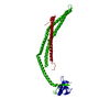
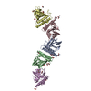
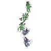
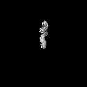
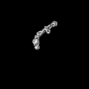
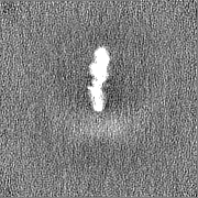
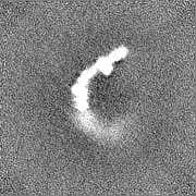
Journal: J Cell Biol / Year: 2020 Title: ULK complex organization in autophagy by a C-shaped FIP200 N-terminal domain dimer. Authors: Xiaoshan Shi / Adam L Yokom / Chunxin Wang / Lindsey N Young / Richard J Youle / James H Hurley / Abstract: The autophagy-initiating human ULK complex consists of the kinase ULK1/2, FIP200, ATG13, and ATG101. Hydrogen-deuterium exchange mass spectrometry was used to map their mutual interactions. The N- ...The autophagy-initiating human ULK complex consists of the kinase ULK1/2, FIP200, ATG13, and ATG101. Hydrogen-deuterium exchange mass spectrometry was used to map their mutual interactions. The N-terminal 640 residues (NTD) of FIP200 interact with the C-terminal IDR of ATG13. Mutations in these regions abolish their interaction. Negative stain EM and multiangle light scattering showed that FIP200 is a dimer, while a single molecule each of the other subunits is present. The FIP200NTD is flexible in the absence of ATG13, but in its presence adopts the shape of the letter C ∼20 nm across. The ULK1 EAT domain interacts loosely with the NTD dimer, while the ATG13:ATG101 HORMA dimer does not contact the NTD. Cryo-EM of the NTD dimer revealed a structural similarity to the scaffold domain of TBK1, suggesting an evolutionary similarity between the autophagy-initiating TBK1 kinase and the ULK1 kinase complex.
History
Deposition
Feb 4, 2020
-
Header (metadata) release
Mar 18, 2020
-
Map release
Jun 24, 2020
-
Update
Dec 2, 2020
-
Current status
Dec 2, 2020
Processing site: RCSB / Status: Released
-
Structure visualization
Movie
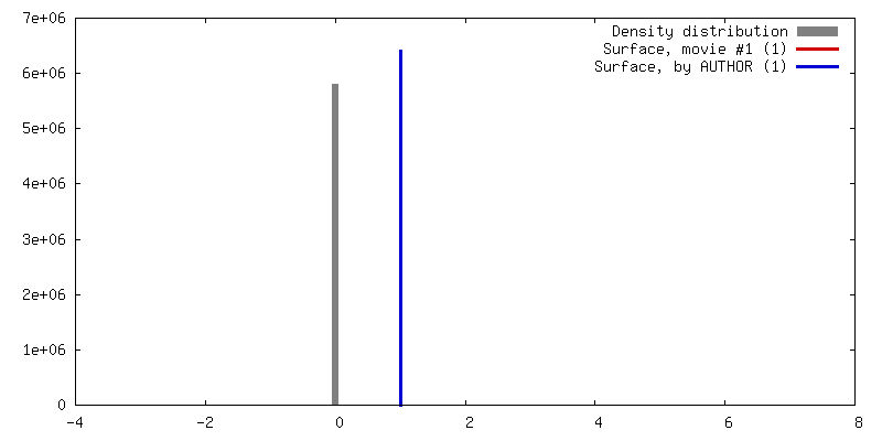
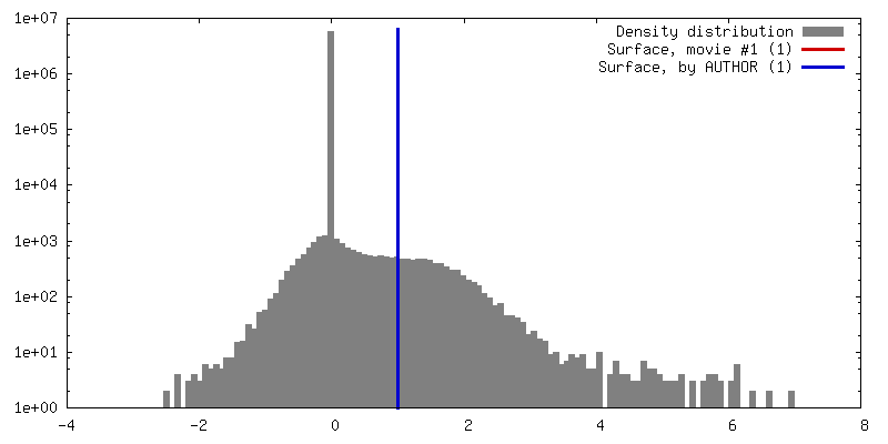
Surface view with section colored by density value
In the structure databanks used in Yorodumi, some data are registered as the other names, "COVID-19 virus" and "2019-nCoV". Here are the details of the virus and the list of structure data.
Jan 31, 2019. EMDB accession codes are about to change! (news from PDBe EMDB page)
EMDB accession codes are about to change! (news from PDBe EMDB page)
The allocation of 4 digits for EMDB accession codes will soon come to an end. Whilst these codes will remain in use, new EMDB accession codes will include an additional digit and will expand incrementally as the available range of codes is exhausted. The current 4-digit format prefixed with “EMD-” (i.e. EMD-XXXX) will advance to a 5-digit format (i.e. EMD-XXXXX), and so on. It is currently estimated that the 4-digit codes will be depleted around Spring 2019, at which point the 5-digit format will come into force.
The EM Navigator/Yorodumi systems omit the EMD- prefix.
Related info.:Q: What is EMD? / ID/Accession-code notation in Yorodumi/EM Navigator
Yorodumi is a browser for structure data from EMDB, PDB, SASBDB, etc.
This page is also the successor to EM Navigator detail page, and also detail information page/front-end page for Omokage search.
The word "yorodu" (or yorozu) is an old Japanese word meaning "ten thousand". "mi" (miru) is to see.
Related info.:EMDB / PDB / SASBDB / Comparison of 3 databanks / Yorodumi Search / Aug 31, 2016. New EM Navigator & Yorodumi / Yorodumi Papers / Jmol/JSmol / Function and homology information / Changes in new EM Navigator and Yorodumi
 Movie
Movie Controller


 Open data
 Basic information
 Map data
 Sample
 Function and homology information
 Homo sapiens (human)
 Authors
 Citation
 Structure visualization
 Downloads & links
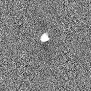
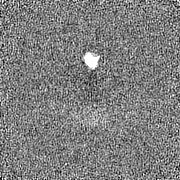
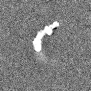
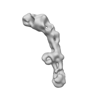 emd_21325.png
 http://ftp.pdbj.org/pub/emdb/structures/EMD-21325

 Z (Sec.)
 Y (Row.)
 X (Col.)


 Sample components
 Processing
 Electron microscopy
 FIELD EMISSION GUN
