 Movie
Movie Controller


+ Open data

- Basic information
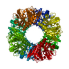
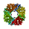
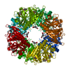
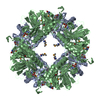
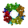
| Entry | Database: PDB / ID: 2wl9 | ||||||
|---|---|---|---|---|---|---|---|
| Title | Crystal structure of catechol 2,3-dioxygenase | ||||||
 Components Components | CATECHOL 2,3-DIOXYGENASE | ||||||
 Keywords Keywords | OXIDOREDUCTASE / AROMATIC HYDROCARBONS CATABOLISM / IRON | ||||||
| Function / homology |  Function and homology information Function and homology informationdioxygenase activity / xenobiotic catabolic process / ferrous iron binding Similarity search - Function | ||||||
| Biological species |  RHODOCOCCUS SP. DK17 (bacteria) RHODOCOCCUS SP. DK17 (bacteria) | ||||||
| Method |  X-RAY DIFFRACTION / SYNCHROTRON / MOLECULAR REPLACEMENT / Resolution: 1.9 Å X-RAY DIFFRACTION / SYNCHROTRON / MOLECULAR REPLACEMENT / Resolution: 1.9 Å | ||||||
 Authors Authors | Cho, H.J. / Kim, K.J. / Kang, B.S. | ||||||
 Citation Citation | Journal: J.Biol.Chem. / Year: 2010 Title: Substrate-Binding Mechanism of a Type I Extradiol Dioxygenase. Authors: Cho, H.J. / Kim, K. / Sohn, S.Y. / Cho, H.Y. / Kim, K.J. / Kim, M.H. / Kim, D. / Kim, E. / Kang, B.S. | ||||||
| History |
|
- Structure visualization
| Structure viewer | Molecule: MolmilJmol/JSmol |
|---|
- Downloads & links
-Download
| PDBx/mmCIF format | 2wl9.cif.gz | 452.8 KB | Display | PDBx/mmCIF format |
|---|---|---|---|---|
| PDB format | pdb2wl9.ent.gz | 376 KB | Display | PDB format |
| PDBx/mmJSON format | 2wl9.json.gz | Tree view | PDBx/mmJSON format | |
| Others |  Other downloads Other downloads |
-Validation report
| Arichive directory | https://data.pdbj.org/pub/pdb/validation_reports/wl/2wl9ftp://data.pdbj.org/pub/pdb/validation_reports/wl/2wl9 | HTTPS FTP |
|---|
-Related structure data
| Related structure data |  2wl3SC S: Starting model for refinement C: citing same article ( |
|---|---|
| Similar structure data |


-Links
 PDBj
PDBj- Assembly
| Deposited unit | 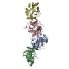
| ||||||||
|---|---|---|---|---|---|---|---|---|---|
| 1 | 
| ||||||||
| 2 | 
| ||||||||
| Unit cell |
|
-Components
-Protein , 1 types, 4 molecules ABCD
| #1: Protein | Mass: 34068.184 Da / Num. of mol.: 4 Source method: isolated from a genetically manipulated source Source: (gene. exp.) RHODOCOCCUS SP. DK17 (bacteria) / Production host: |
|---|
-Non-polymers , 5 types, 312 molecules 


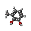

| #2: Chemical | ChemComp-FE /  Mass: 55.845 Da / Num. of mol.: 4 / Source method: obtained synthetically / Formula: Fe Mass: 55.845 Da / Num. of mol.: 4 / Source method: obtained synthetically / Formula: Fe#3: Chemical | ChemComp-MG / |  Mass: 24.305 Da / Num. of mol.: 1 / Source method: obtained synthetically / Formula: Mg Mass: 24.305 Da / Num. of mol.: 1 / Source method: obtained synthetically / Formula: Mg#4: Chemical |  Mass: 92.094 Da / Num. of mol.: 3 / Source method: obtained synthetically / Formula: C3H8O3 Mass: 92.094 Da / Num. of mol.: 3 / Source method: obtained synthetically / Formula: C3H8O3#5: Chemical |  Mass: 124.137 Da / Num. of mol.: 2 / Source method: obtained synthetically / Formula: C7H8O2 Mass: 124.137 Da / Num. of mol.: 2 / Source method: obtained synthetically / Formula: C7H8O2#6: Water | ChemComp-HOH / | Mass: 18.015 Da / Num. of mol.: 302 / Source method: isolated from a natural source / Formula: H2O |
|---|
-Experimental details
-Experiment
| Experiment | Method: X-RAY DIFFRACTION |
|---|
- Sample preparation
| Crystal | Density Matthews: 2.74 Å3/Da / Density % sol: 55 % / Description: NONE |
|---|---|
| Crystal grow | Details: 30% PEG 400. 0.1M HEPES PH7.5, 0.2M MAGNESIUM CHLORIDE |
-Data collection
| Diffraction | Mean temperature: 294 K | |||||||||||||||
|---|---|---|---|---|---|---|---|---|---|---|---|---|---|---|---|---|
| Diffraction source | Source: SYNCHROTRON / Site: APS  / Type: APS / Wavelength: 1 / Type: APS / Wavelength: 1 | |||||||||||||||
| Detector | Date: Aug 12, 2005 | |||||||||||||||
| Radiation | Protocol: SINGLE WAVELENGTH / Monochromatic (M) / Laue (L): M / Scattering type: x-ray | |||||||||||||||
| Radiation wavelength | Wavelength: 1 Å / Relative weight: 1 | |||||||||||||||
| Reflection twin |
| |||||||||||||||
| Reflection | Resolution: 1.9→40 Å / Num. obs: 114402 / % possible obs: 99.2 % / Observed criterion σ(I): 1.9 / Redundancy: 3.7 % / Rmerge(I) obs: 0.06 / Net I/σ(I): 23.7 | |||||||||||||||
| Reflection shell | Resolution: 1.9→1.97 Å / Redundancy: 3.6 % / Rmerge(I) obs: 0.33 / Mean I/σ(I) obs: 3.6 / % possible all: 98.9 |
- Processing
| Software |
| ||||||||||||||||||||||||||||||||||||||||||||||||||||||||||||||||||||||||||||||||||||||||||||||||||||||||||||||||||||||||||||||||||||||||||||||||||||||||||||||||||||||||||||||||||||||
|---|---|---|---|---|---|---|---|---|---|---|---|---|---|---|---|---|---|---|---|---|---|---|---|---|---|---|---|---|---|---|---|---|---|---|---|---|---|---|---|---|---|---|---|---|---|---|---|---|---|---|---|---|---|---|---|---|---|---|---|---|---|---|---|---|---|---|---|---|---|---|---|---|---|---|---|---|---|---|---|---|---|---|---|---|---|---|---|---|---|---|---|---|---|---|---|---|---|---|---|---|---|---|---|---|---|---|---|---|---|---|---|---|---|---|---|---|---|---|---|---|---|---|---|---|---|---|---|---|---|---|---|---|---|---|---|---|---|---|---|---|---|---|---|---|---|---|---|---|---|---|---|---|---|---|---|---|---|---|---|---|---|---|---|---|---|---|---|---|---|---|---|---|---|---|---|---|---|---|---|---|---|---|---|
| Refinement | Method to determine structure: MOLECULAR REPLACEMENT Starting model: PDB ENTRY 2WL3 Resolution: 1.9→142.17 Å / Cor.coef. Fo:Fc: 0.962 / Cor.coef. Fo:Fc free: 0.951 / SU B: 4.922 / SU ML: 0.063 / Cross valid method: THROUGHOUT / ESU R: 0.023 / ESU R Free: 0.022 / Stereochemistry target values: MAXIMUM LIKELIHOOD Details: HYDROGENS HAVE BEEN ADDED IN THE RIDING POSITIONS. CHAIN B AND D RESIDUES 178-183 ARE DISORDERD
| ||||||||||||||||||||||||||||||||||||||||||||||||||||||||||||||||||||||||||||||||||||||||||||||||||||||||||||||||||||||||||||||||||||||||||||||||||||||||||||||||||||||||||||||||||||||
| Solvent computation | Ion probe radii: 0.8 Å / Shrinkage radii: 0.8 Å / VDW probe radii: 1.4 Å / Solvent model: MASK | ||||||||||||||||||||||||||||||||||||||||||||||||||||||||||||||||||||||||||||||||||||||||||||||||||||||||||||||||||||||||||||||||||||||||||||||||||||||||||||||||||||||||||||||||||||||
| Displacement parameters | Biso mean: 18.827 Å2
| ||||||||||||||||||||||||||||||||||||||||||||||||||||||||||||||||||||||||||||||||||||||||||||||||||||||||||||||||||||||||||||||||||||||||||||||||||||||||||||||||||||||||||||||||||||||
| Refinement step | Cycle: LAST / Resolution: 1.9→142.17 Å
| ||||||||||||||||||||||||||||||||||||||||||||||||||||||||||||||||||||||||||||||||||||||||||||||||||||||||||||||||||||||||||||||||||||||||||||||||||||||||||||||||||||||||||||||||||||||
| Refine LS restraints |
|