National Institutes of Health/National Institute of General Medical Sciences (NIH/NIGMS)
R01 GM29169
United States
National Institutes of Health/National Institute of General Medical Sciences (NIH/NIGMS)
HL68744
United States
National Institutes of Health/National Institute of General Medical Sciences (NIH/NIGMS)
GM64779
United States
National Institutes of Health/National Institute of General Medical Sciences (NIH/NIGMS)
CA098131
United States
National Institutes of Health/National Institute of General Medical Sciences (NIH/NIGMS)
ES11993
United States
Citation
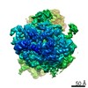
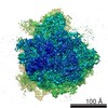
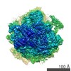
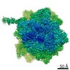
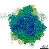
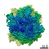
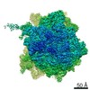
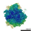
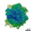
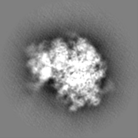
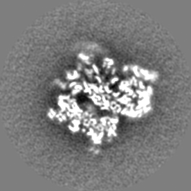
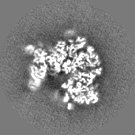
Journal: Proteomics / Year: 2021 Title: A Time-Resolved Cryo-EM Study of Saccharomyces cerevisiae 80S Ribosome Protein Composition in Response to a Change in Carbon Source. Authors: Ming Sun / Bingxin Shen / Wen Li / Parimal Samir / Christopher M Browne / Andrew J Link / Joachim Frank / Abstract: The role of the ribosome in the regulation of gene expression has come into increased focus. It is proposed that ribosomes are catalytic engines capable of changing their protein composition in ...The role of the ribosome in the regulation of gene expression has come into increased focus. It is proposed that ribosomes are catalytic engines capable of changing their protein composition in response to environmental stimuli. Time-resolved cryo-electron microscopy (cryo-EM) techniques are employed to identify quantitative changes in the protein composition and structure of the Saccharomyces cerevisiae 80S ribosomes after shifting the carbon source from glucose to glycerol. Using cryo-EM combined with the computational classification approach, it is found that a fraction of the yeast cells' 80S ribosomes lack ribosomal proteins at the entrance and exit sites for tRNAs, including uL16(RPL10), eS1(RPS1), uS11(RPS14A/B), and eS26(RPS26A/B). This fraction increased after a change from glucose to glycerol medium. The quantitative structural analysis supports the hypothesis that ribosomes are dynamic complexes that alter their composition in response to changes in growth or environmental conditions.
History
Deposition
Jan 16, 2020
-
Header (metadata) release
Feb 19, 2020
-
Map release
Oct 14, 2020
-
Update
Jan 20, 2021
-
Current status
Jan 20, 2021
Processing site: RCSB / Status: Released
-
Structure visualization
Movie
Surface view with section colored by density value
In the structure databanks used in Yorodumi, some data are registered as the other names, "COVID-19 virus" and "2019-nCoV". Here are the details of the virus and the list of structure data.
Jan 31, 2019. EMDB accession codes are about to change! (news from PDBe EMDB page)
EMDB accession codes are about to change! (news from PDBe EMDB page)
The allocation of 4 digits for EMDB accession codes will soon come to an end. Whilst these codes will remain in use, new EMDB accession codes will include an additional digit and will expand incrementally as the available range of codes is exhausted. The current 4-digit format prefixed with “EMD-” (i.e. EMD-XXXX) will advance to a 5-digit format (i.e. EMD-XXXXX), and so on. It is currently estimated that the 4-digit codes will be depleted around Spring 2019, at which point the 5-digit format will come into force.
The EM Navigator/Yorodumi systems omit the EMD- prefix.
Related info.:Q: What is EMD? / ID/Accession-code notation in Yorodumi/EM Navigator
Yorodumi is a browser for structure data from EMDB, PDB, SASBDB, etc.
This page is also the successor to EM Navigator detail page, and also detail information page/front-end page for Omokage search.
The word "yorodu" (or yorozu) is an old Japanese word meaning "ten thousand". "mi" (miru) is to see.
Related info.:EMDB / PDB / SASBDB / Comparison of 3 databanks / Yorodumi Search / Aug 31, 2016. New EM Navigator & Yorodumi / Yorodumi Papers / Jmol/JSmol / Function and homology information / Changes in new EM Navigator and Yorodumi
 Movie
Movie Controller

 Yorodumi
 Open data
 Basic information
 Map data
 Sample
 Authors
 United States, 5 items
  Citation
 Structure visualization
 Movie viewer
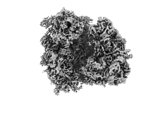
 Downloads & links
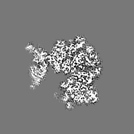
 emd_21213.png
 http://ftp.pdbj.org/pub/emdb/structures/EMD-21213


 Z (Sec.)
 Y (Row.)
 X (Col.)


 Sample components
 Processing
 Electron microscopy
 FIELD EMISSION GUN
