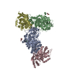
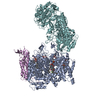
+ Open data
- Basic information
| Entry | Database: EMDB / ID: EMD-21134 | |||||||||
|---|---|---|---|---|---|---|---|---|---|---|
| Title | Structure of the Fanconi Anemia ID complex bound to ICL DNA | |||||||||
 Map data Map data | consensus reconstruction | |||||||||
 Sample Sample |
| |||||||||
 Keywords Keywords | DNA repair / DNA BINDING PROTEIN / DNA BINDING PROTEIN-DNA complex | |||||||||
| Function / homology |  Function and homology information Function and homology informationregulation of CD40 signaling pathway / gamete generation / regulation of regulatory T cell differentiation / double-strand break repair involved in meiotic recombination / homologous chromosome pairing at meiosis / neuronal stem cell population maintenance / brain morphogenesis / DNA repair complex / mitotic intra-S DNA damage checkpoint signaling / interstrand cross-link repair ...regulation of CD40 signaling pathway / gamete generation / regulation of regulatory T cell differentiation / double-strand break repair involved in meiotic recombination / homologous chromosome pairing at meiosis / neuronal stem cell population maintenance / brain morphogenesis / DNA repair complex / mitotic intra-S DNA damage checkpoint signaling / interstrand cross-link repair / condensed chromosome / DNA polymerase binding / positive regulation of protein ubiquitination / response to gamma radiation / TP53 Regulates Transcription of DNA Repair Genes / Fanconi Anemia Pathway / regulation of inflammatory response / cellular response to oxidative stress / nuclear body / DNA repair / chromatin / nucleolus / DNA binding / nucleoplasm / membrane / nucleus / cytoplasm / cytosol Similarity search - Function | |||||||||
| Biological species |  Homo sapiens (human) Homo sapiens (human) | |||||||||
| Method | single particle reconstruction / cryo EM / Resolution: 3.4 Å | |||||||||
 Authors Authors | Pavletich NP | |||||||||
| Funding support |  United States, 1 items United States, 1 items
| |||||||||
 Citation Citation |  Journal: Nature / Year: 2020 Journal: Nature / Year: 2020Title: DNA clamp function of the monoubiquitinated Fanconi anaemia ID complex. Authors: Renjing Wang / Shengliu Wang / Ankita Dhar / Christopher Peralta / Nikola P Pavletich /  Abstract: The ID complex, involving the proteins FANCI and FANCD2, is required for the repair of DNA interstrand crosslinks (ICL) and related lesions. These proteins are mutated in Fanconi anaemia, a disease ...The ID complex, involving the proteins FANCI and FANCD2, is required for the repair of DNA interstrand crosslinks (ICL) and related lesions. These proteins are mutated in Fanconi anaemia, a disease in which patients are predisposed to cancer. The Fanconi anaemia pathway of ICL repair is activated when a replication fork stalls at an ICL; this triggers monoubiquitination of the ID complex, in which one ubiquitin molecule is conjugated to each of FANCI and FANCD2. Monoubiquitination of ID is essential for ICL repair by excision, translesion synthesis and homologous recombination; however, its function remains unknown. Here we report a cryo-electron microscopy structure of the monoubiquitinated human ID complex bound to DNA, and reveal that it forms a closed ring that encircles the DNA. By comparison with the structure of the non-ubiquitinated ID complex bound to ICL DNA-which we also report here-we show that monoubiquitination triggers a complete rearrangement of the open, trough-like ID structure through the ubiquitin of one protomer binding to the other protomer in a reciprocal fashion. These structures-together with biochemical data-indicate that the monoubiquitinated ID complex loses its preference for ICL and related branched DNA structures, and becomes a sliding DNA clamp that can coordinate the subsequent repair reactions. Our findings also reveal how monoubiquitination in general can induce an alternative protein structure with a new function. | |||||||||
| History |
|
- Structure visualization
| Movie |
 Movie viewer Movie viewer |
|---|---|
| Structure viewer | EM map:  SurfView SurfView Molmil Molmil Jmol/JSmol Jmol/JSmol |
| Supplemental images |
- Downloads & links
-EMDB archive
| Map data |  emd_21134.map.gz emd_21134.map.gz | 59.9 MB |  EMDB map data format EMDB map data format | |
|---|---|---|---|---|
| Header (meta data) |  emd-21134-v30.xml emd-21134-v30.xml emd-21134.xml emd-21134.xml | 23 KB 23 KB | Display Display |  EMDB header EMDB header |
| Images |  emd_21134.png emd_21134.png | 67.4 KB | ||
| Filedesc metadata |  emd-21134.cif.gz emd-21134.cif.gz | 7.5 KB | ||
| Others |  emd_21134_additional_1.map.gz emd_21134_additional_1.map.gz emd_21134_additional_2.map.gz emd_21134_additional_2.map.gz emd_21134_additional_3.map.gz emd_21134_additional_3.map.gz | 1.5 MB 59.9 MB 59.6 MB | ||
| Archive directory |  http://ftp.pdbj.org/pub/emdb/structures/EMD-21134 http://ftp.pdbj.org/pub/emdb/structures/EMD-21134 ftp://ftp.pdbj.org/pub/emdb/structures/EMD-21134 ftp://ftp.pdbj.org/pub/emdb/structures/EMD-21134 | HTTPS FTP |
-Related structure data
| Related structure data |  6vaaMC  6vadC  6vaeC  6vafC M: atomic model generated by this map C: citing same article ( |
|---|---|
| Similar structure data |
- Links
Links
| EMDB pages |  EMDB (EBI/PDBe) / EMDB (EBI/PDBe) /  EMDataResource EMDataResource |
|---|---|
| Related items in Molecule of the Month |
- Map
Map
| File |  Download / File: emd_21134.map.gz / Format: CCP4 / Size: 64 MB / Type: IMAGE STORED AS FLOATING POINT NUMBER (4 BYTES) Download / File: emd_21134.map.gz / Format: CCP4 / Size: 64 MB / Type: IMAGE STORED AS FLOATING POINT NUMBER (4 BYTES) | ||||||||||||||||||||||||||||||||||||||||||||||||||||||||||||||||||||
|---|---|---|---|---|---|---|---|---|---|---|---|---|---|---|---|---|---|---|---|---|---|---|---|---|---|---|---|---|---|---|---|---|---|---|---|---|---|---|---|---|---|---|---|---|---|---|---|---|---|---|---|---|---|---|---|---|---|---|---|---|---|---|---|---|---|---|---|---|---|
| Annotation | consensus reconstruction | ||||||||||||||||||||||||||||||||||||||||||||||||||||||||||||||||||||
| Projections & slices | Image control
Images are generated by Spider. | ||||||||||||||||||||||||||||||||||||||||||||||||||||||||||||||||||||
| Voxel size | X=Y=Z: 1.09 Å | ||||||||||||||||||||||||||||||||||||||||||||||||||||||||||||||||||||
| Density |
| ||||||||||||||||||||||||||||||||||||||||||||||||||||||||||||||||||||
| Symmetry | Space group: 1 | ||||||||||||||||||||||||||||||||||||||||||||||||||||||||||||||||||||
| Details | EMDB XML:
CCP4 map header:
| ||||||||||||||||||||||||||||||||||||||||||||||||||||||||||||||||||||
-Supplemental data
-Additional map: refmac composite map
| File | emd_21134_additional_1.map | ||||||||||||
|---|---|---|---|---|---|---|---|---|---|---|---|---|---|
| Annotation | refmac composite map | ||||||||||||
| Projections & Slices |
| ||||||||||||
| Density Histograms |
-Additional map: Fanconi Anemia ID complex bound to ICL DNA
| File | emd_21134_additional_2.map | ||||||||||||
|---|---|---|---|---|---|---|---|---|---|---|---|---|---|
| Annotation | Fanconi Anemia ID complex bound to ICL DNA | ||||||||||||
| Projections & Slices |
| ||||||||||||
| Density Histograms |
-Additional map: Fanconi Anemia ID complex bound to ICL DNA
| File | emd_21134_additional_3.map | ||||||||||||
|---|---|---|---|---|---|---|---|---|---|---|---|---|---|
| Annotation | Fanconi Anemia ID complex bound to ICL DNA | ||||||||||||
| Projections & Slices |
| ||||||||||||
| Density Histograms |
- Sample components
-Entire : FANCI-FANCD2-DNA complex
| Entire | Name: FANCI-FANCD2-DNA complex |
|---|---|
| Components |
|
-Supramolecule #1: FANCI-FANCD2-DNA complex
| Supramolecule | Name: FANCI-FANCD2-DNA complex / type: complex / ID: 1 / Parent: 0 / Macromolecule list: all |
|---|---|
| Source (natural) | Organism:  Homo sapiens (human) Homo sapiens (human) |
-Macromolecule #1: Fanconi anemia, complementation group I
| Macromolecule | Name: Fanconi anemia, complementation group I / type: protein_or_peptide / ID: 1 / Number of copies: 1 / Enantiomer: LEVO |
|---|---|
| Source (natural) | Organism:  Homo sapiens (human) Homo sapiens (human) |
| Molecular weight | Theoretical: 149.566047 KDa |
| Recombinant expression | Organism:  |
| Sequence | String: MDQKILSLAA EKTADKLQEF LQTLREGDLT NLLQNQAVKG KVAGALLRAI FKGSPCSEEA GTLRRRKIYT CCIQLVESGD LQKEIVSEI IGLLMLEAHH FPGPLLVELA NEFISAVREG SLVNGKSLEL LPIILTVLAT KKENLAYGKG VLSGEECKKQ L INTLCSGR ...String: MDQKILSLAA EKTADKLQEF LQTLREGDLT NLLQNQAVKG KVAGALLRAI FKGSPCSEEA GTLRRRKIYT CCIQLVESGD LQKEIVSEI IGLLMLEAHH FPGPLLVELA NEFISAVREG SLVNGKSLEL LPIILTVLAT KKENLAYGKG VLSGEECKKQ L INTLCSGR WDQQYVIQLT SMFKDVPLTA EEVEFVVEKA LSMFSKMNLQ EIPPLVYQLL VLSSKGSRKS VLEGIIAFFS AL DKQHNEE QSGDELLDVV TVPSGELRHV EGTIILHIVF AIKLDYELGR ELVKHLKVGQ QGDSNNNLSP FSIALLLSVT RIQ RFQDQV LDLLKTSVVK SFKDLQLLQG SKFLQNLVPH RSYVSTMILE VVKNSVHSWD HVTQGLVELG FILMDSYGPK KVLD GKTIE TSPSLSRMPN QHACKLGANI LLETFKIHEM IRQEILEQVL NRVVTRASSP ISHFLDLLSN IVMYAPLVLQ NCSSK VTEA FDYLSFLPLQ TVQRLLKAVQ PLLKVSMSMR DCLILVLRKA MFANQLDARK SAVAGFLLLL KNFKVLGSLS SSQCSQ SLS VSQVHVDVHS HYNSVANETF CLEIMDSLRR CLSQQADVRL MLYEGFYDVL RRNSQLANSV MQTLLSQLKQ FYEPEPD LL PPLKLEACIL TQGDQISLQE PLDYLLCCIQ HCLAWYKNTV IPLQQGEEEE EEEEAFYEDL DDILESITNR MIKSELED F ELDKSADFSQ STSIGIKNNI SAFLVMGVCE VLIEYNFSIS SFSKNRFEDI LSLFMCYKKL SDILNEKAGK AKTKMANKT SDSLLSMKFV SSLLTALFRD SIQSHQESLS VLRSSNEFMR YAVNVALQKV QQLKETGHVS GPDGQNPEKI FQNLCDLTRV LLWRYTSIP TSVEESGKKE KGKSISLLCL EGLQKIFSAV QQFYQPKIQQ FLRALDVTDK EGEEREDADV SVTQRTAFQI R QFQRSLLN LLSSQEEDFN SKEALLLVTV LTSLSKLLEP SSPQFVQMLS WTSKICKENS REDALFCKSL MNLLFSLHVS YK SPVILLR DLSQDIHGHL GDIDQDVEVE KTNHFAIVNL RTAAPTVCLL VLSQAEKVLE EVDWLITKLK GQVSQETLSE EAS SQATLP NQPVEKAIIM QLGTLLTFFH ELVQTALPSG SCVDTLLKDL CKMYTTLTAL VRYYLQVCQS SGGIPKNMEK LVKL SGSHL TPLCYSFISY VQNKSKSLNY TGEKKEKPAV VATAMARVLR ETKPIPNLIF AIEQYEKFLI HLSKKSKVSL MQHMK LSTS RDFKIKGNIL DMVLREDGED ENEEGTASEH GGQNKEPAKK KRKK UniProtKB: Fanconi anemia, complementation group I |
-Macromolecule #2: Fanconi anemia group D2 protein
| Macromolecule | Name: Fanconi anemia group D2 protein / type: protein_or_peptide / ID: 2 / Number of copies: 1 / Enantiomer: LEVO |
|---|---|
| Source (natural) | Organism:  Homo sapiens (human) Homo sapiens (human) |
| Molecular weight | Theoretical: 164.314516 KDa |
| Recombinant expression | Organism:  |
| Sequence | String: MVSKRRLSKS EDKESLTEDA SKTRKQPLSK KTKKSHIANE VEENDSIFVK LLKISGIILK TGESQNQLAV DQIAFQKKLF QTLRRHPSY PKIIEEFVSG LESYIEDEDS FRNCLLSCER LQDEEASMGA SYSKSLIKLL LGIDILQPAI IKTLFEKLPE Y FFENKNSD ...String: MVSKRRLSKS EDKESLTEDA SKTRKQPLSK KTKKSHIANE VEENDSIFVK LLKISGIILK TGESQNQLAV DQIAFQKKLF QTLRRHPSY PKIIEEFVSG LESYIEDEDS FRNCLLSCER LQDEEASMGA SYSKSLIKLL LGIDILQPAI IKTLFEKLPE Y FFENKNSD EINIPRLIVS QLKWLDRVVD GKDLTTKIMQ LISIAPENLQ HDIITSLPEI LGDSQHADVG KELSDLLIEN TS LTVPILD VLSSLRLDPN FLLKVRQLVM DKLSSIRLED LPVIIKFILH SVTAMDTLEV ISELREKLDL QHCVLPSRLQ ASQ VKLKSK GRASSSGNQE SSGQSCIILL FDVIKSAIRY EKTISEAWIK AIENTASVSE HKVFDLVMLF IIYSTNTQTK KYID RVLRN KIRSGCIQEQ LLQSTFSVHY LVLKDMCSSI LSLAQSLLHS LDQSIISFGS LLYKYAFKFF DTYCQQEVVG ALVTH ICSG NEAEVDTALD VLLELVVLNP SAMMMNAVFV KGILDYLDNI SPQQIRKLFY VLSTLAFSKQ NEASSHIQDD MHLVIR KQL SSTVFKYKLI GIIGAVTMAG IMAADRSESP SLTQERANLS DEQCTQVTSL LQLVHSCSEQ SPQASALYYD EFANLIQ HE KLDPKALEWV GQTICNDFQD AFVVDSCVVP EGDFPFPVKA LYGLEEYDTQ NGIAINLLPL LFSQDFAKDG GPVTSQES G QKLVSPLCLA PYFRLLRLCV ERQHNGNLEE IDGLLDCPIF LTDLEPGEKL ESMSAKERSF MCSLIFLTLN WFREIVNAF CQETSPEMKG KVLTRLKHIV ELQIILEKYL AVTPDYVPPL GNFDVETLDI TPHTVTAISA KIRKKGKIER KQKTDGSKTS SSDTLSEEK NSECDPTPSH RGQLNKEFTG KEEKTSLLLH NSHAFFRELD IEVFSILHCG LVTKFILDTE MHTEATEVVQ L GPPELLFL LEDLSQKLES MLTPPIARRV PFLKNKGSRN IGFSHLQQRS AQEIVHCVFQ LLTPMCNHLE NIHNYFQCLA AE NHGVVDG PGVKVQEYHI MSSCYQRLLQ IFHGLFAWSG FSQPENQNLL YSALHVLSSR LKQGEHSQPL EELLSQSVHY LQN FHQSIP SFQCALYLIR LLMVILEKST ASAQNKEKIA SLARQFLCRV WPSGDKEKSN ISNDQLHALL CIYLEHTESI LKAI EEIAG VGVPELINSP KDASSSTFPT LTRHTFVVFF RVMMAELEKT VKKIEPGTAA DSQQIHEEKL LYWNMAVRDF SILIN LIKV FDSHPVLHVC LKYGRLFVEA FLKQCMPLLD FSFRKHREDV LSLLETFQLD TRLLHHLCGH SKIHQDTRLT QHVPLL KKT LELLVCRVKA MLTLNNCREA FWLGNLKNRD LQGEEIKSQN SQESTADESE DDMSSQASKS KATEDGEEDE VSAGEKE QD SDESYDDSD UniProtKB: Fanconi anemia group D2 protein |
-Macromolecule #3: DNA (26-MER)
| Macromolecule | Name: DNA (26-MER) / type: dna / ID: 3 / Number of copies: 1 / Classification: DNA |
|---|---|
| Source (natural) | Organism:  Homo sapiens (human) Homo sapiens (human) |
| Molecular weight | Theoretical: 8.411627 KDa |
| Sequence | String: (DA)(DA)(DA)(DA)(DA)(DA)(DA)(DA)(DA)(DA) (DA)(DA)(DA)(DA)(DA)(DA)(DA)(DA)(DA)(DA) (DA)(DA)(DA)(DA)(DA)(DA)(DA) |
-Macromolecule #4: DNA (5'-D(P*TP*TP*TP*TP*TP*TP*TP*TP*TP*TP*TP*TP*TP*TP*TP*TP*TP*T)-3')
| Macromolecule | Name: DNA (5'-D(P*TP*TP*TP*TP*TP*TP*TP*TP*TP*TP*TP*TP*TP*TP*TP*TP*TP*T)-3') type: dna / ID: 4 / Number of copies: 1 / Classification: DNA |
|---|---|
| Source (natural) | Organism:  Homo sapiens (human) Homo sapiens (human) |
| Molecular weight | Theoretical: 5.430513 KDa |
| Sequence | String: (DT)(DT)(DT)(DT)(DT)(DT)(DT)(DT)(DT)(DT) (DT)(DT)(DT)(DT)(DT)(DT)(DT)(DT) |
-Macromolecule #5: DNA (5'-D(P*AP*AP*AP*AP*AP*AP*AP*AP*AP*AP*AP*AP*AP*AP*A)-3')
| Macromolecule | Name: DNA (5'-D(P*AP*AP*AP*AP*AP*AP*AP*AP*AP*AP*AP*AP*AP*AP*A)-3') type: dna / ID: 5 / Number of copies: 1 / Classification: DNA |
|---|---|
| Source (natural) | Organism:  Homo sapiens (human) Homo sapiens (human) |
| Molecular weight | Theoretical: 4.653145 KDa |
| Sequence | String: (DA)(DA)(DA)(DA)(DA)(DA)(DA)(DA)(DA)(DA) (DA)(DA)(DA)(DA)(DA) |
-Macromolecule #6: DNA (5'-D(*TP*TP*TP*TP*TP*TP*TP*TP*TP*TP*TP*TP*TP*TP*TP*T)-3')
| Macromolecule | Name: DNA (5'-D(*TP*TP*TP*TP*TP*TP*TP*TP*TP*TP*TP*TP*TP*TP*TP*T)-3') type: dna / ID: 6 / Number of copies: 1 / Classification: DNA |
|---|---|
| Source (natural) | Organism:  Homo sapiens (human) Homo sapiens (human) |
| Molecular weight | Theoretical: 4.822127 KDa |
| Sequence | String: (DT)(DT)(DT)(DT)(DT)(DT)(DT)(DT)(DT)(DT) (DT)(DT)(DT)(DT)(DT)(DT) |
-Experimental details
-Structure determination
| Method | cryo EM |
|---|---|
 Processing Processing | single particle reconstruction |
| Aggregation state | particle |
- Sample preparation
| Buffer | pH: 8 |
|---|---|
| Grid | Details: unspecified |
| Vitrification | Cryogen name: ETHANE |
- Electron microscopy
| Microscope | FEI TITAN KRIOS |
|---|---|
| Image recording | Film or detector model: GATAN K2 SUMMIT (4k x 4k) / Average electron dose: 51.0 e/Å2 |
| Electron beam | Acceleration voltage: 300 kV / Electron source:  FIELD EMISSION GUN FIELD EMISSION GUN |
| Electron optics | Illumination mode: FLOOD BEAM / Imaging mode: BRIGHT FIELD |
| Experimental equipment |  Model: Titan Krios / Image courtesy: FEI Company |
- Image processing
| Startup model | Type of model: PDB ENTRY |
|---|---|
| Final reconstruction | Applied symmetry - Point group: C1 (asymmetric) / Resolution.type: BY AUTHOR / Resolution: 3.4 Å / Resolution method: FSC 0.143 CUT-OFF / Number images used: 231943 |
| Initial angle assignment | Type: MAXIMUM LIKELIHOOD / Software - Name: RELION (ver. 3) |
| Final angle assignment | Type: MAXIMUM LIKELIHOOD |
 Movie
Movie Controller


 Z (Sec.)
 Y (Row.)
 X (Col.)


