ムービー
ムービー コントローラー

[日本語] English
 万見
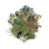
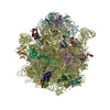
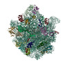
万見- EMDB-21034: Cryo-EM structure of the Acinetobacter baumannii Ribosome: 30S subunit -
+ データを開く

- 基本情報
基本情報
| 登録情報 | データベース: EMDB / ID: EMD-21034 | |||||||||
|---|---|---|---|---|---|---|---|---|---|---|
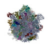
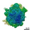
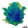
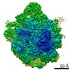
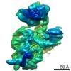
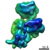
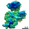
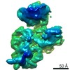
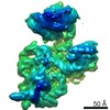
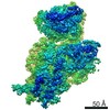
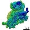
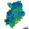
| タイトル | Cryo-EM structure of the Acinetobacter baumannii Ribosome: 30S subunit | |||||||||
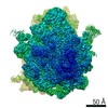
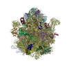
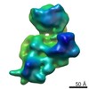
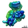
 マップデータ マップデータ | 30S composite map created with two separate masked refinements (30S head and 30S core). | |||||||||
 試料 試料 |
| |||||||||
 キーワード キーワード | Ribosome / Acinetobacter baumannii | |||||||||
| 機能・相同性 |  機能・相同性情報 機能・相同性情報ribosomal small subunit assembly / ribosome biogenesis / ribosomal small subunit biogenesis / small ribosomal subunit / small ribosomal subunit rRNA binding / cytosolic small ribosomal subunit / tRNA binding / rRNA binding / structural constituent of ribosome / ribosome ...ribosomal small subunit assembly / ribosome biogenesis / ribosomal small subunit biogenesis / small ribosomal subunit / small ribosomal subunit rRNA binding / cytosolic small ribosomal subunit / tRNA binding / rRNA binding / structural constituent of ribosome / ribosome / translation / ribonucleoprotein complex / mRNA binding / RNA binding / cytoplasm / cytosol 類似検索 - 分子機能 | |||||||||
| 生物種 |  Acinetobacter baumannii AB0057 (バクテリア) / Acinetobacter baumannii (バクテリア) / Acinetobacter baumannii (strain AB0057) (バクテリア) / Acinetobacter sp. ANC 5600 (バクテリア) / Acinetobacter sp. 263903-1 (バクテリア) / Acinetobacter venetianus (バクテリア) Acinetobacter baumannii AB0057 (バクテリア) / Acinetobacter baumannii (バクテリア) / Acinetobacter baumannii (strain AB0057) (バクテリア) / Acinetobacter sp. ANC 5600 (バクテリア) / Acinetobacter sp. 263903-1 (バクテリア) / Acinetobacter venetianus (バクテリア) | |||||||||
| 手法 | 単粒子再構成法 / クライオ電子顕微鏡法 / 解像度: 4.4 Å | |||||||||
 データ登録者 データ登録者 | Morgan CE / Yu EW | |||||||||
| 資金援助 |  米国, 1件 米国, 1件
| |||||||||
 引用 引用 | ジャーナル: mBio / 年: 2020 タイトル: Cryo-electron Microscopy Structure of the Acinetobacter baumannii 70S Ribosome and Implications for New Antibiotic Development. 著者: Christopher E Morgan / Wei Huang / Susan D Rudin / Derek J Taylor / James E Kirby / Robert A Bonomo / Edward W Yu / 要旨: Antimicrobial resistance is a major health threat as it limits treatment options for infection. At the forefront of this serious issue is , a Gram-negative opportunistic pathogen that exhibits the ...Antimicrobial resistance is a major health threat as it limits treatment options for infection. At the forefront of this serious issue is , a Gram-negative opportunistic pathogen that exhibits the remarkable ability to resist antibiotics through multiple mechanisms. As bacterial ribosomes represent a target for multiple distinct classes of existing antimicrobial agents, we here use single-particle cryo-electron microscopy (cryo-EM) to elucidate five different structural states of the ribosome, including the 70S, 50S, and 30S forms. We also determined interparticle motions of the 70S ribosome in different tRNA bound states using three-dimensional (3D) variability analysis. Together, our structural data further our understanding of the ribosome from and other Gram-negative pathogens and will enable structure-based drug discovery to combat antibiotic-resistant bacterial infections. is a severe nosocomial threat largely due to its intrinsic antibiotic resistance and remarkable ability to acquire new resistance determinants. The bacterial ribosome serves as a major target for modern antibiotics and the design of new therapeutics. Here, we present cryo-EM structures of the 70S ribosome, revealing several unique species-specific structural features that may facilitate future drug development to combat this recalcitrant bacterial pathogen. | |||||||||
| 履歴 |
|
- 構造の表示
構造の表示
| ムービー |
ムービービューア |
|---|---|
| 構造ビューア | EMマップ: SurfViewMolmilJmol/JSmol |
| 添付画像 |

- ダウンロードとリンク
-EMDBアーカイブ
| マップデータ | emd_21034.map.gz | 7.1 MB | EMDBマップデータ形式 | |
|---|---|---|---|---|
| ヘッダ (付随情報) | emd-21034-v30.xmlemd-21034.xml | 30.8 KB 30.8 KB | 表示 表示 | EMDBヘッダ |
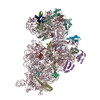
| 画像 | 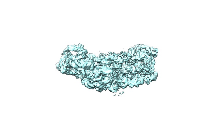 emd_21034.png emd_21034.png | 115.5 KB | ||
| Filedesc metadata | emd-21034.cif.gz | 8.4 KB | ||
| アーカイブディレクトリ |  http://ftp.pdbj.org/pub/emdb/structures/EMD-21034ftp://ftp.pdbj.org/pub/emdb/structures/EMD-21034 http://ftp.pdbj.org/pub/emdb/structures/EMD-21034ftp://ftp.pdbj.org/pub/emdb/structures/EMD-21034 | HTTPS FTP |
-関連構造データ
-リンク
| EMDBのページ | EMDB (EBI/PDBe) / EMDataResource |
|---|---|
| 「今月の分子」の関連する項目 |


-マップ
| ファイル | ダウンロード / ファイル: emd_21034.map.gz / 形式: CCP4 / 大きさ: 512 MB / タイプ: IMAGE STORED AS FLOATING POINT NUMBER (4 BYTES) | ||||||||||||||||||||||||||||||||||||||||||||||||||||||||||||||||||||
|---|---|---|---|---|---|---|---|---|---|---|---|---|---|---|---|---|---|---|---|---|---|---|---|---|---|---|---|---|---|---|---|---|---|---|---|---|---|---|---|---|---|---|---|---|---|---|---|---|---|---|---|---|---|---|---|---|---|---|---|---|---|---|---|---|---|---|---|---|---|
| 注釈 | 30S composite map created with two separate masked refinements (30S head and 30S core). | ||||||||||||||||||||||||||||||||||||||||||||||||||||||||||||||||||||
| 投影像・断面図 | 画像のコントロール
画像は Spider により作成 | ||||||||||||||||||||||||||||||||||||||||||||||||||||||||||||||||||||
| ボクセルのサイズ | X=Y=Z: 1.064 Å | ||||||||||||||||||||||||||||||||||||||||||||||||||||||||||||||||||||
| 密度 |
| ||||||||||||||||||||||||||||||||||||||||||||||||||||||||||||||||||||
| 対称性 | 空間群: 1 | ||||||||||||||||||||||||||||||||||||||||||||||||||||||||||||||||||||
| 詳細 | EMDB XML:
CCP4マップ ヘッダ情報:
| ||||||||||||||||||||||||||||||||||||||||||||||||||||||||||||||||||||
 Z (Sec.)
 Y (Row.)
 X (Col.)


-添付データ
- 試料の構成要素
+全体 : Acinetobacter baumannii Small ribosomal subunit
+超分子 #1: Acinetobacter baumannii Small ribosomal subunit
+分子 #1: 16s Ribosomal RNA
+分子 #2: 30S ribosomal protein S2
+分子 #3: 30S ribosomal protein S3
+分子 #4: 30S ribosomal protein S4
+分子 #5: 30S ribosomal protein S5
+分子 #6: 30S ribosomal protein S6
+分子 #7: 30S ribosomal protein S7
+分子 #8: 30S ribosomal protein S8
+分子 #9: 30S ribosomal protein S9
+分子 #10: 30S ribosomal protein S10
+分子 #11: 30S ribosomal protein S11
+分子 #12: 30S ribosomal protein S12
+分子 #13: 30S ribosomal protein S13
+分子 #14: 30S ribosomal protein S14
+分子 #15: 30S ribosomal protein S15
+分子 #16: 30S ribosomal protein S16
+分子 #17: 30S ribosomal protein S17
+分子 #18: 30S ribosomal protein S18
+分子 #19: 30S ribosomal protein S19
+分子 #20: 30S ribosomal protein S20
-実験情報
-構造解析
| 手法 | クライオ電子顕微鏡法 |
|---|---|
 解析 解析 | 単粒子再構成法 |
| 試料の集合状態 | particle |
-試料調製
| 緩衝液 | pH: 7.6 |
|---|---|
| 凍結 | 凍結剤: ETHANE |
- 電子顕微鏡法
| 顕微鏡 | FEI TITAN KRIOS |
|---|---|
| 撮影 | フィルム・検出器のモデル: GATAN K2 SUMMIT (4k x 4k) 検出モード: SUPER-RESOLUTION / 平均電子線量: 40.0 e/Å2 |
| 電子線 | 加速電圧: 300 kV / 電子線源:  FIELD EMISSION GUN FIELD EMISSION GUN |
| 電子光学系 | 照射モード: FLOOD BEAM / 撮影モード: BRIGHT FIELD |
| 実験機器 |  モデル: Titan Krios / 画像提供: FEI Company |